Cohen-Haguenauer O et al. (FEB 2006)
Proceedings of the National Academy of Sciences of the United States of America 103 7 2340--5
In vivo repopulation ability of genetically corrected bone marrow cells from Fanconi anemia patients.
Fanconi anemia (FA) is a rare inherited genomic instability syndrome representing one of the best examples of hematopoietic stem cell deficiency. Although FA might be an excellent candidate for bone marrow (BM) genetic correction ex vivo,knockout animal models are not sufficient to guide preclinical steps,and gene therapy attempts have proven disappointing so far. Contributing to these poor results is a characteristic and dramatic early BM-cells die-off when placed in culture. We show here that human primary FA BM cell survival can be ameliorated by using specific culture conditions that limit oxidative stress. When coupled with retrovirus-mediated transfer of the main complementation group FANCA-cDNA,we could achieve long-term reconstitution of the stem cell compartment both in vitro and in vivo. Gene-corrected BM cultures grew for textgreater120 days,and after cultured cell transplantation into NOD/SCID mice,clonogenic human cells carrying the FANCA transgene could be detected 6 months after transduction. By comparison,untransduced cells died in culture by 15 days. Of necessity for ethical reasons,experiments were conducted on a very limited number of primary BM cells. By using low cytokine regimen and conditions matching regulatory requirements,a contingent of gene-corrected cells slowly emerges with an unmet potential for in vivo engraftment. Future therapeutic applications of stem cells might be expanding from these data. In addition,we provide a model of gene-corrected human primary cell growth that carries the potential to better delineate the combined role of both DNA damage and oxidative stress in the pathogenesis of FA.
View Publication
产品类型:
产品号#:
04436
产品名:
MethoCult™ SF H4436
Xu X-L et al. (FEB 2010)
Carcinogenesis 31 2 167--74
The properties of tumor-initiating cells from a hepatocellular carcinoma patient's primary and recurrent tumor.
Hepatocellular carcinoma (HCC) is associated with a high morbidity and mortality due to its high rate of recurrence. However,little is known about the biological characteristics of recurrent HCC cells. A single patient's primary and recurrent HCC-derived cell lines,Hep-11 and Hep-12,respectively,were established by primary culture. These two cell lines have the same hepatitis B virus integration site and share many common amplifications and deletions,which suggest that they have the same clonal origin. While Hep-11 cells were non-tumorigenic at 16 weeks following injection of up to 10 000 cells,injection of only 100 Hep-12 cells was sufficient to initiate tumor growth,and all single Hep-12 clones were tumorigenic in immunodeficient mice. Compared with Hep-11,Hep-12 cells expressed the oval cell markers AFP,NCAM/CD56,c-kit/CD117,as well as multiple stem cell markers such as Nanog,OCT4 and SOX2. In addition,textgreater90% of Hep-12 cells were aldehyde dehydrogenase positive. They were also less resistant to paclitaxel,but more resistant to doxorubicin,cisplatin and hydroxycamptothecin (HCPT),which had been administrated to the patient. Furthermore,Hep-12 cells expressed higher levels of poly (adenosine diphosphate-ribose) polymerase-1 (PARP-1) than Hep-11,and PARP-1 inhibition potentiated the sensitivity to HCPT in Hep-12 cells but not in Hep-11 cells. These results indicate that a large population of the recurrent HCC-derived Hep-12 cells were tumor-initiating cells and that elevated expression of PARP-1 was related to their resistance to HCPT.
View Publication
产品类型:
产品号#:
01700
01702
01705
产品名:
ALDEFLUOR™ 试剂盒
ALDEFLUOR™测定缓冲液
ALDEFLUOR™ DEAB试剂
Kö et al. (JUL 2010)
Cancer letters 293 1 117--23
Circulating tumor cells in metastatic colorectal cancer: efficacy and feasibility of different enrichment methods.
Comprehensive in vitro and in vivo studies comparing EpCAM-based methods with other cytometric CTC enrichment technologies in metastatic colorectal cancer (mCRC) patients are lacking. We compare four manual cytometric methods to detect CTCs in vitro and in mCRC patients. The EpCAM-based technology,MACS HEA MicroBeads((R)),showed a significant better tumor cell recovery rate compared to other cytometric methods (p-valuetextless0.0001). CTCs of 38 mCRC patients were enriched with MACS HEA MicroBeads(R). Progression-free survival did significantly differ between mCRC patients without detectable and with textgreateror= 1 CTCs (p=0.007). CTC enrichment with EpCAM coupled antibodies is superior to other cytometric methods and is a feasible method for CTC detection in mCRC patients.
View Publication
产品类型:
产品号#:
产品名:
Calcagno AM et al. (NOV 2010)
Journal of the National Cancer Institute 102 21 1637--52
Prolonged drug selection of breast cancer cells and enrichment of cancer stem cell characteristics.
BACKGROUND: Cancer stem cells are presumed to have virtually unlimited proliferative and self-renewal abilities and to be highly resistant to chemotherapy,a feature that is associated with overexpression of ATP-binding cassette transporters. We investigated whether prolonged continuous selection of cells for drug resistance enriches cultures for cancer stem-like cells. METHODS: Cancer stem cells were defined as CD44+/CD24�?� cells that could self-renew (ie,generate cells with the tumorigenic CD44+/CD24�?� phenotype),differentiate,invade,and form tumors in vivo. We used doxorubicin-selected MCF-7/ADR cells,weakly tumorigenic parental MCF-7 cells,and MCF-7/MDR,an MCF-7 subline with forced expression of ABCB1 protein. Cells were examined for cell surface markers and side-population fractions by microarray and flow cytometry,with in vitro invasion assays,and for ability to form mammospheres. Xenograft tumors were generated in mice to examine tumorigenicity (n = 52). The mRNA expression of multidrug resistance genes was examined in putative cancer stem cells and pathway analysis of statistically significantly differentially expressed genes was performed. All statistical tests were two-sided. RESULTS: Pathway analysis showed that MCF-7/ADR cells express mRNAs from ABCB1 and other genes also found in breast cancer stem cells (eg,CD44,TGFB1,and SNAI1). MCF-7/ADR cells were highly invasive,formed mammospheres,and were tumorigenic in mice. In contrast to parental MCF-7 cells,more than 30% of MCF-7/ADR cells had a CD44+/CD24�?� phenotype,could self-renew,and differentiate (ie,produce CD44+/CD24�?� and CD44+/CD24+ cells) and overexpressed various multidrug resistance-linked genes (including ABCB1,CCNE1,and MMP9). MCF-7/ADR cells were statistically significantly more invasive in Matrigel than parental MCF-7 cells (MCF-7 cells = 0.82 cell per field and MCF-7/ADR = 7.51 cells per field,difference = 6.69 cells per field,95% confidence interval = 4.82 to 8.55 cells per field,P textless .001). No enrichment in the CD44+/CD24�?� or CD133+ population was detected in MCF-7/MDR. CONCLUSION: The cell population with cancer stem cell characteristics increased after prolonged continuous selection for doxorubicin resistance.
View Publication
产品类型:
产品号#:
01700
01705
01702
产品名:
ALDEFLUOR™ 试剂盒
ALDEFLUOR™ DEAB试剂
ALDEFLUOR™测定缓冲液
Cheng Y et al. ( 2013)
BMC cell biology 14 1 44
Physiological β-catenin signaling controls self-renewal networks and generation of stem-like cells from nasopharyngeal carcinoma.
BACKGROUND: A few reports suggested that low levels of Wnt signaling might drive cell reprogramming,but these studies could not establish a clear relationship between Wnt signaling and self-renewal networks. There are ongoing debates as to whether and how the Wnt/β-catenin signaling is involved in the control of pluripotency gene networks. Additionally,whether physiological β-catenin signaling generates stem-like cells through interactions with other pathways is as yet unclear. The nasopharyngeal carcinoma HONE1 cells have low expression of β-catenin and wild-type expression of p53,which provided a possibility to study regulatory mechanism of stemness networks induced by physiological levels of Wnt signaling in these cells.backslashnbackslashnRESULTS: Introduction of increased β-catenin signaling,haploid expression of β-catenin under control by its natural regulators in transferred chromosome 3,resulted in activation of Wnt/β-catenin networks and dedifferentiation in HONE1 hybrid cell lines,but not in esophageal carcinoma SLMT1 hybrid cells that had high levels of endogenous β-catenin expression. HONE1 hybrid cells displayed stem cell-like properties,including enhancement of CD24(+) and CD44(+) populations and generation of spheres that were not observed in parental HONE1 cells. Signaling cascades were detected in HONE1 hybrid cells,including activation of p53- and RB1-mediated tumor suppressor pathways,up-regulation of Nanog-,Oct4-,Sox2-,and Klf4-mediated pluripotency networks,and altered E-cadherin expression in both in vitro and in vivo assays. qPCR array analyses further revealed interactions of physiological Wnt/β-catenin signaling with other pathways such as epithelial-mesenchymal transition,TGF-β,Activin,BMPR,FGFR2,and LIFR- and IL6ST-mediated cell self-renewal networks. Using β-catenin shRNA inhibitory assays,a dominant role for β-catenin in these cellular network activities was observed. The expression of cell surface markers such as CD9,CD24,CD44,CD90,and CD133 in generated spheres was progressively up-regulated compared to HONE1 hybrid cells. Thirty-four up-regulated components of the Wnt pathway were identified in these spheres.backslashnbackslashnCONCLUSIONS: Wnt/β-catenin signaling regulates self-renewal networks and plays a central role in the control of pluripotency genes,tumor suppressive pathways and expression of cancer stem cell markers. This current study provides a novel platform to investigate the interaction of physiological Wnt/β-catenin signaling with stemness transition networks.
View Publication
Targeting NF-kappaB activation via pharmacologic inhibition of IKK2-induced apoptosis of human acute myeloid leukemia cells.
Acute myeloid leukemia (AML) cells are characterized by a constitutive and abnormal activation of the nuclear factor-kappaB (NF-kappaB) transcription factor. This study,conducted in vitro on 18 patients,shows that targeting the IKB kinase 2 (IKK2) kinase with the specific pharmacologic inhibitor AS602868 to block NF-kappaB activation led to apoptosis of human primary AML cells. Moreover,AS602868 potentiated the apoptotic response induced by the current chemotherapeutic drugs doxorubicin,cytarabine,or etoposide (VP16). AS602868-induced cell death was associated with rupture of the mitochondrial transmembrane potential and activation of cellular caspases. NF-kappaB inhibition did not affect normal CD34+ hematopoietic precursors,suggesting that it could represent a new adjuvant strategy for AML treatment.
View Publication
产品类型:
产品号#:
15026
15066
产品名:
RosetteSep™ 人造血祖细胞富集抗体混合物
RosetteSep™人造血祖细胞富集抗体混合物
Ben-David U et al. (SEP 2014)
Nature communications 5 4825
Aneuploidy induces profound changes in gene expression, proliferation and tumorigenicity of human pluripotent stem cells.
Human pluripotent stem cells (hPSCs) tend to acquire genomic aberrations in culture,the most common of which is trisomy of chromosome 12. Here we dissect the cellular and molecular implications of this trisomy in hPSCs. Global gene expression analyses reveal that trisomy 12 profoundly affects the gene expression profile of hPSCs,inducing a transcriptional programme similar to that of germ cell tumours. Comparison of proliferation,differentiation and apoptosis between diploid and aneuploid hPSCs shows that trisomy 12 significantly increases the proliferation rate of hPSCs,mainly as a consequence of increased replication. Furthermore,trisomy 12 increases the tumorigenicity of hPSCs in vivo,inducing transcriptionally distinct teratomas from which pluripotent cells can be recovered. Last,a chemical screen of 89 anticancer drugs discovers that trisomy 12 raises the sensitivity of hPSCs to several replication inhibitors. Together,these findings demonstrate the extensive effect of trisomy 12 and highlight its perils for successful hPSC applications.
View Publication
产品类型:
产品号#:
07909
85850
85857
产品名:
IV型胶原酶(1mg /mL)
mTeSR™1
mTeSR™1
Sloand EM et al. (SEP 2006)
Proceedings of the National Academy of Sciences of the United States of America 103 39 14483--8
Granulocyte colony-stimulating factor preferentially stimulates proliferation of monosomy 7 cells bearing the isoform IV receptor.
Granulocyte colony-stimulating factor (GCSF) administration has been linked to the development of monosomy 7 in severe congenital neutropenia and aplastic anemia. We assessed the effect of pharmacologic doses of GCSF on monosomy 7 cells to determine whether this chromosomal abnormality developed de novo or arose as a result of favored expansion of a preexisting clone. Fluorescence in situ hybridization (FISH) of chromosome 7 was used to identify small populations of aneuploid cells. When bone marrow mononuclear cells from patients with monosomy 7 were cultured with 400 ng/ml GCSF,all samples showed significant increases in the proportion of monosomy 7 cells. In contrast,bone marrow from karyotypically normal aplastic anemia,myelodysplastic syndrome,or healthy individuals did not show an increase in monosomy 7 cells in culture. In bone marrow CD34 cells of patients with myelodysplastic syndrome and monosomy 7,GCSF receptor (GCSFR) protein was increased. Although no mutation was found in genomic GCSFR DNA,CD34 cells showed increased expression of the GCSFR class IV mRNA isoform,which is defective in signaling cellular differentiation. GCSFR signal transduction via the Jak/Stat system was abnormal in monosomy 7 CD34 cells,with increased phosphorylated signal transducer and activation of transcription protein,STAT1-P,and increased STAT5-P relative to STAT3-P. Our results suggest that pharmacologic doses of GCSF increase the proportion of preexisting monosomy 7 cells. The abnormal response of monosomy 7 cells to GCSF would be explained by the expansion of undifferentiated monosomy 7 clones expressing the class IV GCSFR,which is defective in signaling cell maturation.
View Publication
产品类型:
产品号#:
05150
产品名:
MyeloCult™H5100
Zhang F-Q et al. ( 2015)
Oncotarget
JAK2 inhibitor TG101348 overcomes erlotinib-resistance in non-small cell lung carcinoma cells with mutated EGF receptor.
Non-small cell lung cancer (NSCLC) patients with epidermal growth factor receptor (EGFR) mutations are responsive to EGFR-tyrosine kinase inhibitor (EGFR-TKI). However,NSCLC patients with secondary somatic EGFR mutations are resistant to EGFR-TKI treatment. In this study,we investigated the effect of TG101348 (a JAK2 inhibitor) on the tumor growth of erlotinib-resistant NSCLC cells. Cell proliferation,apoptosis,gene expression and tumor growth were evaluated by diphenyltetrazolium bromide (MTT) assay,flow cytometry,terminal deoxynucleotidyl transferase biotin-dUTP nick end labeling (TUNEL) staining,Western Blot and a xenograft mouse model,respectively. Results showed that erlotinib had a stronger impact on the induction of apoptosis in erlotinib-sensitive PC-9 cells but had a weaker effect on erlotinib-resistant H1975 and H1650 cells than TG101348. TG101348 significantly enhanced the cytotoxicity of erlotinib to erlotinib-resistant NSCLC cells,stimulated erlotinib-induced apoptosis and downregulated the expressions of EGFR,p-EGFR,p-STAT3,Bcl-xL and survivin in erlotinib-resistant NSCLC cells. Moreover,the combined treatment of TG101348 and erlotinib induced apoptosis,inhibited the activation of p-EGFR and p-STAT3,and inhibited tumor growth of erlotinib-resistant NSCLC cells in vivo. Our results indicate that TG101348 is a potential adjuvant for NSCLC patients during erlotinib treatment.
View Publication
产品类型:
产品号#:
73472
73474
产品名:
TG101348
TG101348
Carpentino JE et al. (OCT 2009)
Cancer research 69 20 8208--15
Aldehyde dehydrogenase-expressing colon stem cells contribute to tumorigenesis in the transition from colitis to cancer.
Patients with chronic ulcerative colitis are at increased risk of developing colorectal cancer. Although current hypotheses suggest that sporadic colorectal cancer is due to inability to control cancer stem cells,the cancer stem cell hypothesis has not yet been validated in colitis-associated cancer. Furthermore,the identification of the colitis to cancer transition is challenging. We recently showed that epithelial cells with the increased expression of aldehyde dehydrogenase in sporadic colon cancer correlate closely with tumor-initiating ability. We sought to determine whether ALDH can be used as a marker to isolate tumor-initiating populations from patients with chronic ulcerative colitis. We used fluorescence-activated cell sorting to identify precursor colon cancer stem cells from colitis patients and report both their transition to cancerous stem cells in xenografting studies as well as their ability to generate spheres in vitro. Similar to sporadic colon cancer,these colitis-derived tumors were capable of propagation as sphere cultures. However,unlike the origins of sporadic colon cancer,the primary colitic tissues did not express any histologic evidence of dysplasia. To elucidate a potential mechanism for our findings,we compared the stroma of these different environments and determined that at least one paracrine factor is up-regulated in the inflammatory and malignant stroma compared with resting,normal stroma. These data link colitis and cancer identifying potential tumor-initiating cells from colitic patients,suggesting that sphere and/or xenograft formation will be useful to survey colitic patients at risk of developing cancer.
View Publication
产品类型:
产品号#:
01700
01702
01705
产品名:
ALDEFLUOR™ 试剂盒
ALDEFLUOR™测定缓冲液
ALDEFLUOR™ DEAB试剂
Zhou L et al. (AUG 2010)
Breast cancer research and treatment 122 3 795--801
The prognostic role of cancer stem cells in breast cancer: a meta-analysis of published literatures.
CD44+/CD24-/low tumor cells or aldehyde dehydrogenase 1 (ALDH1) positive tumor cells are considered cancer stem cells (CSCs) that possess the properties of self-renewal and tumorigenicity. However,their clinical value and significance in breast cancer remain controversial. A meta-analysis based on published studies was performed with the aim of obtaining an accurate evaluation of the association between the presence of CSCs in clinical samples and clinical outcome. A total of 12 eligible studies with 898 cases and 1,853 controls were included. CSC positive breast cancers,in particular those positive for ALDH1,were significantly associated with high histological grade,estrogen receptor (ER) negativity,progesterone receptor (PR) negativity,and human epidermal growth factor receptor type 2 (HER2) positivity. However,the presence of cancer stem cells was not associated with tumor size or nodal status. ALDH1 positive (RR = 2.83,95% CI: 2.16-3.67,P textless 0.001) and CD44+/CD24-/low tumor cells (RR = 2.32,95% CI: 1.51-3.60,P textless 0.001) were significantly associated with poor overall survival (OS). The stem cell markers are prognostic factors in breast cancer. Larger clinical studies are required to further evaluate the role of these markers in clinical practice.
View Publication

 EasySep™小鼠TIL(CD45)正选试剂盒


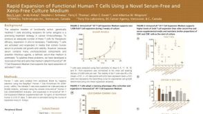 科学海报Rapid Expansion of Functional Human T Cells Using a Novel Serum-Free and Xeno-Free Culture Medium
 沪公网安备31010102008431号