Chang M-J et al. (DEC 2010)
Cancer research 70 24 10234--42
Histone H3 lysine 79 methyltransferase Dot1 is required for immortalization by MLL oncogenes.
Chimeric oncoproteins resulting from fusion of MLL to a wide variety of partnering proteins cause biologically distinctive and clinically aggressive acute leukemias. However,the mechanism of MLL-mediated leukemic transformation is not fully understood. Dot1,the only known histone H3 lysine 79 (H3K79) methyltransferase,has been shown to interact with multiple MLL fusion partners including AF9,ENL,AF10,and AF17. In this study,we utilize a conditional Dot1l deletion model to investigate the role of Dot1 in hematopoietic progenitor cell immortalization by MLL fusion proteins. Western blot and mass spectrometry show that Dot1-deficient cells are depleted of the global H3K79 methylation mark. We find that loss of Dot1 activity attenuates cell viability and colony formation potential of cells immortalized by MLL oncoproteins but not by the leukemic oncoprotein E2a-Pbx1. Although this effect is most pronounced for MLL-AF9,we find that Dot1 contributes to the viability of cells immortalized by other MLL oncoproteins that are not known to directly recruit Dot1. Cells immortalized by MLL fusions also show increased apoptosis,suggesting the involvement of Dot1 in survival pathways. In summary,our data point to a pivotal requirement for Dot1 in MLL fusion protein-mediated leukemogenesis and implicate Dot1 as a potential therapeutic target.
View Publication
产品类型:
产品号#:
03234
18757
18757RF
产品名:
MethoCult™M3234
EasySep™小鼠CD117(cKIT)正选试剂盒
RoboSep™ 小鼠CD117(cKIT)正选试剂盒含滤芯吸头
Wunderlich M et al. (SEP 2006)
Blood 108 5 1690--7
Human CD34+ cells expressing the inv(16) fusion protein exhibit a myelomonocytic phenotype with greatly enhanced proliferative ability.
The t(16:16) and inv(16) are associated with FAB M4Eo myeloid leukemias and result in fusion of the CBFB gene to the MYH11 gene (encoding smooth muscle myosin heavy chain [SMMHC]). Knockout of CBFbeta causes embryonic lethality due to lack of definitive hematopoiesis. Although knock-in of CBFB-MYH11 is not sufficient to cause disease,expression increases the incidence of leukemia when combined with cooperating events. Although mouse models are valuable tools in the study of leukemogenesis,little is known about the contribution of CBFbeta-SMMHC to human hematopoietic stem and progenitor cell self-renewal. We introduced the CBFbeta-MYH11 cDNA into human CD34+ cells via retroviral transduction. Transduced cells displayed an initial repression of progenitor activity but eventually dominated the culture,resulting in the proliferation of clonal populations for up to 7 months. Long-term cultures displayed a myelomonocytic morphology while retaining multilineage progenitor activity and engraftment in NOD/SCID-B2M-/- mice. Progenitor cells from long-term cultures showed altered expression of genes defining inv(16) identified in microarray studies of human patient samples. This system will be useful in examining the effects of CBFbeta-SMMHC on gene expression in the human preleukemic cell,in characterizing the effect of this oncogene on human stem cell biology,and in defining its contribution to the development of leukemia.
View Publication
产品类型:
产品号#:
04100
18056
18056RF
产品名:
MethoCult™ H4100
Maes C et al. (MAY 2006)
The Journal of clinical investigation 116 5 1230--42
Placental growth factor mediates mesenchymal cell development, cartilage turnover, and bone remodeling during fracture repair.
Current therapies for delayed- or nonunion bone fractures are still largely ineffective. Previous studies indicated that the VEGF homolog placental growth factor (PlGF) has a more significant role in disease than in health. Therefore we investigated the role of PlGF in a model of semi-stabilized bone fracture healing. Fracture repair in mice lacking PlGF was impaired and characterized by a massive accumulation of cartilage in the callus,reminiscent of delayed- or nonunion fractures. PlGF was required for the early recruitment of inflammatory cells and the vascularization of the fracture wound. Interestingly,however,PlGF also played a role in the subsequent stages of the repair process. Indeed in vivo and in vitro findings indicated that PlGF induced the proliferation and osteogenic differentiation of mesenchymal progenitors and stimulated cartilage turnover by particular MMPs. Later in the process,PlGF was required for the remodeling of the newly formed bone by stimulating osteoclast differentiation. As PlGF expression was increased throughout the process of bone repair and all the important cell types involved expressed its receptor VEGFR-1,the present data suggest that PlGF is required for mediating and coordinating the key aspects of fracture repair. Therefore PlGF may potentially offer therapeutic advantages for fracture repair.
View Publication
Application of the pMHC Array to Characterise Tumour Antigen Specific T Cell Populations in Leukaemia Patients at Disease Diagnosis.
Immunotherapy treatments for cancer are becoming increasingly successful,however to further improve our understanding of the T-cell recognition involved in effective responses and to encourage moves towards the development of personalised treatments for leukaemia immunotherapy,precise antigenic targets in individual patients have been identified. Cellular arrays using peptide-MHC (pMHC) tetramers allow the simultaneous detection of different antigen specific T-cell populations naturally circulating in patients and normal donors. We have developed the pMHC array to detect CD8+ T-cell populations in leukaemia patients that recognise epitopes within viral antigens (cytomegalovirus (CMV) and influenza (Flu)) and leukaemia antigens (including Per Arnt Sim domain 1 (PASD1),MelanA,Wilms' Tumour (WT1) and tyrosinase). We show that the pMHC array is at least as sensitive as flow cytometry and has the potential to rapidly identify more than 40 specific T-cell populations in a small sample of T-cells (0.8-1.4 x 10(6)). Fourteen of the twenty-six acute myeloid leukaemia (AML) patients analysed had T cells that recognised tumour antigen epitopes,and eight of these recognised PASD1 epitopes. Other tumour epitopes recognised were MelanA (n = 3),tyrosinase (n = 3) and WT1(126-134) (n = 1). One of the seven acute lymphocytic leukaemia (ALL) patients analysed had T cells that recognised the MUC1(950-958) epitope. In the future the pMHC array may be used provide point of care T-cell analyses,predict patient response to conventional therapy and direct personalised immunotherapy for patients.
View Publication
产品类型:
产品号#:
19053
19053RF
产品名:
EasySep™人CD8+ T细胞富集试剂盒
RoboSep™ 人CD8+ T细胞富集试剂盒含滤芯吸头
Biswas S et al. (OCT 2009)
Journal of immunology (Baltimore,Md. : 1950) 183 8 5050--8
Elevated levels of select gangliosides in T cells from renal cell carcinoma patients is associated with T cell dysfunction.
Increased expression of gangliosides by different tumor types including renal cell carcinoma (RCC) is thought to contribute to the immune suppression observed in cancer patients. In this study,we report an increase in apoptotic T cells from RCC patients compared with T cells from normal donors that coincided with the detection of T cells staining positive for GM2 and that the apoptosis was predominantly observed in the GM2(+) but not the GM2(-) T cell population. Ganglioside shedding from tumor rather than endogenous production accounts for GM2(+) T cells since there was no detectable level of mRNA for GM2 synthase in RCC patient T cells and in T cells from normal healthy donors after incubation with either purified GM2 or supernatant from RCC cell lines despite their staining positive for GM2. Moreover,reactive oxygen species as well as activated caspase 3,8,and 9 were predominantly elevated in GM2(+) but not GM2(-) T cells. Similarly,increased staining for GD2 and GD3 but not GD1a was detected with patient T cells with elevated levels of apoptosis in the GD2(+) and GD3(+) cells. These findings suggest that GM2,GD2,and GD3 play a significant role in immune dysfunction observed in RCC patient T cells.
View Publication
Eric Song
Priming the Immune System Against Brain Tumors
产品类型:
研究方向:
免疫学,癌症研究
产品号#:
产品名:
发布日期: 04/09/2020
Y. Zhang et al. ( 2015)
The Journal of Immunology 194 5937-5947
Genetic Vaccines To Potentiate the Effective CD103+ Dendritic Cell-Mediated Cross-Priming of Antitumor Immunity
The development of effective cancer vaccines remains an urgent,but as yet unmet,clinical need. This deficiency is in part due to an incomplete understanding of how to best invoke dendritic cells (DC) that are crucial for the induction of tumor-specific CD8(+) T cells capable of mediating durable protective immunity. In this regard,elevated expression of the transcription factor X box-binding protein 1 (XBP1) in DC appears to play a decisive role in promoting the ability of DC to cross-present Ags to CD8(+) T cells in the therapeutic setting. Delivery of DNA vaccines encoding XBP1 and tumor Ag to skin DC resulted in increased IFN-? production by plasmacytoid DC (pDC) from skin/tumor draining lymph nodes and the cross-priming of Ag-specific CD8(+) T cell responses associated with therapeutic benefit. Antitumor protection was dependent on cross-presenting Batf3(+) DC,pDC,and CD8(+) T cells. CD103(+) DC from the skin/tumor draining lymph nodes of the immunized mice appeared responsible for activation of Ag-specific naive CD8(+) T cells,but were dependent on pDC for optimal effectiveness. Similarly,human XBP1 improved the capacity of human blood- and skin-derived DC to activate human T cells. These data support an important intrinsic role for XBP1 in DC for effective cross-priming and orchestration of Batf3(+) DC-pDC interactions,thereby enabling effective vaccine induction of protective antitumor immunity.
View Publication

 EasySep™小鼠TIL(CD45)正选试剂盒


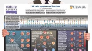 挂图Natural Killer Cells Overview of NK cell receptors, subsets, activation and function
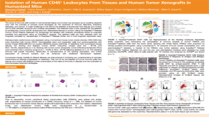 科学海报Isolation of Human CD45+ Leukocytes From Tissues and Human Tumor Xenografts in Humanized Mice
 专家访谈Eric Song Priming the Immune System Against Brain Tumors


 沪公网安备31010102008431号