Regulation of mir-196b by MLL and its overexpression by MLL fusions contributes to immortalization.
Chromosomal translocations involving the Mixed Lineage Leukemia (MLL) gene produce chimeric proteins that cause abnormal expression of a subset of HOX genes and leukemia development. Here,we show that MLL normally regulates expression of mir-196b,a hematopoietic microRNA located within the HoxA cluster,in a pattern similar to that of the surrounding 5' Hox genes,Hoxa9 and Hoxa10,during embryonic stem (ES) cell differentiation. Within the hematopoietic lineage,mir-196b is most abundant in short-term hematopoietic stem cells and is down-regulated in more differentiated hematopoietic cells. Leukemogenic MLL fusion proteins cause overexpression of mir-196b,while treatment of MLL-AF9 transformed bone marrow cells with mir-196-specific antagomir abrogates their replating potential in methylcellulose. This demonstrates that mir-196b function is necessary for MLL fusion-mediated immortalization. Furthermore,overexpression of mir-196b was found specifically in patients with MLL associated leukemias as determined from analysis of 55 primary leukemia samples. Overexpression of mir-196b in bone marrow progenitor cells leads to increased proliferative capacity and survival,as well as a partial block in differentiation. Our results suggest a mechanism whereby increased expression of mir-196b by MLL fusion proteins significantly contributes to leukemia development.
View Publication
产品类型:
产品号#:
03534
19756
19756RF
产品名:
MethoCult™GF M3534
Arendt BK et al. (SEP 2008)
Blood 112 5 1931--41
Biologic and genetic characterization of the novel amyloidogenic lambda light chain-secreting human cell lines, ALMC-1 and ALMC-2.
Primary systemic amyloidosis (AL) is a rare monoclonal plasma cell (PC) disorder characterized by the deposition of misfolded immunoglobulin (Ig) light chains (LC) in vital organs throughout the body. To our knowledge,no cell lines have ever been established from AL patients. Here we describe the establishment of the ALMC-1 and ALMC-2 cell lines from an AL patient. Both cell lines exhibit a PC phenotype and display cytokine-dependent growth. Using a comprehensive genetic approach,we established the genetic relationship between the cell lines and the primary patient cells,and we were also able to identify new genetic changes accompanying tumor progression that may explain the natural history of this patient's disease. Importantly,we demonstrate that free lambda LC secreted by both cell lines contained a beta structure and formed amyloid fibrils. Despite absolute Ig LC variable gene sequence identity,the proteins show differences in amyloid formation kinetics that are abolished by the presence of Na(2)SO(4). The formation of amyloid fibrils from these naturally secreting human LC cell lines is unprecedented. Moreover,these cell lines will provide an invaluable tool to better understand AL,from the combined perspectives of amyloidogenic protein structure and amyloid formation,genetics,and cell biology.
View Publication
产品类型:
产品号#:
18357
18357RF
21000
20119
20155
18387
18387RF
产品名:
RoboSep™- S
RoboSep™ 吸头组件抛光剂
RoboSep™分选管套装(9个塑料管)
Cammenga J et al. (JAN 2007)
Cancer research 67 2 537--45
Mutations in the RUNX1 gene are found at high frequencies in minimally differentiated acute myelogenous leukemia. In addition to null mutations,many of the mutations generate Runx1 DNA-binding (RDB) mutants. To determine if these mutants antagonize wild-type protein activity,cDNAs were transduced into murine bone marrow or human cord blood cells using retroviral vectors. Significantly,the RDB mutants did not act in a transdominant fashion in vivo to disrupt Runx1 activity in either T-cell or platelet development,which are highly sensitive to Runx1 dosage. However,RDB mutant expression impaired expansion and differentiation of the erythroid compartment in which Runx1 expression is normally down-regulated,showing that a RDB-independent function is incompatible with erythroid differentiation. Significantly,both bone marrow progenitors expressing RDB mutants or deficient for Runx1 showed increased replating efficiencies in vitro,accompanied by the accumulation of myeloblasts and dysplastic progenitors,but the effect was more pronounced in RDB cultures. Disruption of the interface that binds CBFbeta,an important cofactor of Runx1,did not impair RDB mutant replating activity,arguing against inactivation of Runx1 function by CBFbeta sequestration. We propose that RDB mutants antagonize Runx1 function in early progenitors by disrupting a critical balance between DNA-binding-independent and DNA-binding-dependent signaling.
View Publication
产品类型:
产品号#:
03434
03444
09500
09600
09650
18096
18096RF
84434
84444
产品名:
MethoCult™GF M3434
MethoCult™GF M3434
BIT 9500血清替代物
StemSpan™ SFEM
StemSpan™ SFEM
Timm MM et al. (OCT 2006)
Leukemia 20 10 1863--9
Thymoglobulin targets multiple plasma cell antigens and has in vitro and in vivo activity in multiple myeloma.
Multiple myeloma is characterized by the proliferation of clonal plasma cells that have a heterogeneous expression of various cell surface markers,precluding successful use of monoclonal antibodies for therapeutic targeting of the tumor cell. Thymoglobulin (rabbit-derived polyclonal anti-thymocyte globulin),by virtue of its method of preparation,contains antibodies against several B-cell and plasma cell antigens and offers an attractive option for immunotherapy of myeloma. Here,we demonstrate potent anti-myeloma activity of the rabbit anti-thymocyte globulin preparation Thymoglobulin in vitro and in vivo in an animal model of myeloma. Thymoglobulin was able to induce dose- and time-dependent apoptosis of several myeloma cell lines,including those resistant to conventional anti-myeloma agents. Importantly,the anti-myeloma activity was preserved even when myeloma cells were grown with different cytokines demonstrating the ability to overcome microenvironment-mediated resistance. Thymoglobulin induced apoptosis of freshly isolated primary myeloma cells from patients. Using a competitive flow cytometric analysis,we were able to identify the potential antigen targets for Thymoglobulin preparation. Finally,in a plasmacytoma mouse model of myeloma,Thymoglobulin delayed the tumor growth in a dose-dependent manner providing convincing evidence for continued evaluation of this agent in the clinic in patients with myeloma,either alone or in combination with other agents.
View Publication

 EasySep™小鼠TIL(CD45)正选试剂盒


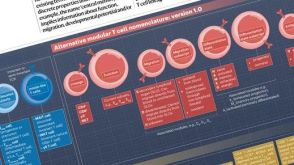 挂图T Cell Nomenclature: From Subsets to Modules A modular framework for classifying T cells by lineage, function, migration, differentiation, and antigen context.发布日期: 12/05/2025
 实验方案How to Collect Plasma from Whole Blood Before Cell Isolation

 沪公网安备31010102008431号