
 EasySep™小鼠TIL(CD45)正选试剂盒


搜索结果: 'methocult media formulations for mouse hematopoietic cells serum containing'
-
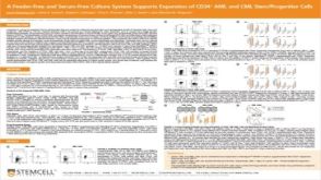 科学海报A Feeder-Free and Serum-Free Culture System Supports Expansion of CD34+ AML and CML Stem/Progenitor Cells
产品类型:
Conference:
ISEH 2019
产品号#:
产品名:
发布日期: 09/10/2019 -

产品类型:
产品号#:
01700
01705
01701
01702
产品名:
ALDEFLUOR™ 试剂盒
ALDEFLUOR™ DEAB试剂
ALDEFLUOR™测定缓冲液
-
产品类型:
产品号#:
01700
01705
01702
产品名:
ALDEFLUOR™ 试剂盒
ALDEFLUOR™ DEAB试剂
ALDEFLUOR™测定缓冲液
-
产品类型:
产品号#:
15129
15169
产品名:
RosetteSep™人多发性骨髓瘤细胞富集抗体混合物
RosetteSep™人多发性骨髓瘤细胞富集抗体混合物
-
产品类型:
产品号#:
09600
09650
产品名:
StemSpan™ SFEM
StemSpan™ SFEM
-
产品类型:
产品号#:
15122
15162
产品名:
RosetteSep™ 人CD45去除抗体混合物
RosetteSep™人CD45去除抗体混合物
-
产品类型:
产品号#:
04100
产品名:
MethoCult™ H4100
-
产品类型:
产品号#:
05700
05701
05702
产品名:
NeuroCult™ 基础培养基(小鼠&大鼠)
NeuroCult™ 扩增添加物 (小鼠&大鼠)
NeuroCult™ 扩增试剂盒 (小鼠&大鼠)
-
产品类型:
产品号#:
05620
产品名:
MammoCult™人培养基试剂盒
-
 产品手册Reliable Antibodies for Your Research
产品类型:
品牌:
EasySep
产品号#:
60100
60100.1
60100AD
60100AD.1
60102
60102FI
60104
60104AD
60104AD.1
60104AZ
60104AZ.1
60104BT
60104BT.1
60104BT.2
60104FI
60104FI.1
60104PE
60104PE.1
60106
60106.1
60106AZ
60106AZ.1
60106BT
60106BT.1
60106FI
60106FI.1
60106PE
60106PE.1
60107
60107.1
601
产品名:
抗β-微管蛋白III抗体,克隆AA10
抗β-微管蛋白III抗体,克隆AA10
抗β-微管蛋白III抗体,克隆AA10,Alexa Fluor® 488
抗β-微管蛋白III抗体,clone AA10,Alexa Fluor® 488
抗小鼠TCR Gamma/Delta抗体,clone GL3
抗小鼠TCR Gamma/Delta抗体,clone GL3,Alexa Fluor® 488
抗小鼠TCR Gamma/Delta抗体,clone GL3,APC
抗小鼠TCR Gamma/Delta抗体,clone GL3,APC
抗小鼠TCR Gamma/Delta抗体,clone GL3,Biotin
抗小鼠TCR Gamma/Delta抗体,clone GL3,Biotin
抗小鼠TCR Gamma/Delta抗体,clone GL3,FITC
抗小鼠TCR Gamma/Delta抗体,clone GL3,PE
抗小鼠TCR Gamma/Delta抗体,clone GL3,PE
抗人CD71(转铁蛋白受体)抗体,clone OKT9
抗人CD71(转铁蛋白受体)抗体,clone OKT9
抗人CD71(转铁蛋白受体)抗体,clone OKT9,APC
抗人CD71(转铁蛋白受体)抗体,clone OKT9,APC
抗人CD71(转铁蛋白受体)抗体,clone OKT9,Biotin
抗人CD71(转铁蛋白受体)抗体,clone OKT9,FITC
抗人CD71(转铁蛋白受体)抗体,clone OKT9,FITC
抗人CD71(转铁蛋白受体)抗体,clone OKT9,PE
抗人CD71(转铁蛋白受体)抗体,clone OKT9,PE
-
产品类型:
产品号#:
01700
01705
01702
产品名:
ALDEFLUOR™ 试剂盒
ALDEFLUOR™ DEAB试剂
ALDEFLUOR™测定缓冲液

 沪公网安备31010102008431号