Gazi E et al. (AUG 2007)
Journal of lipid research 48 8 1846--56
Direct evidence of lipid translocation between adipocytes and prostate cancer cells with imaging FTIR microspectroscopy.
Various epidemiological studies show a positive correlation between high intake of dietary FAs and metastatic prostate cancer (CaP). Moreover,CaP metastasizes to the bone marrow,which harbors a rich source of lipids stored within adipocytes. Here,we use Fourier transform infrared (FTIR) microspectroscopy to study adipocyte biochemistry and to demonstrate that PC-3 cells uptake isotopically labeled FA [deuterated palmitic acid (D(31)-PA)] from an adipocyte. Using this vibrational spectroscopic technique,we detected subcellular locations in a single adipocyte enriched with D(31)-PA using the upsilon(as+s)(C-D)(2+3) (D(31)-PA): upsilon(as+s)(C-H)(2+3) (lipid hydrocarbon) signal. In addition,larger adipocytes were found to consist of a higher percentage of D(31)-PA of the total lipid found within the adipocyte. Following background subtraction,the upsilon(as)(C-D)(2+3) signal illuminated starved PC-3 cells cocultured with D(31)-PA-loaded adipocytes,indicating translocation of the labeled FA. This study demonstrates lipid-specific translocation between adipocytes and tumor cells and the use of FTIR microspectroscopy to characterize various biomolecular features of a single adipocyte without the requirement for cell isolation and lipid extraction.
View Publication
产品类型:
产品号#:
15128
15168
产品名:
RosetteSep™人间充质干细胞富集抗体混合物
RosetteSep™人间充质干细胞富集抗体混合物
Jimeno A et al. (FEB 2009)
Molecular cancer therapeutics 8 2 310--4
A direct pancreatic cancer xenograft model as a platform for cancer stem cell therapeutic development.
There is an enormous gap between the antiproliferative and in vivo antitumor efficacy of gemcitabine in cell line-based models and its clinical efficacy. This may be due to insensitiveness of the precursor,cancer stem cell (CSC) compartment to cytotoxic agents. The hedgehog pathway is associated with CSC signaling and control. We used a direct xenograft model of pancreatic cancer and a two-stage approach was used to test the hypotheses that targeting CSC could increase the efficacy of gemcitabine. Tumors from a gemcitabine-sensitive xenograft were treated with gemcitabine first,and randomized,after tumor regression to continuing treatment with gemcitabine,a hedgehog inhibitor alone or in combination with gemcitabine. We tested markers described as associated with CSC such as CD24,CD44,ALDH,nestin,and the hedgehog pathway. After induction with gemcitabine,treated tumor showed an enrichment in CSC markers such as ALDH and CD24. Subsequently,a release from gemcitabine prompted a repopulation of proliferating cells and a decrease in such markers to equilibrate from pretreatment levels. Combined treatment with gemcitabine and cyclopamine induced tumor regression and decrease in CSC markers and hedgehog signaling. Cytoplasmic CD24 and ALDH were inversely and strongly associated with growth and were expressed in a minority of cells that we propose constitute the CSC compartment. Hedgehog inhibitors as part of a dual compartment therapeutic approach were able to further reduce tumor growth and decreased both static and dynamic markers of CSC. Direct tumor xenografts are a valid platform to test multicompartment therapeutic approaches in pancreatic cancer.
View Publication
Ungefroren H et al. ( 2011)
Current cancer drug targets 11 4 524--535
The Src family kinase inhibitors PP2 and PP1 block TGF-beta1-mediated cellular responses by direct and differential inhibition of type I and type II TGF-beta receptors.
Both the nonreceptor tyrosine kinase Src and the receptors for transforming growth factor (TGF)-β (TβRI,TβRII) play major roles during tumorigenesis by regulating cell growth,migration/invasion and metastasis. The common Src family kinase inhibitors PP2 and PP1 effectively block Src activity in vitro and in vivo,however,they may exert non-specific effects on other kinases. In this study,we have evaluated PP2 and PP1 for their ability to inhibit TGFβ1-mediated responses in the TGF-β-responsive pancreatic adenocarcinoma cell line Panc1. We show that PP2 and PP1 but not the more specific Src inhibitor SU6656 effectively relieved TGF-b1-induced growth arrest and p21(WAF1) induction,while basal growth was enhanced by PP2 and PP1,and suppressed by SU6656. PP2 and PP1 but not SU6656 also suppressed TGF-β1-induced epithelial-to-mesenchymal transition (EMT) as evidenced by their ability to inhibit downregulation of the epithelial marker E-cadherin,and upregulation of the EMT-associated transcription factor Slug. Likewise,PP2 and PP1 but not SU6656 effectively blocked TGF-β1-induced activation of Smad2 and p38 MAPK and partially suppressed Smad activation and transcriptional activity on TGF-β/Smad-responsive reporters of a kinase-active TβRI mutant ectopically expressed in Panc1 cells. Interestingly,PP2 and PP1 strongly inhibited recombinant TβRI in an in vitro kinase assay,with PP1 being more potent and PP2 being nearly as potent as the established TβRI inhibitor SB431542. PP2 but not PP1 also weakly inhibited the TβRII kinase. Together,these data provide evidence that PP2 and PP1 are powerful inhibitors of TβR function that can block TGF-β/Smad signaling in a Src-unrelated fashion. Both agents may be useful as dual TGF-β/Src inhibitors in experimental therapeutics of late stage metastatic disease.
View Publication
Noninvasive MR imaging of magnetically labeled stem cells to directly identify neovasculature in a glioma model.
Bone marrow-derived endothelial precursor cells incorporate into neovasculature and have been successfully used as vehicles for gene delivery to brain tumors. To determine whether systemically administered Sca1+ bone marrow cells labeled with superparamagnetic iron oxide nanoparticles can be detected by in vivo magnetic resonance imaging in a mouse brain tumor model,mouse Sca1+ cells were labeled in vitro with ferumoxides-poly-L-lysine complexes. Labeled or control cells were administered intravenously to glioma-bearing severe combined immunodeficient (SCID) mice. Magnetic resonance imaging (MRI) was performed during tumor growth. Mice that received labeled cells demonstrated hypointense regions within the tumor that evolved over time and developed a continuous dark hypointense ring at a consistent time point. This effect was not cleared by administration of a gadolinium contrast agent. Histology showed iron-labeled cells around the tumor rim in labeled mice,which expressed CD31 and von Willebrand factor,indicating the transplanted cells detected in the tumor have differentiated into endothelial-like cells. These results demonstrate that MRI can detect the incorporation of magnetically labeled bone marrow-derived precursor cells into tumor vasculature as part of ongoing angiogenesis and neovascularization. This technique can be used to directly identify neovasculature in vivo and to facilitate gene therapy by noninvasively monitoring these cells as gene delivery vectors.
View Publication
产品类型:
产品号#:
09600
09650
09850
产品名:
StemSpan™ SFEM
StemSpan™ SFEM
Eguchi M et al. (JAN 2005)
Proceedings of the National Academy of Sciences of the United States of America 102 4 1133--8
Directing oncogenic fusion genes into stem cells via an SCL enhancer.
TEL-TRKC is a fusion gene generated by chromosomal translocation and encodes an activated tyrosine kinase. Uniquely,it is found in both solid tumors and leukemia. However,a single exon difference (in TEL) in TEL-TRKC fusions is associated with the two sets of cancer phenotypes. We expressed the two TEL-TRKC variants in vivo by using the 3' regulatory element of SCL that is selectively active in a subset of mesodermal cell lineages,including endothelial and hematopoietic stem cells and progenitors. The leukemia form of TEL-TRKC (-exon 5 of TEL) enhanced hematopoietic stem cell renewal and initiated leukemia. In contrast,the TEL-TRKC solid tumor variant (+ TEL exon 5) elicited an embryonic lethal phenotype with impairment of both angiogenesis and hematopoiesis indicative of an effect at the level of the hemangioblasts. The ability of TEL-TRKC to repress expression of Flk1,a critical regulator of early endothelial and hematopoietic cells,depended on TEL exon 5. These data indicate that related oncogenic fusion proteins similarly expressed in a hierarchy of early stem cells can have selective,cell type-specific developmental impacts.
View Publication
产品类型:
产品号#:
03231
产品名:
MethoCult™M3231
Morabito A et al. ( 2009)
The oncologist 14 4 378--390
Vandetanib (ZD6474), a dual inhibitor of vascular endothelial growth factor receptor (VEGFR) and epidermal growth factor receptor (EGFR) tyrosine kinases: current status and future directions.
Vandetanib is a novel,orally available inhibitor of different intracellular signaling pathways involved in tumor growth,progression,and angiogenesis: vascular endothelial growth factor receptor-2,epidermal growth factor receptor,and REarranged during Transfection tyrosine kinase activity. Phase I clinical trials have shown that vandetanib is well tolerated as a single agent at daily doses textless or =300 mg. In the phase II setting,negative results were observed with vandetanib in small cell lung cancer,metastatic breast cancer,and multiple myeloma. In contrast,three randomized phase II studies showed that vandetanib prolonged the progression-free survival (PFS) time of patients with non-small cell lung cancer (NSCLC) as a single agent when compared with gefitinib or when added to chemotherapy. Rash,diarrhea,hypertension,fatigue,and asymptomatic QTc prolongation were the most common adverse events. Antitumor activity was also observed in medullary thyroid cancer. Four randomized phase III clinical trials in NSCLC are exploring the efficacy of vandetanib in combination with docetaxel,the Zactima in cOmbination with Docetaxel In non-small cell lung Cancer (ZODIAC) trial,or with pemetrexed,the Zactima Efficacy with Alimta in Lung cancer (ZEAL) trial,or as a single agent,the Zactima Efficacy when Studied versus Tarceva (ZEST) and the Zactima Efficacy trial for NSCLC Patients with History of EGFR-TKI chemo-Resistance (ZEPHYR) trials. Based on a press release by the sponsor of these trials,the PFS time was longer with vandetanib in the ZODIAC and ZEAL trials; the ZEST trial was negative for its primary superiority analysis,but was successful according to a preplanned noninferiority analysis of PFS. Ongoing phase II and III clinical trials will better define the appropriate schedule,the optimal setting of evaluation,and the safety of long-term use of vandetanib.
View Publication

 EasySep™小鼠TIL(CD45)正选试剂盒


 EasySep™小鼠/人嵌合体分选试剂盒

 技术公告Achieve Scalable, High-Quality Nucleic Acid Extractions with the EasySep™ Total Nucleic Acid Extraction Kit
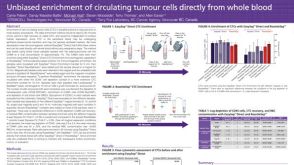 科学海报Unbiased Enrichment of Circulating Tumour Cells Directly from Whole Blood
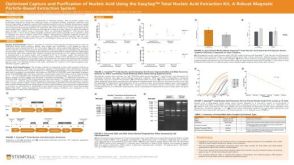


 沪公网安备31010102008431号