Lee Y-KK et al. (JAN 2016)
International journal of cardiology 203 964--971
Efficient attenuation of Friedreich's ataxia (FRDA) cardiomyopathy by modulation of iron homeostasis-human induced pluripotent stem cell (hiPSC) as a drug screening platform for FRDA.
BACKGROUND Friedreich's ataxia (FRDA),a recessive neurodegenerative disorder commonly associated with hypertrophic cardiomyopathy,is caused by silencing of the frataxin (FXN) gene encoding the mitochondrial protein involved in iron-sulfur cluster biosynthesis. METHODS Application of our previously established FRDA human induced pluripotent stem cell (hiPSC) derived cardiomyocytes model as a platform to assess the efficacy of treatment with either the antioxidant coenzyme Q10 analog,idebenone (IDE) or the iron chelator,deferiprone (DFP),which are both under clinical trial. RESULTS DFP was able to more significantly suppress synthesis of reactive oxygen species (ROS) than IDE at the dosages of 25 $\$ and 10nM respectively which agreed with the reduced rate of intracellular accumulation of iron by DFP treatment from 25 to 50 $\$ With regard to cardiac electrical-contraction (EC) coupling function,decay velocity of calcium handling kinetics in FRDA-hiPSC-cardiomyocytes was significantly improved by DFP treatment but not by IDE. Further mechanistic studies revealed that DFP also modulated iron induced mitochondrial stress as reflected by mitochondria network disorganization and decline level of respiratory chain protein,succinate dehydrogenase (CxII) and cytochrome c oxidase (COXIV). In addition,iron-response protein (IRP-1) regulatory loop was overridden by DFP as reflected by resumed level of ferritin (FTH) back to basal level and the attenuated transferrin receptor (TSFR) mRNA level suppression thereby reducing further iron uptake. CONCLUSIONS DFP modulated iron homeostasis in FRDA-hiPSC-cardiomyocytes and effectively relieved stress-stimulation related to cardiomyopathy. The resuming of redox condition led to the significantly improved cardiac prime events,cardiac electrical-coupling during contraction.
View Publication
Song DH et al. (AUG 2000)
Journal of Biological Chemistry 275 31 23790--97
Endogenous protein kinase CK2 participates in Wnt signaling in mammary epithelial cells
Protein kinase CK2 (formerly casein kinase II) is a serine/threonine kinase overexpressed in many human tumors,transformed cell lines,and rapidly proliferating tissues. Recent data have shown that many cancers involve inappropriate reactivation of Wnt signaling through ectopic expression of Wnts themselves,as has been seen in a number of human breast cancers,or through mutation of intermediates in the Wnt pathway,such as adenomatous polyposis coli or beta-catenin,as described in colon and other cancers. Wnts are secreted factors that are important in embryonic development,but overexpression of certain Wnts,such as Wnt-1,leads to proliferation and transformation of cells. We report that upon stable transfection of Wnt-1 into the mouse mammary epithelial cell line C57MG,morphological changes and increased proliferation are accompanied by increased levels of CK2,as well as of beta-catenin. CK2 and beta-catenin co-precipitate with the Dvl proteins,which are Wnt signaling intermediates. A major phosphoprotein of the size of beta-catenin appears in in vitro kinase reactions performed on the Dvl immunoprecipitates. In vitro translated beta-catenin,Dvl-2,and Dvl-3 are phosphorylated by CK2. The selective CK2 inhibitor apigenin blocks proliferation of Wnt-1-transfected cells,abrogates phosphorylation of beta-catenin,and reduces beta-catenin and Dvl protein levels. These results demonstrate that endogenous CK2 is a positive regulator of Wnt signaling and growth of mammary epithelial cells.
View Publication
产品类型:
产品号#:
03800
03801
03802
03803
03804
03805
03806
产品名:
ClonaCell™-HY 杂交瘤试剂盒
ClonaCell™-HY Medium
ClonaCell™-HY Medium
ClonaCell™-HY Medium
ClonaCell™-HY Medium
ClonaCell™-HY Medium
ClonaCell™-HY PEG (融合)
Pei Y et al. (MAY 2016)
Brain research 1638 Pt A 57--73
Comparative neurotoxicity screening in human iPSC-derived neural stem cells, neurons and astrocytes.
Induced pluripotent stem cells (iPSC) and their differentiated derivatives offer a unique source of human primary cells for toxicity screens. Here,we report on the comparative cytotoxicity of 80 compounds (neurotoxicants,developmental neurotoxicants,and environmental compounds) in iPSC as well as isogenic iPSC-derived neural stem cells (NSC),neurons,and astrocytes. All compounds were tested over a 24-h period at 10 and 100$\$,in duplicate,with cytotoxicity measured using the MTT assay. Of the 80 compounds tested,50 induced significant cytotoxicity in at least one cell type; per cell type,32,38,46,and 41 induced significant cytotoxicity in iPSC,NSC,neurons,and astrocytes,respectively. Four compounds (valinomycin,3,3',5,5'-tetrabromobisphenol,deltamethrin,and triphenyl phosphate) were cytotoxic in all four cell types. Retesting these compounds at 1,10,and 100$\$ using the same exposure protocol yielded consistent results as compared with the primary screen. Using rotenone,we extended the testing to seven additional iPSC lines of both genders; no substantial difference in the extent of cytotoxicity was detected among the cell lines. Finally,the cytotoxicity assay was simplified by measuring luciferase activity using lineage-specific luciferase reporter iPSC lines which were generated from the parental iPSC line. This article is part of a Special Issue entitled SI: PSC and the brain.
View Publication
产品类型:
产品号#:
05850
05857
05870
05875
85850
85857
85870
85875
产品名:
mTeSR™1
mTeSR™1
Graham B et al. (JUL 2014)
International Journal of Environmental Research and Public Health 11 7 7524--7536
Enhancement of arsenic trioxide-mediated changes in human induced pluripotent stem cells (IPS)
Induced pluripotent stem cells (IPS) are an artificially derived type of pluripotent stem cell,showing many of the same characteristics as natural pluripotent stem cells. IPS are a hopeful therapeutic model; however there is a critical need to determine their response to environmental toxins. Effects of arsenic on cells have been studied extensively; however,its effect on IPS is yet to be elucidated. Arsenic trioxide (ATO) has been shown to inhibit cell proliferation,induce apoptosis and genotoxicity in many cells. Based on ATOs action in other cells,we hypothesize that it will induce alterations in morphology,inhibit cell viability and induce a genotoxic effect on IPS. Cells were treated for 24 hours with ATO (0-9 µg/mL). Cell morphology,viability and DNA damage were documented. Results indicated sufficient changes in morphology of cell colonies mainly in cell ability to maintain grouping and ability to remain adherent. Cell viability decreased in a dose dependent manner. There were significant increases in tail length and moment as well as destruction of intact DNA as concentration increased. Exposure to ATO resulted in a reproducible dose dependent sequence of events marked by changes in morphology,decrease of cell viability,and induction of genotoxicity in IPS.
View Publication
Chen C et al. (JUL 2014)
Nature communications 5 4430
Role of astroglia in Down's syndrome revealed by patient-derived human-induced pluripotent stem cells.
Down's syndrome (DS),caused by trisomy of human chromosome 21,is the most common genetic cause of intellectual disability. Here we use induced pluripotent stem cells (iPSCs) derived from DS patients to identify a role for astrocytes in DS pathogenesis. DS astroglia exhibit higher levels of reactive oxygen species and lower levels of synaptogenic molecules. Astrocyte-conditioned medium collected from DS astroglia causes toxicity to neurons,and fails to promote neuronal ion channel maturation and synapse formation. Transplantation studies show that DS astroglia do not promote neurogenesis of endogenous neural stem cells in vivo. We also observed abnormal gene expression profiles from DS astroglia. Finally,we show that the FDA-approved antibiotic drug,minocycline,partially corrects the pathological phenotypes of DS astroglia by specifically modulating the expression of S100B,GFAP,inducible nitric oxide synthase,and thrombospondins 1 and 2 in DS astroglia. Our studies shed light on the pathogenesis and possible treatment of DS by targeting astrocytes with a clinically available drug.
View Publication

 EasySep™小鼠TIL(CD45)正选试剂盒


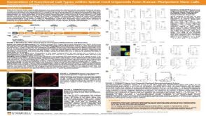 科学海报Generation of Functional 3D Spinal Cord Organoids from Human Pluripotent Stem Cells
 实验方案Gene Editing Human Pluripotent Stem Cells (hPSCs) Using the CellPore™ Transfection System
 科学海报Generation of a Glia-Midbrain Neuron Co-Culture System Derived From Human Pluripotent Stem Cells

 沪公网安备31010102008431号