Lopez-Izquierdo A et al. (NOV 2014)
American journal of physiology. Heart and circulatory physiology 307 9 H1370--7
A near-infrared fluorescent voltage-sensitive dye allows for moderate-throughput electrophysiological analyses of human induced pluripotent stem cell-derived cardiomyocytes.
Human induced pluripotent stem cell-derived cardiomyocyte (iPSC-CM)-based assays are emerging as a promising tool for the in vitro preclinical screening of QT interval-prolonging side effects of drugs in development. A major impediment to the widespread use of human iPSC-CM assays is the low throughput of the currently available electrophysiological tools. To test the precision and applicability of the near-infrared fluorescent voltage-sensitive dye 1-(4-sulfanatobutyl)-4-β[2-(di-n-butylamino)-6-naphthyl]butadienylquinolinium betaine (di-4-ANBDQBS) for moderate-throughput electrophysiological analyses,we compared simultaneous transmembrane voltage and optical action potential (AP) recordings in human iPSC-CM loaded with di-4-ANBDQBS. Optical AP recordings tracked transmembrane voltage with high precision,generating nearly identical values for AP duration (AP durations at 10%,50%,and 90% repolarization). Human iPSC-CMs tolerated repeated laser exposure,with stable optical AP parameters recorded over a 30-min study period. Optical AP recordings appropriately tracked changes in repolarization induced by pharmacological manipulation. Finally,di-4-ANBDQBS allowed for moderate-throughput analyses,increasing throughput textgreater10-fold over the traditional patch-clamp technique. We conclude that the voltage-sensitive dye di-4-ANBDQBS allows for high-precision optical AP measurements that markedly increase the throughput for electrophysiological characterization of human iPSC-CMs.
View Publication
Amelioration of murine beta-thalassemia through drug selection of hematopoietic stem cells transduced with a lentiviral vector encoding both gamma-globin and the MGMT drug-resistance gene.
Correction of murine models of beta-thalassemia has been achieved through high-level globin lentiviral vector gene transfer into mouse hematopoietic stem cells (HSCs). However,transduction of human HSCs is less robust and may be inadequate to achieve therapeutic levels of genetically modified erythroid cells. We therefore developed a double gene lentiviral vector encoding both human gamma-globin under the transcriptional control of erythroid regulatory elements and methylguanine methyltransferase (MGMT),driven by a constitutive cellular promoter. MGMT expression provides cellular resistance to alkylator drugs,which can be administered to kill residual untransduced,diseased HSCs,whereas transduced cells are protected. Mice transplanted with beta-thalassemic HSCs transduced with a gamma-globin/MGMT vector initially had subtherapeutic levels of red cells expressing gamma-globin. To enrich gamma-globin-expressing cells,transplanted mice were treated with the alkylator agent 1,3-bis-chloroethyl-1-nitrosourea. This resulted in significant increases in the number of gamma-globin-expressing red cells and the amount of fetal hemoglobin,leading to resolution of anemia. Selection of transduced HSCs was also obtained when cells were drug-treated before transplantation. Mice that received these cells demonstrated reconstitution with therapeutic levels of gamma-globin-expressing cells. These data suggest that MGMT-based drug selection holds promise as a modality to improve gene therapy for beta-thalassemia.
View Publication
产品类型:
产品号#:
09600
09650
产品名:
StemSpan™ SFEM
StemSpan™ SFEM
Pei Y et al. (MAY 2016)
Brain research 1638 Pt A 57--73
Comparative neurotoxicity screening in human iPSC-derived neural stem cells, neurons and astrocytes.
Induced pluripotent stem cells (iPSC) and their differentiated derivatives offer a unique source of human primary cells for toxicity screens. Here,we report on the comparative cytotoxicity of 80 compounds (neurotoxicants,developmental neurotoxicants,and environmental compounds) in iPSC as well as isogenic iPSC-derived neural stem cells (NSC),neurons,and astrocytes. All compounds were tested over a 24-h period at 10 and 100$\$,in duplicate,with cytotoxicity measured using the MTT assay. Of the 80 compounds tested,50 induced significant cytotoxicity in at least one cell type; per cell type,32,38,46,and 41 induced significant cytotoxicity in iPSC,NSC,neurons,and astrocytes,respectively. Four compounds (valinomycin,3,3',5,5'-tetrabromobisphenol,deltamethrin,and triphenyl phosphate) were cytotoxic in all four cell types. Retesting these compounds at 1,10,and 100$\$ using the same exposure protocol yielded consistent results as compared with the primary screen. Using rotenone,we extended the testing to seven additional iPSC lines of both genders; no substantial difference in the extent of cytotoxicity was detected among the cell lines. Finally,the cytotoxicity assay was simplified by measuring luciferase activity using lineage-specific luciferase reporter iPSC lines which were generated from the parental iPSC line. This article is part of a Special Issue entitled SI: PSC and the brain.
View Publication
产品类型:
产品号#:
05850
05857
05870
05875
85850
85857
85870
85875
产品名:
mTeSR™1
mTeSR™1
Graham B et al. (JUL 2014)
International Journal of Environmental Research and Public Health 11 7 7524--7536
Enhancement of arsenic trioxide-mediated changes in human induced pluripotent stem cells (IPS)
Induced pluripotent stem cells (IPS) are an artificially derived type of pluripotent stem cell,showing many of the same characteristics as natural pluripotent stem cells. IPS are a hopeful therapeutic model; however there is a critical need to determine their response to environmental toxins. Effects of arsenic on cells have been studied extensively; however,its effect on IPS is yet to be elucidated. Arsenic trioxide (ATO) has been shown to inhibit cell proliferation,induce apoptosis and genotoxicity in many cells. Based on ATOs action in other cells,we hypothesize that it will induce alterations in morphology,inhibit cell viability and induce a genotoxic effect on IPS. Cells were treated for 24 hours with ATO (0-9 µg/mL). Cell morphology,viability and DNA damage were documented. Results indicated sufficient changes in morphology of cell colonies mainly in cell ability to maintain grouping and ability to remain adherent. Cell viability decreased in a dose dependent manner. There were significant increases in tail length and moment as well as destruction of intact DNA as concentration increased. Exposure to ATO resulted in a reproducible dose dependent sequence of events marked by changes in morphology,decrease of cell viability,and induction of genotoxicity in IPS.
View Publication

 EasySep™小鼠TIL(CD45)正选试剂盒


 科学海报Efficient Differentiation of Human Pluripotent Stem Cells to Sensory Neurons
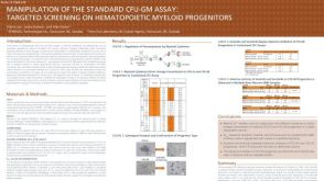 科学海报Manipulation of the Standard CFU-GM Assay Targeted Screening of Hematopoietic Myeloid Progenitors
 科学海报Rapid, High-Efficiency Differentiation of Motor Neurons from Human Pluripotent Stem Cells
 实验方案How to Culture Human Pluripotent Stem Cell (hPSC)-Derived Forebrain Neurons for MEA Analysis Using the Maestro MEA™ System
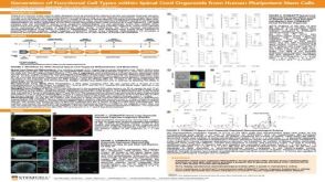 科学海报Generation of Functional 3D Spinal Cord Organoids from Human Pluripotent Stem Cells
 实验方案Gene Editing Human Pluripotent Stem Cells (hPSCs) Using the CellPore™ Transfection System


 沪公网安备31010102008431号