Usta S et al. (OCT 2014)
Annals of translational medicine 2 10 97
Chemically defined serum-free and xeno-free media for multiple cell lineages.
Cell culture is one of the most common methods used to recapitulate a human disease environment in a laboratory setting. Cell culture techniques are used to grow and maintain cells of various types including those derived from primary tissues,such as stem cells and cancer tumors. However,a major confounding factor with cell culture is the use of serum and animal (xeno) products in the media. The addition of animal products introduces batch and lot variations that lead to experimental variability,confounds studies with therapeutic outcomes for cultured cells,and represents a major cost associated with cell culture. Here we report a commercially available serum-free,albumin-free,and xeno free (XF) media (Neuro-Pure(TM)) that is more cost-effective than other commercial medias. Neuro-Pure was used to maintain and differentiate various cells of neuronal lineages,fibroblasts,as well as specific cancer cell lines; without the use of contaminants such serum,albumin,and animal products. Neuro-Pure allows for a controlled and reproducible cell culture environment that is applicable to translational medicine and general tissue culture.
View Publication
Ferreira JS et al. (JUN 2015)
The Journal of neuroscience : the official journal of the Society for Neuroscience 35 22 8462--79
GluN2B-Containing NMDA Receptors Regulate AMPA Receptor Traffic through Anchoring of the Synaptic Proteasome.
NMDA receptors play a central role in shaping the strength of synaptic connections throughout development and in mediating synaptic plasticity mechanisms that underlie some forms of learning and memory formation in the CNS. In the hippocampus and the neocortex,GluN1 is combined primarily with GluN2A and GluN2B,which are differentially expressed during development and confer distinct molecular and physiological properties to NMDA receptors. The contribution of each subunit to the synaptic traffic of NMDA receptors and therefore to their role during development and in synaptic plasticity is still controversial. We report a critical role for the GluN2B subunit in regulating NMDA receptor synaptic targeting. In the absence of GluN2B,the synaptic levels of AMPA receptors are increased and accompanied by decreased constitutive endocytosis of GluA1-AMPA receptor. We used quantitative proteomic analysis to identify changes in the composition of postsynaptic densities from GluN2B(-/-) mouse primary neuronal cultures and found altered levels of several ubiquitin proteasome system components,in particular decreased levels of proteasome subunits. Enhancing the proteasome activity with a novel proteasome activator restored the synaptic levels of AMPA receptors in GluN2B(-/-) neurons and their endocytosis,revealing that GluN2B-mediated anchoring of the synaptic proteasome is responsible for fine tuning AMPA receptor synaptic levels under basal conditions.
View Publication
产品类型:
产品号#:
05711
产品名:
NeuroCult™SM1神经元补充剂
Reference
Zhang L et al. (APR 2016)
Human Reproduction 31 4 832--843
Protein kinase A inhibitor, H89, enhances survival and clonogenicity of dissociated human embryonic stem cells through Rho-associated coiled-coil containing protein kinase (ROCK) inhibition
H89 inhibits the dissociation-induced phosphorylation of PKA and two substrates of Rho-associated coiled-coil containing protein kinase (ROCK),myosin light chain (MLC2) and myosin phosphatase target subunit 1 (MYPT1),significantly increases cell survival and colony formation,and strongly depresses dissociation-induced cell death and cell blebbing without affecting the pluripotency of hESCs and their differentiation in vitro.
View Publication
产品类型:
产品号#:
05835
05839
产品名:
STEMdiff™神经诱导培养基
STEMdiff™神经诱导培养基
Reference
Walker TL et al. (JAN 2012)
PloS one 7 9 e44371
Prolactin stimulates precursor cells in the adult mouse hippocampus.
In the search for ways to combat degenerative neurological disorders,neurogenesis-stimulating factors are proving to be a promising area of research. In this study,we show that the hormonal factor prolactin (PRL) can activate a pool of latent precursor cells in the adult mouse hippocampus. Using an in vitro neurosphere assay,we found that the addition of exogenous PRL to primary adult hippocampal cells resulted in an approximate 50% increase in neurosphere number. In addition,direct infusion of PRL into the adult dentate gyrus also resulted in a significant increase in neurosphere number. Together these data indicate that exogenous PRL can increase hippocampal precursor numbers both in vitro and in vivo. Conversely,PRL null mice showed a significant reduction (approximately 80%) in the number of hippocampal-derived neurospheres. Interestingly,no deficit in precursor proliferation was observed in vivo,indicating that in this situation other niche factors can compensate for a loss in PRL. The PRL loss resulted in learning and memory deficits in the PRL null mice,as indicated by significant deficits in the standard behavioral tests requiring input from the hippocampus. This behavioral deficit was rescued by direct infusion of recombinant PRL into the hippocampus,indicating that a lack of PRL in the adult mouse hippocampus can be correlated with impaired learning and memory.
View Publication
产品类型:
产品号#:
05700
05701
05702
产品名:
NeuroCult™基础培养基(小鼠和大鼠)
NeuroCult™增殖补充剂(小鼠和大鼠)
NeuroCult™扩增试剂盒(小鼠和大鼠)
Reference
Zhou F-W et al. ( 2015)
PloS one 10 3 e0120281
Functional integration of human neural precursor cells in mouse cortex.
This study investigates the electrophysiological properties and functional integration of different phenotypes of transplanted human neural precursor cells (hNPCs) in immunodeficient NSG mice. Postnatal day 2 mice received unilateral injections of 100,000 GFP+ hNPCs into the right parietal cortex. Eight weeks after transplantation,1.21% of transplanted hNPCs survived. In these hNPCs,parvalbumin (PV)-,calretinin (CR)-,somatostatin (SS)-positive inhibitory interneurons and excitatory pyramidal neurons were confirmed electrophysiologically and histologically. All GFP+ hNPCs were immunoreactive with anti-human specific nuclear protein. The proportions of PV-,CR-,and SS-positive cells among GFP+ cells were 35.5%,15.7%,and 17.1%,respectively; around 15% of GFP+ cells were identified as pyramidal neurons. Those electrophysiologically and histological identified GFP+ hNPCs were shown to fire action potentials with the appropriate firing patterns for different classes of neurons and to display spontaneous excitatory and inhibitory postsynaptic currents (sEPSCs and sIPSCs). The amplitude,frequency and kinetic properties of sEPSCs and sIPSCs in different types of hNPCs were comparable to host cells of the same type. In conclusion,GFP+ hNPCs produce neurons that are competent to integrate functionally into host neocortical neuronal networks. This provides promising data on the potential for hNPCs to serve as therapeutic agents in neurological diseases with abnormal neuronal circuitry such as epilepsy.
View Publication
产品类型:
产品号#:
05750
05751
产品名:
NeuroCult™NS-A基础培养基(人)
NeuroCult™NS-A增殖试剂盒(人)
Reference
Wang F et al. (DEC 2017)
Stem Cell Research & Therapy 8 1 26
CCL11 promotes migration and proliferation of mouse neural progenitor cells
BACKGROUND Neonatal hypoxia-ischemia induces massive brain damage during the perinatal period,resulting in long-term consequences to central nervous system structural and functional maturation. Although neural progenitor cells (NPCs) migrate through the parenchyma and home in to injury sites in the rodent brain,the molecular mechanisms are unknown. We examined the role of chemokines in mediating NPC migration after neonatal hypoxic-ischemic brain injury. METHODS Nine-day-old mice were exposed to a 120-minute hypoxia following unilateral carotid occlusion. Chemokine levels were quantified in mouse brain extract. Migration and proliferation assays were performed using embryonic and infant mouse NPCs. RESULTS The neonatal hypoxic-ischemic brain injury resulted in an ipsilateral lesion,which was extended to the cortical and striatal areas. NPCs migrated toward an injured area,where a marked increase of CC chemokines was detected. In vitro studies showed that incubation of NPCs with recombinant mouse CCL11 promoted migration and proliferation. These effects were partly inhibited by a CCR3 antagonist,SB297006. CONCLUSIONS Our data implicate an important effect of CCL11 for mouse NPCs. The effective activation of NPCs may offer a promising strategy for neuroregeneration in neonatal hypoxic-ischemic brain injury.
View Publication
产品类型:
产品号#:
05700
05701
05702
产品名:
NeuroCult™基础培养基(小鼠和大鼠)
NeuroCult™增殖补充剂(小鼠和大鼠)
NeuroCult™扩增试剂盒(小鼠和大鼠)
Reference
Lee S-HH et al. (JUN 2000)
Nature biotechnology 18 6 675--9
Efficient generation of midbrain and hindbrain neurons from mouse embryonic stem cells.
Embryonic stem (ES) cells are clonal cell lines derived from the inner cell mass of the developing blastocyst that can proliferate extensively in vitro and are capable of adopting all the cell fates in a developing embryo. Clinical interest in the use of ES cells has been stimulated by studies showing that isolated human cells with ES properties from the inner cell mass or developing germ cells can provide a source of somatic precursors. Previous studies have defined in vitro conditions for promoting the development of specific somatic fates,specifically,hematopoietic,mesodermal,and neurectodermal. In this study,we present a method for obtaining dopaminergic (DA) and serotonergic neurons in high yield from mouse ES cells in vitro. Furthermore,we demonstrate that the ES cells can be obtained in unlimited numbers and that these neuron types are generated efficiently. We generated CNS progenitor populations from ES cells,expanded these cells and promoted their differentiation into dopaminergic and serotonergic neurons in the presence of mitogen and specific signaling molecules. The differentiation and maturation of neuronal cells was completed after mitogen withdrawal from the growth medium. This experimental system provides a powerful tool for analyzing the molecular mechanisms controlling the functions of these neurons in vitro and in vivo,and potentially for understanding and treating neurodegenerative and psychiatric diseases.
View Publication
Daynac M et al. (DEC 2014)
STEM CELLS 32 12 3257--3265
TGFβ Lengthens the G1 Phase of Stem Cells in Aged Mouse Brain
Neurogenesis decreases during aging causing a progressive cognitive decline but it is still controversial whether proliferation defects in neurogenic niches result from a loss of neural stem cells or from an impairment of their progression through the cell cycle. Using an accurate fluorescence-activated cell sorting technique,we show that the pool of neural stem cells is maintained in the subventricular zone of middle-aged mice while they have a reduced proliferative potential eventually leading to the subsequent decrease of their progeny. In addition,we demonstrate that the G1 phase is lengthened during aging specifically in activated stem cells,but not in transit-amplifying cells,and directly impacts on neurogenesis. Finally,we report that inhibition of TGFβ signaling restores cell cycle progression defects in stem cells. Our data highlight the significance of cell cycle dysregulation in stem cells in the aged brain and provide an attractive foundation for the development of anti-TGFβ regenerative therapies based on stimulating endogenous neural stem cells.
View Publication
产品类型:
产品号#:
05700
05701
05702
产品名:
NeuroCult™基础培养基(小鼠和大鼠)
NeuroCult™增殖补充剂(小鼠和大鼠)
NeuroCult™扩增试剂盒(小鼠和大鼠)
Reference
Li Z-H et al. (MAR 2014)
PLoS ONE 9 3 e91260
Nardosinone Improves the Proliferation, Migration and Selective Differentiation of Mouse Embryonic Neural Stem Cells
In this study,we investigated the impact of Nardosinone,a bioactive component in Nardostachys root,on the proliferation and differentiation of neural stem cells. The neural stem cells were isolated from cerebrums of embryonic day 14 CD1 mice. The proliferation of cells was monitored using the cell counting kit-8 assay,bromodeoxyuridine incorporation and cell cycle analysis. Cell migration and differentiation were investigated with the neurosphere assay and cell specific markers,respectively. The results showed that Nardosinone promotes cells proliferation and increases cells migration distance in a dose-dependent manner. Nardosinone also induces the selective differentiation of neural stem cells to neurons and oligodendrocytes,as indicated by the expression of microtubule-associated protein-2 and myelin basic protein,respectively. Nardosinone also increases the expression of phospho-extracellular signal-regulated kinase and phospho-cAMP response element binding protein during proliferation and differentiation. In conclusion,this study reveals the regulatory effects of Nardosinone on neural stem cells,which may have significant implications for the treatment of brain injury and neurodegenerative diseases.
View Publication

 EasySep™小鼠TIL(CD45)正选试剂盒


 Reference
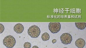 BrochureNeural Stem Cells: Standardized Media and Reagents