Hu Y-L et al. (SEP 2010)
Nucleic acids research 38 16 5472--8
HOXA9 regulates miR-155 in hematopoietic cells.
HOXA9-mediated up-regulation of miR-155 was noted during an array-based analysis of microRNA expression in Hoxa9(-/-)bone marrow (BM) cells. HOXA9 induction of miR-155 was confirmed in these samples,as well as in wild-type versus Hoxa9-deficient marrow,using northern analysis and qRT-PCR. Infection of wild-type BM with HOXA9 expressing or GFP(+) control virus further confirmed HOXA9-mediated regulation of miR-155. miR-155 expression paralleled Hoxa9 mRNA expression in fractionated BM progenitors,being highest in the stem cell enriched pools. HOXA9 capacity to induce myeloid colony formation was blunted in miR-155-deficient BM cells,indicating that miR-155 is a downstream mediator of HOXA9 function in blood cells. Pu.1,an important regulator of myelopoiesis,was identified as a putative down stream target for miR-155. Although miR-155 was shown to down-regulate the Pu.1 protein,HOXA9 did not appear to modulate Pu.1 expression in murine BM cells.
View Publication
Slug deficiency enhances self-renewal of hematopoietic stem cells during hematopoietic regeneration.
Both extrinsic and intrinsic mechanisms tightly govern hematopoietic stem cell (HSC) decisions of self-renewal and differentiation. However,transcription factors that can selectively regulate HSC self-renewal division after stress remain to be identified. Slug is an evolutionarily conserved zinc-finger transcription factor that is highly expressed in primitive hematopoietic cells and is critical for the radioprotection of these key cells. We studied the effect of Slug in the regulation of HSCs in Slug-deficient mice under normal and stress conditions using serial functional assays. Here,we show that Slug deficiency does not disturb hematopoiesis or alter HSC homeostasis and differentiation in bone marrow but increases the numbers of primitive hematopoietic cells in the extramedullary spleen site. Deletion of Slug enhances HSC repopulating potential but not its homing and differentiation ability. Furthermore,Slug deficiency increases HSC proliferation and repopulating potential in vivo after myelosuppression and accelerates HSC expansion during in vitro culture. Therefore,we propose that Slug is essential for controlling the transition of HSCs from relative quiescence under steady-state condition to rapid proliferation under stress conditions. Our data suggest that inhibition of Slug in HSCs may present a novel strategy for accelerating hematopoietic recovery,thus providing therapeutic benefits for patients after clinical myelosuppressive treatment.
View Publication
产品类型:
产品号#:
09600
09650
28600
产品名:
StemSpan™ SFEM
StemSpan™ SFEM
L-Calc™有限稀释软件
文献
Allan LL et al. (SEP 2009)
Blood 114 12 2411--6
Apolipoprotein-mediated lipid antigen presentation in B cells provides a pathway for innate help by NKT cells.
Natural killer T (NKT) cells are innate-like lymphocytes that recognize lipid antigens and have been shown to enhance B-cell activation and antibody production. B cells typically recruit T-cell help by presenting internalized antigens recognized by their surface antigen receptor. Here,we demonstrate a highly efficient means whereby human B cells present lipid antigens to NKT cells,capturing the antigen using apolipoprotein E (apoE) and the low-density lipoprotein receptor (LDL-R). ApoE dramatically enhances B-cell presentation of alpha-galactosylceramide (alphaGalCer),an exogenous CD1d presented antigen,inducing activation of NKT cells and the subsequent activation of B cells. B cells express the LDL-R on activation,and the activation of NKT cells by B cells is completely LDL-R dependent,as shown by blocking experiments and the complete lack of presentation when using apoE2,an isoform of apoE incapable of LDL-R binding. The dependence on apoE and the LDL-R is much more pronounced in B cells than we had previously seen in dendritic cells,which can apparently use alternate pathways of lipid antigen uptake. Thus,B cells use an apolipoprotein-mediated pathway of lipid antigen presentation,which constitutes a form of innate help for B cells by NKT cells.
View Publication
Fibroblast growth factor-1 and -2 preserve long-term repopulating ability of hematopoietic stem cells in serum-free cultures.
In this study,we demonstrate that extended culture of unfractionated mouse bone marrow (BM) cells,in serum-free medium,supplemented only with fibroblast growth factor (FGF)-1,FGF-2,or FGF-1 +2 preserves long-term repopulating hematopoietic stem cells (HSCs). Using competitive repopulation assays,high levels of stem cell activity were detectable at 1,3,and 5 weeks after initiation of culture. FGFs as single growth factors failed to support cultures of highly purified Lin(-)Sca-1(+)c-Kit(+)(LSK) cells. However,cocultures of purified CD45.1 LSK cells with whole BM CD45.2 cells provided high levels of CD45.1 chimerism after transplant,showing that HSC activity originated from LSK cells. Subsequently,we tested the reconstituting potential of cells cultured in FGF-1 + 2 with the addition of early acting stimulatory molecules,stem cell factor +interleukin-11 + Flt3 ligand. The addition of these growth factors resulted in a strong mitogenic response,inducing rapid differentiation and thereby completely overriding FGF-dependent stem cell conservation. Importantly,although HSC activity is typically rapidly lost after short-term culture in vitro,our current protocol allows us to sustain stem cell repopulation potential for periods up to 5 weeks.
View Publication
产品类型:
产品号#:
09600
09650
产品名:
StemSpan™ SFEM
StemSpan™ SFEM
文献
Chang M-YY et al. (NOV 2015)
Stem cell research 15 3 608--613
Doxycycline supplementation allows for the culture of human ESCs/iPSCs with media changes at 3-day intervals.
Culturing human embryonic stem and induced pluripotent stem cells (hESCs/iPSCs) is one of the most costly and labor-intensive tissue cultures,as media containing expensive factors/cytokines should be changed every day to maintain and propagate undifferentiated hESCs/iPSCs in vitro. We recently reported that doxycycline,an anti-bacterial agent,had dramatic effects on hESC/iPSC survival and promoted self-renewal. In this study,we extended the effects of doxycycline to a more practical issue to save cost and labor in hESC/iPSC cultures. Regardless of cultured cell conditions,hESCs/iPSCs in doxycycline-supplemented media were viable and proliferating for at least 3 days without media change,while none or few viable cells were detected in the absence of doxycycline in the same conditions. Thus,hESCs/iPSCs supplemented with doxycycline can be cultured for a long period of time with media changes at 3-day intervals without altering their self-renewal and pluripotent properties,indicating that doxycycline supplementation can reduce the frequency of media changes and the amount of media required by 1/3. These findings strongly encourage the use of doxycycline to save cost and labor in culturing hESCs/iPSCs.
View Publication
Phosphoinositide 3-kinase signaling is essential for ABL oncogene-mediated transformation of B-lineage cells.
BCR-ABL and v-ABL are oncogenic forms of the Abl tyrosine kinase that can cause leukemias in mice and humans. ABL oncogenes trigger multiple signaling pathways whose contribution to transformation varies among cell types. Activation of phosphoinositide 3-kinase (PI3K) is essential for ABL-dependent proliferation and survival in some cell types,and global PI3K inhibitors can enhance the antileukemia effects of the Abl kinase inhibitor imatinib. Although a significant fraction of BCR-ABL-induced human leukemias are of B-cell origin,little is known about PI3K signaling mechanisms in B-lineage cells transformed by ABL oncogenes. Here we show that activation of class I(A) PI3K and downstream inactivation of FOXO transcription factors are essential for survival of murine pro/pre-B cells transformed by v-ABL or BCR-ABL. In addition,analysis of mice lacking individual PI3K genes indicates that products of the Pik3r1 gene contribute to transformation efficiency by BCR-ABL. These findings establish a role for PI3K signaling in B-lineage transformation by ABL oncogenes.
View Publication

 EasySep™小鼠TIL(CD45)正选试剂盒


 文献
文献 1:58
NeuroCult™ Proliferation Media
1:58
NeuroCult™ Proliferation Media
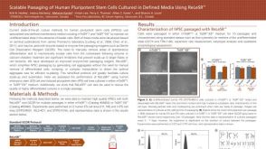 Scalable Passaging of Human Pluripotent Stem Cells Cultured in Defined Media Using ReLeSR™
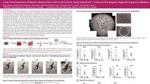 科学海报Long-Term Expansion of Mouse Hepatic Stem Cells in 3D Culture Using HepatiCult™: A Serum-Free Hepatic Organoid Expansion Medium
 沪公网安备31010102008431号