Hockemeyer D et al. (SEP 2008)
Cell stem cell 3 3 346--53
A drug-inducible system for direct reprogramming of human somatic cells to pluripotency.
Current approaches to reprogram human somatic cells to pluripotent iPSCs utilize viral transduction of different combinations of transcription factors. These protocols are highly inefficient because only a small fraction of cells carry the appropriate number and stoichiometry of proviral insertions to initiate the reprogramming process. Here we have generated genetically homogeneous secondary" somatic cells�
View Publication
产品类型:
产品号#:
72742
产品名:
Doxycycline (Hyclate)
文献
Pahwa R et al. (DEC 2010)
Journal of immunological methods 363 1 67--79
Isolation and expansion of human natural T regulatory cells for cellular therapy.
Natural T regulatory cells (nTregs) play a key role in inducing and maintaining immunological tolerance. Cell-based therapy using purified nTregs is under consideration for several conditions,but procedures employed to date have resulted in cell populations that are contaminated with cytokine secreting effector cells. We have established a method for isolation and ex vivo expansion of human nTregs from healthy blood donors for cellular therapy aimed at preventing allograft rejection in organ transplants. The Robosep instrument was used for initial nTreg isolation and rapamycin was included in the expansion phase of cell cultures. The resulting cell population exhibited a stable CD4(+)CD25(++bright)Foxp3(+) phenotype,had potent functional ability to suppress CD4(+)CD25(negative) T cells without evidence of conversion to effector T cells including TH17 cells,and manifested little to no production of pro-inflammatory cytokines upon in vitro stimulation. Boolean gating analysis of cytokine-expressing cells by flow cytometry for 32 possible profile end points revealed that 96% of expanded nTregs did not express any cytokine. From a single buffy coat,approximately 80 million pure nTregs were harvested after expansion under cGMP conditions; these cell numbers are adequate for infusion of approximately one million cells kg�?�¹ for cell therapy in clinical trials.
View Publication
Dobo I et al. (JAN 2001)
The hematology journal : the official journal of the European Haematology Association / EHA 2 6 396--403
Comparison of four serum-free, cytokine-free media for analysis of endogenous erythroid colony growth in polycythemia vera and essential thrombocythemia.
INTRODUCTION: The assay of endogenous erythroid colony formation (EEC),a characteristic of polycythemia vera and essential thrombocythemia,is not standardized. In this multicentric study,we tested four semisolid,serum-free,cytokine-free media based on either methylcellulose (M1,M2) or collagen (C1,C2) commercialized for the EEC assay. MATERIALS AND METHODS: Bone marrow mononuclear cells (BMMC) from 73 individuals (62 patients with either polycythemia vera (26),essential thrombocythemia (19),secondary polyglobuly (17) or chronic myeloid leukemia (2) and 11 healthy donors) were grown in parallel in the four media without,or with 0.01 U/ml erythropoietin (EPo). RESULTS: In all four media EEC formation was specific,as it was not observed in cultures of patients with secondary polyglobuly or chronic myeloid leukemia,nor of healthy donors. Analysis of fresh or MGG-stained collagen gel cultures allowed detection of EEC formation significantly more frequently than methylcellulose-based media; addition of 0.01 U/ml of EPo had little or no effect on EEC formation. Collagen-based medium C1 gave better results than the other media tested: the 'C1' EEC assay was positive for 68.2% of polycythemia vera cultures with significantly higher median EEC numbers (6.5/10(5) BMMC for patients with one major criteria of polycythemia vera and 19 and 21/10(5) BMMC for patients with two or three major criteria,respectively). Medium C1 was also better for essential thrombocythemia cultures with 47.4% of positive results but with a low median EEC number (6.7/10(5) BMMC). When associated with the ELISA dosage of serum EPo,the 'C1' EEC assay allowed confirmation or elimination of the diagnosis of polycythemia vera for 91% (20/22) of polyglobulic patients. CONCLUSION: We propose that serum-free collagen-based culture systems be considered to standardize the EEC assay,now part of the new criteria of polycythemia vera.
View Publication
产品类型:
产品号#:
04961
04962
04915
04807
04809
04906
04913
04803
04804
04905
04850
04974
04902
04960
04900
04901
04963
04970
04971
产品名:
MegaCult™-C胶原蛋白和细胞因子培养基
MegaCult™-C cfu染色试剂盒
MegaCult-C 10% BSA, 6mL
MegaCult-C Human Serum, 6mL
Alkaline Phosphatase Substrate Tabs, pk
Biotin/Conjugate Goat Anti-Mu lgG, 125uL
MegaCult-C Evans Blue Stain, 5mL
Primary Ab, Anti-HuAnti-GPIIb/IIIa 360uL
MegaCult-C Control Antibody, 100 µL
Avidin-Alk Phosphatase Conjugate, 200 uL
MegaCult™-C含脂培养基
MegaCult™-C胶原蛋白和脂质培养基
胶原蛋白溶液
MegaCult™-C胶原蛋白和不含细胞因子的培养基
MegaCult™-C培养基无细胞因子
MegaCult™-C细胞因子培养基
双室载玻片试剂盒
MegaCult™-C不含细胞因子完整试剂盒
MegaCult™-C细胞因子完整试剂盒
文献
Hess DA et al. (MAR 2006)
Blood 107 5 2162--9
Selection based on CD133 and high aldehyde dehydrogenase activity isolates long-term reconstituting human hematopoietic stem cells.
The development of novel cell-based therapies requires understanding of distinct human hematopoietic stem and progenitor cell populations. We recently isolated reconstituting hematopoietic stem cells (HSCs) by lineage depletion and purification based on high aldehyde dehydrogenase activity (ALDH(hi)Lin- cells). Here,we further dissected the ALDH(hi)-Lin- population by selection for CD133,a surface molecule expressed on progenitors from hematopoietic,endothelial,and neural lineages. ALDH(hi)CD133+Lin- cells were primarily CD34+,but also included CD34-CD38-CD133+ cells,a phenotype previously associated with repopulating function. Both ALDH(hi)CD133-Lin- and ALDH(hi)CD133+Lin- cells demonstrated distinct clonogenic progenitor function in vitro,whereas only the ALDH(hi)CD133+Lin- population seeded the murine bone marrow 48 hours after transplantation. Significant human cell repopulation was observed only in NOD/SCID and NOD/SCID beta2M-null mice that received transplants of ALDH(hi)CD133+Lin- cells. Limiting dilution analysis demonstrated a 10-fold increase in the frequency of NOD/SCID repopulating cells compared with CD133+Lin- cells,suggesting that high ALDH activity further purified cells with repopulating function. Transplanted ALDH(hi)CD133+Lin- cells also maintained primitive hematopoietic phenotypes (CD34+CD38-) and demonstrated enhanced repopulating function in recipients of serial,secondary transplants. Cell selection based on ALDH activity and CD133 expression provides a novel purification of HSCs with long-term repopulating function and may be considered an alternative to CD34 cell selection for stem cell therapies.
View Publication
产品类型:
产品号#:
01700
01705
01702
产品名:
ALDEFLUOR™工具
ALDEFLUOR™ DEAB试剂
ALDEFLUOR™测定缓冲液
文献
Haniffa M et al. (FEB 2009)
The Journal of experimental medicine 206 2 371--85
Differential rates of replacement of human dermal dendritic cells and macrophages during hematopoietic stem cell transplantation.
Animal models of hematopoietic stem cell transplantation have been used to analyze the turnover of bone marrow-derived cells and to demonstrate the critical role of recipient antigen-presenting cells (APC) in graft versus host disease (GVHD). In humans,the phenotype and lineage relationships of myeloid-derived tissue APC remain incompletely understood. It has also been proposed that the risk of acute GVHD,which extends over many months,is related to the protracted survival of certain recipient APC. Human dermis contains three principal subsets of CD45(+)HLA-DR(+) cells: CD1a(+)CD14(-) DC,CD1a(-)CD14(+) DC,and CD1a(-)CD14(+)FXIIIa(+) macrophages. In vitro,each subset has characteristic properties. After transplantation,both CD1a(+) and CD14(+) DC are rapidly depleted and replaced by donor cells,but recipient macrophages can be found in GVHD lesions and may persist for many months. Macrophages isolated from normal dermis secrete proinflammatory cytokines. Although they stimulate little proliferation of naive or memory CD4(+) T cells,macrophages induce cytokine expression in memory CD4(+) T cells and activation and proliferation of CD8(+) T cells. These observations suggest that dermal macrophages and DC are from distinct lineages and that persistent recipient macrophages,although unlikely to initiate alloreactivity,may contribute to GVHD by sustaining the responses of previously activated T cells.
View Publication
产品类型:
产品号#:
产品名:
文献
Larochelle A et al. (FEB 2011)
Blood 117 5 1550--4
Human and rhesus macaque hematopoietic stem cells cannot be purified based only on SLAM family markers.
Various combinations of antibodies directed to cell surface markers have been used to isolate human and rhesus macaque hematopoietic stem cells (HSCs). These protocols result in poor enrichment or require multiple complex steps. Recently,a simple phenotype for HSCs based on cell surface markers from the signaling lymphocyte activation molecule (SLAM) family of receptors has been reported in the mouse. We examined the possibility of using the SLAM markers to facilitate the isolation of highly enriched populations of HSCs in humans and rhesus macaques. We isolated SLAM (CD150(+)CD48(-)) and non-SLAM (not CD150(+)CD48(-)) cells from human umbilical cord blood CD34(+) cells as well as from human and rhesus macaque mobilized peripheral blood CD34(+) cells and compared their ability to form colonies in vitro and reconstitute immune-deficient (nonobese diabetic/severe combined immunodeficiency/interleukin-2 γc receptor(null),NSG) mice. We found that the CD34(+) SLAM population contributed equally or less to colony formation in vitro and to long-term reconstitution in NSG mice compared with the CD34(+) non-SLAM population. Thus,SLAM family markers do not permit the same degree of HSC enrichment in humans and rhesus macaques as in mice.
View Publication
产品类型:
产品号#:
04435
04445
产品名:
MethoCult™H4435富集
MethoCult™H4435富集
文献
Romanov YA et al. (JAN 2003)
Stem cells (Dayton,Ohio) 21 1 105--10
Searching for alternative sources of postnatal human mesenchymal stem cells: candidate MSC-like cells from umbilical cord.
Mesenchymal stem cells (MSCs) have the capability for renewal and differentiation into various lineages of mesenchymal tissues. These features of MSCs attract a lot of attention from investigators in the context of cell-based therapies of several human diseases. Despite the fact that bone marrow represents the main available source of MSCs,the use of bone marrow-derived cells is not always acceptable due to the high degree of viral infection and the significant drop in cell number and proliferative/differentiation capacity with age. Thus,the search for possible alternative MSC sources remains to be validated. Umbilical cord blood is a rich source of hematopoietic stem/progenitor cells and does not contain mesenchymal progenitors. However,MSCs circulate in the blood of preterm fetuses and may be successfully isolated and expanded. Where these cells home at the end of gestation is not clear. In this investigation,we have made an attempt to isolate MSCs from the subendothelial layer of umbilical cord vein using two standard methodological approaches: the routine isolation of human umbilical vein endothelial cell protocol and culture of isolated cells under conditions appropriate for bone-marrow-derived MSCs. Our results suggest that cord vasculature contains a high number of MSC-like elements forming colonies of fibroblastoid cells that may be successfully expanded in culture. These MSC-like cells contain no endothelium- or leukocyte-specific antigens but express alpha-smooth muscle actin and several mesenchymal cell markers. Therefore,umbilical cord/placenta stroma could be regarded as an alternative source of MSCs for experimental and clinical needs.
View Publication
Akatsuka A et al. (SEP 2010)
International immunology 22 9 783--90
Tumor cells of non-hematopoietic and hematopoietic origins express activation-induced C-type lectin, the ligand for killer cell lectin-like receptor F1.
Killer cell lectin-like receptor F1 (KLRF1) is an activating C-type lectin-like receptor expressed on human NK cells and subsets of T cells. In this study,we show that activation-induced C-type lectin (AICL) is a unique KLRF1 ligand expressed on tumor cell lines of hematopoietic and non-hematopoietic origins. We screened a panel of human tumor cell lines using the KLRF1 reporter cells and found that several tumor lines expressed KLRF1 ligands. We characterized a putative KLRF1 ligand expressed on the U937 cell line. The molecular mass for the deglycosylated ligand was 28 kDa under non-reducing condition and 17 kDa under reducing condition,suggesting that the KLRF1 ligand is a homodimer. By expression cloning from a U937 cDNA library,we identified AICL as a KLRF1 ligand. We generated mAbs against AICL to identify the KLRF1 ligands on non-hematopoietic tumor lines. The anti-AICL mAbs stained the tumor lines that express the KLRF1 ligands and importantly the interaction of KLRF1 with the KLRF1 ligand on non-hematopoietic tumors was completely blocked by the two anti-AICL mAbs. Moreover,NK cell degranulation triggered by AICL-expressing targets was partially inhibited by the anti-AICL mAb. Finally,we demonstrate that AICL is expressed in human primary liver cancers. These results suggest that AICL is expressed on tumor cells of non-hematopoietic origins and raise the possibility that AICL may contribute to NK cell surveillance of tumor cells.
View Publication
产品类型:
产品号#:
18554
18554RF
18564
18564RF
产品名:
文献
Hawley RG et al. (JAN 2006)
Methods in enzymology 419 149--79
Hematopoietic stem cells.
Hematopoietic stem cells (HSCs) have the capacity to self-renew and the potential to differentiate into all of the mature blood cell types. The ability to prospectively identify and isolate HSCs has been the subject of extensive investigation since the first transplantation studies implying their existence almost 50 years ago. Despite significant advances in enrichment protocols,the continuous in vitro propagation of human HSCs has not yet been achieved. This chapter describes current procedures used to phenotypically and functionally characterize candidate human HSCs and initial efforts to derive permanent human HSC lines.
View Publication

 EasySep™小鼠TIL(CD45)正选试剂盒


 文献
文献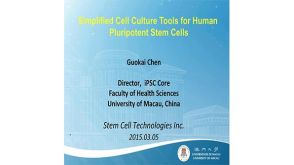 57:55
Development of Simplified and Defined Cell Culture for Human Pluripotent Stem Cells
57:55
Development of Simplified and Defined Cell Culture for Human Pluripotent Stem Cells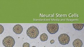 Neural Stem Cells: Standardized Media and Reagents
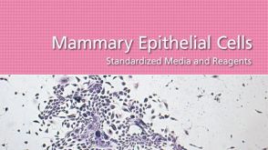 Mammary Epithelial Cells: Standardized Media and Reagents
 沪公网安备31010102008431号