Woods EJ et al. (OCT 2009)
Cryobiology 59 2 150--7
Optimized cryopreservation method for human dental pulp-derived stem cells and their tissues of origin for banking and clinical use.
Dental pulp is a promising source of mesenchymal stem cells with the potential for cell-mediated therapies and tissue engineering applications. We recently reported that isolation of dental pulp-derived stem cells (DPSC) is feasible for at least 120h after tooth extraction,and that cryopreservation of early passage cultured DPSC leads to high-efficiency recovery post-thaw. This study investigated additional processing and cryobiological characteristics of DPSC,ending with development of procedures for banking. First,we aimed to optimize cryopreservation of established DPSC cultures,with regards to optimizing the cryoprotective agent (CPA),the CPA concentration,the concentration of cells frozen,and storage temperatures. Secondly,we focused on determining cryopreservation characteristics of enzymatically digested tissue as a cell suspension. Lastly,we evaluated the growth,surface markers and differentiation properties of DPSC obtained from intact teeth and undigested,whole dental tissue frozen and thawed using the optimized procedures. In these experiments it was determined that Me(2)SO at a concentration between 1 and 1.5M was the ideal cryopreservative of the three studied. It was also determined that DPSC viability after cryopreservation is not limited by the concentration of cells frozen,at least up to 2x10(6) cells/mL. It was further established that DPSC can be stored at -85 degrees C or -196 degrees C for at least six months without loss of functionality. The optimal results with the least manipulation were achieved by isolating and cryopreserving the tooth pulp tissues,with digestion and culture performed post-thaw. A recovery of cells from textgreater85% of the tissues frozen was achieved and cells isolated post-thaw from tissue processed and frozen with a serum free,defined cryopreservation medium maintained morphological and developmental competence and demonstrated MSC-hallmark trilineage differentiation under the appropriate culture conditions.
View Publication
Porayette P et al. (DEC 2007)
Biochemical and biophysical research communications 364 3 522--527
Amyloid-?? precursor protein expression and modulation in human embryonic stem cells: A novel role for human chorionic gonadotropin
The amyloid-beta precursor protein (AbetaPP) is a ubiquitously expressed adhesion and neuritogenic protein whose processing has previously been shown to be regulated by reproductive hormones including the gonadotropin luteinizing hormone (LH) in human neuroblastoma cells. We report for the first time the expression of AbetaPP in human embryonic stem (hES) cells at the mRNA and protein levels. Using N- and C-terminal antibodies against AbetaPP,we detected both the mature and immature forms of AbetaPP as well as truncated variants ( approximately 53kDa,47kDa,and 29kDa) by immunoblot analyses. Expression of AbetaPP is regulated by both the stemness of the cells and pregnancy-associated hormones. Addition of human chorionic gonadotropin,the fetal equivalent of LH that is dramatically elevated during pregnancy,markedly increased the expression of all AbetaPP forms. These results indicate a critical molecular signaling link between the hormonal environment of pregnancy and the expression of AbetaPP in hES cells that is suggestive of an important function for this protein during early human embryogenesis prior to the formation of neural precursor cells.
View Publication
产品类型:
产品号#:
85850
85857
产品名:
mTeSR™1
mTeSR™1
文献
Srour EF et al. (APR 2005)
Blood 105 8 3109--16
Modulation of in vitro proliferation kinetics and primitive hematopoietic potential of individual human CD34+CD38-/lo cells in G0.
Whether cytokines can modulate the fate of primitive hematopoietic progenitor cells (HPCs) through successive in vitro cell divisions has not been established. Single human marrow CD34+CD38-/lo cells in the G0 phase of cell cycle were cultured under 7 different cytokine combinations,monitored for proliferation on days 3,5,and 7,then assayed for long-term culture-initiating cell (LTC-IC) function on day 7. LTC-IC function was then retrospectively correlated with prior number of in vitro cell divisions to determine whether maintenance of LTC-IC function after in vitro cell division is dependent on cytokine exposure. In the presence of proliferation progression signals,initial cell division was independent of cytokine stimulation,suggesting that entry of primitive HPCs into the cell cycle is a stochastic property. However,kinetics of proliferation beyond day 3 and maintenance of LTC-IC function were sensitive to cytokine stimulation,such that LTC-IC underwent an initial long cell cycle,followed by more synchronized shorter cycles varying in length depending on the cytokine combination. Nonobese diabetic/severe combined immunodeficiency (NOD/SCID) transplantation studies revealed analogous results to those obtained with LTC-ICs. These data suggest that although exit from quiescence and commitment to proliferation might be stochastic,kinetics of proliferation,and possibly fate of primitive HPCs,might be modulated by extrinsic factors.
View Publication
ErbB4 Activated p38$$ MAPK Isoform Mediates Early Cardiogenesis Through NKx2.5 in Human Pluripotent Stem Cells
Activation of ErbB4 receptor signaling is instrumental in heart development,lack of which results in embryonic lethality. However,mechanism governing its intracellular signaling remains elusive. Using human pluripotent stem cells,we show that ErbB4 is critical for cardiogenesis whereby its genetic knockdown results in loss of cardiomyocytes. Phospho-proteome profiling and Western blot studies attribute this loss to inactivation of p38$\$ isoform which physically interacts with NKx2.5 and GATA4 transcription factors. Post-cardiomyocyte formation p38$\$/NKx2.5 downregulation is followed by p38$\$/MEF2c upregulation suggesting stage-specific developmental roles of p38 MAPK isoforms. Knockdown of p38$\$ similarly disrupts cardiomyocyte formation in spite of the presence of NKx2.5. Cell fractionation and NKx2.5 phosphorylation studies suggest inhibition of ErbB4-p38$\$ hinders NKx2.5 nuclear translocation during early cardiogenesis. This study reveals a novel pathway that directly links ErbB4 and p38$\$ the transcriptional machinery of NKx2.5-GATA4 complex which is critical for cardiomyocyte formation during mammalian heart development.
View Publication
产品类型:
产品号#:
85850
85857
产品名:
mTeSR™1
mTeSR™1
文献
Wagner W et al. (OCT 2007)
Stem cells (Dayton,Ohio) 25 10 2638--47
Molecular and secretory profiles of human mesenchymal stromal cells and their abilities to maintain primitive hematopoietic progenitors.
Mesenchymal stromal cells (MSC) provide a supportive cellular microenvironment and are able to maintain the self-renewal capacity of hematopoietic progenitor cells (HPC). Isolation procedures for MSC vary extensively,and this may influence their biologic properties. In this study,we have compared human MSC isolated from bone marrow (BM) using two culture conditions,from cord blood (CB),and from adipose tissue (AT). The ability to maintain long-term culture-initiating cell frequency and a primitive CD34(+)CD38(-) immunophenotype was significantly higher for MSC derived from BM and CB compared with those from AT. These results were in line with a significantly higher adhesion of HPC to MSC from BM and CB versus MSC from AT. We have compared the cytokine production of MSC by cytokine antibody arrays,enzyme-linked immunosorbent assay,and a cytometric bead array. There were reproducible differences in the chemokine secretion profiles of various MSC preparations,but there was no clear concordance with differences in their potential to maintain primitive function of HPC. Global gene expression profiles of MSC preparations were analyzed and showed that adhesion proteins including cadherin-11,N-cadherin,vascular cell adhesion molecule 1,neural cell adhesion molecule 1,and integrins were highly expressed in MSC preparations derived from BM and CB. Thus,MSC from BM and CB are superior to MSC from AT for maintenance of primitive HPC. The latter property is associated with specific molecular profiles indicating the significance of cell-cell junctions but not with secretory profiles. Disclosure of potential conflicts of interest is found at the end of this article.
View Publication
产品类型:
产品号#:
05401
05402
05411
产品名:
MesenCult™ MSC基础培养基 (人)
MesenCult™ MSC 刺激补充剂(人)
MesenCult™ 增殖试剂盒(人)
文献
Chen G et al. (AUG 2010)
Cell stem cell 7 2 240--8
Actin-myosin contractility is responsible for the reduced viability of dissociated human embryonic stem cells.
Human ESCs are the pluripotent precursor of the three embryonic germ layers. Human ESCs exhibit basal-apical polarity,junctional complexes,integrin-dependent matrix adhesion,and E-cadherin-dependent cell-cell adhesion,all characteristics shared by the epiblast epithelium of the intact mammalian embryo. After disruption of epithelial structures,programmed cell death is commonly observed. If individualized human ESCs are prevented from reattaching and forming colonies,their viability is significantly reduced. Here,we show that actin-myosin contraction is a critical effector of the cell death response to human ESC dissociation. Inhibition of myosin heavy chain ATPase,downregulation of myosin heavy chain,and downregulation of myosin light chain all increase survival and cloning efficiency of individualized human ESCs. ROCK inhibition decreases phosphorylation of myosin light chain,suggesting that inhibition of actin-myosin contraction is also the mechanism through which ROCK inhibitors increase cloning efficiency of human ESCs.
View Publication
产品类型:
产品号#:
72402
72404
85850
85857
产品名:
(-)-Blebbistatin
(-)-Blebbistatin
mTeSR™1
mTeSR™1
文献
Du S-HH et al. (AUG 2015)
Journal of bioscience and bioengineering 120 2 210--217
Human iPS cell-derived fibroblast-like cells as feeder layers for iPS cell derivation and expansion
Mouse embryonic fibroblasts (MEFs) are commonly used as feeder cells for the generation of human induced pluripotent stem cells (hiPSCs). However,medical applications of cell derivatives of hiPSCs generated with a MEF feeder system run the risk of having xeno-factor contamination due to long-term cell culturing under an animal factor-containing environment. We developed a new method for the derivation of human fibroblast-like cells (FLCs) from a previously established hiPSC line in an FLC differentiation medium. The method was based on direct differentiation of hiPSCs seeded on Matrigel followed by expansion of differentiating cells on gelatin. Using inactivated FLCs as feeder layers,primary human foreskin fibroblasts were successfully reprogrammed into a state of pluripotency by Oct4,Sox2 Klf4,and c-Myc (OSKM) transcription factor genes,with a reprogramming efficiency under an optimized condition superior to that obtained on MEF feeder layers. Furthermore,the FLCs were more effective in supporting the growth of human pluripotent stem cells. The pluripotency and differentiation capability of the cells cultured on FLC feeder layers were well retained. Our results suggest that FLCs are a safe alternative to MEFs for hiPSC generation and expansion,especially in the clinical settings wherein hiPSC derivatives will be used for medical treatment.
View Publication
A. Holtzinger et al. ( 2015)
Development (Cambridge,England) 142 4253-65
New markers for tracking endoderm induction and hepatocyte differentiation from human pluripotent stem cells.
The efficient generation of hepatocytes from human pluripotent stem cells (hPSCs) requires the induction of a proper endoderm population,broadly characterized by the expression of the cell surface marker CXCR4. Strategies to identify and isolate endoderm subpopulations predisposed to the liver fate do not exist. In this study,we generated mouse monoclonal antibodies against human embryonic stem cell-derived definitive endoderm with the goal of identifying cell surface markers that can be used to track the development of this germ layer and its specification to a hepatic fate. Through this approach,we identified two endoderm-specific antibodies,HDE1 and HDE2,which stain different stages of endoderm development and distinct derivative cell types. HDE1 marks a definitive endoderm population with high hepatic potential,whereas staining of HDE2 tracks with developing hepatocyte progenitors and hepatocytes. When used in combination,the staining patterns of these antibodies enable one to optimize endoderm induction and hepatic specification from any hPSC line.
View Publication
产品类型:
产品号#:
产品名:
文献
Behar RZ et al. (NOV 2012)
Journal of Pharmacological and Toxicological Methods 66 3 238--245
A method for rapid dose-response screening of environmental chemicals using human embryonic stem cells
Introduction: Human embryonic stem cells (hESC) provide an invaluable model for assessing the effects of environmental chemicals and drugs on human prenatal development. However,hESC are difficult to adapt to 96-well plate screening assays,because they survive best when plated as colonies,which are difficult to count and plate accurately. The purpose of this study is to present an experimental method and analysis procedure to accomplish reliable screening of toxicants using hESC. Methods: We present a method developed to rapidly and easily determine the number of cells in small colonies of hESC spectrophotometerically and then accurately dispense equivalent numbers of cells in 96-well plates. The MTT assay was used to evaluate plating accuracy,and the method was tested using known toxicants. Results: The quality of the plate set-up and analysis procedure was evaluated with NIH plate validation and assessment software. All statistical parameters measured by the software were acceptable,and no drift or edge effects were observed. The 96-well plate MTT assay with hESC was tested by performing a dose-response screen of commercial products,which contain a variety of chemicals. The screen was done using single wells/dose,and the reliability of this method was demonstrated in a subsequent screen of the same products repeated three times. The single and triple screens were in good agreement,and NOAELs and IC50s could be determined from the single screen. The effects of vapor from volatile chemicals were studied,and methods to monitor and avoid vapor effects were incorporated into the assay. Discussion: Our method overcomes the difficulty of using hESC for reliable quantitative 96-well plate assays. It enables rapid dose-response screening using equipment that is commonly available in laboratories that culture hESC. This method could have a broad application in studies of environmental chemicals and drugs using hESC as models of prenatal development. ?? 2012 Elsevier Inc.
View Publication
产品类型:
产品号#:
85850
85857
产品名:
mTeSR™1
mTeSR™1
文献
Kang L et al. ( 2013)
Frontiers in immunology 4 MAY 101
Characterization and ex vivo Expansion of Human Placenta-Derived Natural Killer Cells for Cancer Immunotherapy.
Recent clinical studies suggest that adoptive transfer of donor-derived natural killer (NK) cells may improve clinical outcome in hematological malignancies and some solid tumors by direct anti-tumor effects as well as by reduction of graft versus host disease (GVHD). NK cells have also been shown to enhance transplant engraftment during allogeneic hematopoietic stem cell transplantation (HSCT) for hematological malignancies. The limited ex vivo expansion potential of NK cells from peripheral blood (PB) or umbilical cord blood (UCB) has however restricted their therapeutic potential. Here we define methods to efficiently generate NK cells from donor-matched,full-term human placenta perfusate (termed Human Placenta-Derived Stem Cell,HPDSC) and UCB. Following isolation from cryopreserved donor-matched HPDSC and UCB units,CD56+CD3- placenta-derived NK cells,termed pNK cells,were expanded in culture for up to 3 weeks to yield an average of 1.2 billion cells per donor that were textgreater80% CD56+CD3-,comparable to doses previously utilized in clinical applications. Ex vivo-expanded pNK cells exhibited a marked increase in anti-tumor cytolytic activity coinciding with the significantly increased expression of NKG2D,NKp46,and NKp44 (p textless 0.001,p textless 0.001,and p textless 0.05,respectively). Strong cytolytic activity was observed against a wide range of tumor cell lines in vitro. pNK cells display a distinct microRNA (miRNA) expression profile,immunophenotype,and greater anti-tumor capacity in vitro compared to PB NK cells used in recent clinical trials. With further development,pNK may represent a novel and effective cellular immunotherapy for patients with high clinical needs and few other therapeutic options.
View Publication

 EasySep™小鼠TIL(CD45)正选试剂盒


 文献
文献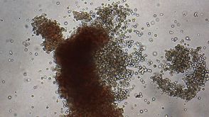 点播Human Hematopoietic CFU Assay Course Learn how to perform the CFU assay with the CFU Assay Starter Kit.
 MesenCult™-hPL Medium For the Derivation and Expansion of Human Mesenchymal Stem and Progenitor Cells
 沪公网安备31010102008431号