Chromosome 13 abnormalities identified by FISH analysis and serum beta2-microglobulin produce a powerful myeloma staging system for patients receiving high-dose therapy.
A careful prognostic evaluation of patients referred for high-dose therapy (HDT) is warranted to identify those who maximally benefit from HDT as well as those who clearly fail current HDT and are candidates for more innovative treatments. In a series of 110 patients with myeloma who received HDT as first-line therapy,times to event (disease progression and death) were studied through proportional hazard models,in relation to different prognostic factors,including a chromosome 13 fluorescence in situ hybridization (FISH) analysis using a D13S319 probe. Delta13 was detected in 42 patients (38%). Follow-up time among surviving patients and survival time were 48 +/- 3 and 51 +/- 7 months,respectively (median +/- SE). In the univariate analysis,Delta13 was the most powerful adverse prognostic factor for all times to event,especially for the survival time (P textless.0001) and was followed by beta2-microglobulin (beta2m) levels 2.5 mg/L or higher (P =.0001). The comparison of survival prognostic models including beta2m 2.5 mg/L or greater and another factor favored the Delta13/beta2m combination. In 22 patients (20%) with no unfavorable factor,the median survival time was not reached at 111 months. In contrast,among 55 patients (50%) with one unfavorable factor and 33 patients (30%) with 2 unfavorable factors,median survival times were 47.3 +/- 4.6 months and 25.3 +/- 3.2 months,respectively (P textless.0001). We conclude that delta13,adequately detected by FISH analysis,is a very strong factor related to poor survival,especially when associated with a beta2m level of 2.5 mg/L or higher. Routine FISH Delta13 assessment is strongly recommended for patients considered for HDT.
View Publication
T. J. Lynch et al. (MAY 2018)
Cell stem cell 22 5 653--667.e5
Submucosal Gland Myoepithelial Cells Are Reserve Stem Cells That Can Regenerate Mouse Tracheal Epithelium.
The mouse trachea is thought to contain two distinct stem cell compartments that contribute to airway repair-basal cells in the surface airway epithelium (SAE) and an unknown submucosal gland (SMG) cell type. Whether a lineage relationship exists between these two stem cell compartments remains unclear. Using lineage tracing of glandular myoepithelial cells (MECs),we demonstrate that MECs can give rise to seven cell types of the SAE and SMGs following severe airway injury. MECs progressively adopted a basal cell phenotype on the SAE and established lasting progenitors capable of further regeneration following reinjury. MECs activate Wnt-regulated transcription factors (Lef-1/TCF7) following injury and Lef-1 induction in cultured MECs promoted transition to a basal cell phenotype. Surprisingly,dose-dependent MEC conditional activation of Lef-1 in vivo promoted self-limited airway regeneration in the absence of injury. Thus,modulating the Lef-1 transcriptional program in MEC-derived progenitors may have regenerative medicine applications for lung diseases.
View Publication
产品类型:
产品号#:
05001
05021
05022
产品名:
PneumaCult™阿里介质
PneumaCult™阿里介质
PneumaCult™阿里介质
文献
Yau WW et al. (JAN 2011)
Proteome science 9 1 3
Cardiogenol C can induce Mouse Hair Bulge Progenitor Cells to Transdifferentiate into Cardiomyocyte-like Cells.
BACKGROUND: Hair bulge progenitor cells (HBPCs) are multipotent stem cells derived from the bulge region of mice vibrissal hairs. The purified HBPCs express CD34,K15 and K14 surface markers. It has been reported that HBPCs could be readily induced to transdifferentiate into adipocytes and osteocytes. However,the ability of HBPCs to transdifferentiate into cardiomyocytes has not yet been investigated. METHODOLOGY/PRINCIPAL FINDINGS: The cardiomyogenic potential of HBPCs was investigated using a small cell-permeable molecule called Cardiogenol C. We established that Cardiogenol C could induce HBPCs to express transcription factors GATA4,Nkx2.5 and Tbx5,which are early specific markers for pre-cardiomyogenic cells. In prolonged cultures,the Cardiogenol C-treated HBPCs can also express muscle proteins,cardiac-specific troponin I and sarcomeric myosin heavy chain. However,we did not observe the ability of these cells to functionally contract. Hence,we called these cells cardiomyocyte-like cells rather than cardiomyocytes. We tried to remedy this deficiency by pre-treating HBPCs with Valproic acid first before exposing them to Cardiogenol C. This pretreatment inhibited,rather than improved,the effectiveness of Cardiogenol C in reprogramming the HBPCs. We used comparative proteomics to determine how Cardiogenol C worked by identifying proteins that were differentially expressed. We identified proteins that were involved in promoting cell differentiation,cardiomyocyte development and for the normal function of striated muscles. From those differentially expressed proteins,we further propose that Cardiogenol C might exert its effect by activating the Wnt signaling pathway through the suppression of Kremen1. In addition,by up-regulating the expression of chromatin remodeling proteins,SIK1 and Smarce1 would initiate cardiac differentiation. CONCLUSIONS/SIGNIFICANCE: In conclusion,our CD34+/K15+ HBPCs could be induced to transdifferentiate into cardiomyocyte-like cells using a small molecule called Cardiogenol C. The process involves activation of the Wnt signaling pathway and altered expression of several key chromatin remodeling proteins. The finding is clinically significant as HBPCs offer a readily accessible and autologous source of progenitor cells for cell-based therapy of heart disease,which is one of major killers in developed countries.
View Publication
A critical role for SHP2 in STAT5 activation and growth factor-mediated proliferation, survival, and differentiation of human CD34+ cells.
SHP2,a cytoplasmic protein-tyrosine phosphatase encoded by the PTPN11 gene,plays a critical role in developmental hematopoiesis in the mouse,and gain-of-function mutations of SHP2 are associated with hematopoietic malignancies. However,the role of SHP2 in adult hematopoiesis has not been addressed in previous studies. In addition,the role of SHP2 in human hematopoiesis has not been described. These questions are of considerable importance given the interest in development of SHP2 inhibitors for cancer treatment. We used shRNA-mediated inhibition of SHP2 expression to investigate the function of SHP2 in growth factor (GF) signaling in normal human CD34(+) cells. SHP2 knockdown resulted in markedly reduced proliferation and survival of cells cultured with GF,and reduced colony-forming cell growth. Cells expressing gain-of-function SHP2 mutations demonstrated increased dependency on SHP2 expression for survival compared with cells expressing wild-type SHP2. SHP2 knockdown was associated with significantly reduced myeloid and erythroid differentiation with retention of CD34(+) progenitors with enhanced proliferative capacity. Inhibition of SHP2 expression initially enhanced and later inhibited STAT5 phosphorylation and reduced expression of the antiapoptotic genes MCL1 and BCLXL. These results indicate an important role for SHP2 in STAT5 activation and GF-mediated proliferation,survival,and differentiation of human progenitor cells.
View Publication
产品类型:
产品号#:
09600
09650
产品名:
StemSpan™ SFEM
StemSpan™ SFEM
文献
Mahdipour E et al. (JAN 2011)
Blood 117 3 815--26
Hoxa3 promotes the differentiation of hematopoietic progenitor cells into proangiogenic Gr-1+CD11b+ myeloid cells.
Injury induces the recruitment of bone marrow-derived cells (BMDCs) that contribute to the repair and regeneration process. The behavior of BMDCs in injured tissue has a profound effect on repair,but the regulation of BMDC behavior is poorly understood. Aberrant recruitment/retention of these cells in wounds of diabetic patients and animal models is associated with chronic inflammation and impaired healing. BMD Gr-1(+)CD11b(+) cells function as immune suppressor cells and contribute significantly to tumor-induced neovascularization. Here we report that Gr-1(+)CD11b(+) cells also contribute to injury-induced neovascularization,but show altered recruitment/retention kinetics in the diabetic environment. Moreover,diabetic-derived Gr-1(+)CD11b(+) cells fail to stimulate neovascularization in vivo and have aberrant proliferative,chemotaxis,adhesion,and differentiation potential. Previously we demonstrated that gene transfer of HOXA3 to wounds of diabetic mice is taken up by and expressed by recruited BMDCs. This is associated with a suppressed inflammatory response,enhanced neovascularization,and accelerated wound healing. Here we show that sustained expression of Hoxa3 in diabetic-derived BMD Gr-1(+)CD11b(+) cells reverses their diabetic phenotype. These findings demonstrate that manipulation of adult stem/progenitor cells ex vivo could be used as a potential therapy in patients with impaired wound healing.
View Publication
Characterization of primitive hematopoietic cells from patients with dyskeratosis congenita.
Dyskeratosis congenita (DC) is an inherited bone marrow (BM) failure syndrome associated with mutations in telomerase genes and the acquisition of shortened telomeres in blood cells. To investigate the basis of the compromised hematopoiesis seen in DC,we analyzed cells from granulocyte colony-stimulating factor mobilized peripheral blood (mPB) collections from 5 members of a family with autosomal dominant DC with a hTERC mutation. Premobilization BM samples were hypocellular,and percentages of CD34(+) cells in marrow and mPB collections were significantly below values for age-matched controls in 4 DC subjects. Directly clonogenic cells,although present at normal frequencies within the CD34(+) subset,were therefore absolutely decreased. In contrast,even the frequency of long-term culture-initiating cells within the CD34(+) DC mPB cells was decreased,and the telomere lengths of these cells were also markedly reduced. Nevertheless,the different lineages of mature cells were produced in normal numbers in vitro. These results suggest that marrow failure in DC is caused by a reduction in the ability of hematopoietic stem cells to sustain their numbers due to telomere impairment rather than a qualitative defect in their commitment to specific lineages or in the ability of their lineage-restricted progeny to execute normal differentiation programs.
View Publication

 EasySep™小鼠TIL(CD45)正选试剂盒


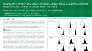 Enhanced Proliferation of Multipotent Human Adipose Tissue-Derived Mesenchymal Progenitor Cells Cultured in a Novel Xeno-Free Media
 文献
文献 Products for Human Pluripotent Stem Cells
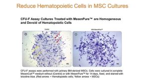 43:13
Tips & Techniques for Highly Enriched Mouse MSC Cultures As Early As Passage 0
43:13
Tips & Techniques for Highly Enriched Mouse MSC Cultures As Early As Passage 0
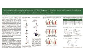 Cell Isolation of Functional CD4+CD25+ Regulatory T Cells from Mouse Strains
 5:34
How to Isolate Mesenchymal Stromal Cells (MSCs) from Mouse Compact Bone
5:34
How to Isolate Mesenchymal Stromal Cells (MSCs) from Mouse Compact Bone
 沪公网安备31010102008431号