G. La Manno et al. (OCT 2016)
Cell 167 2 566--580.e19
Molecular Diversity of Midbrain Development in Mouse, Human, and Stem Cells.
Understanding human embryonic ventral midbrain is of major interest for Parkinson's disease. However,the cell types,their gene expression dynamics,and their relationship to commonly used rodent models remain to be defined. We performed single-cell RNA sequencing to examine ventral midbrain development in human and mouse. We found 25 molecularly defined human cell types,including five subtypes of radial glia-like cells and four progenitors. In the mouse,two mature fetal dopaminergic neuron subtypes diversified into five adult classes during postnatal development. Cell types and gene expression were generally conserved across species,but with clear differences in cell proliferation,developmental timing,and dopaminergic neuron development. Additionally,we developed a method to quantitatively assess the fidelity of dopaminergic neurons derived from human pluripotent stem cells,at a single-cell level. Thus,our study provides insight into the molecular programs controlling human midbrain development and provides a foundation for the development of cell replacement therapies.
View Publication
Brambrink T et al. (FEB 2008)
Cell stem cell 2 2 151--9
Sequential expression of pluripotency markers during direct reprogramming of mouse somatic cells.
Pluripotency can be induced in differentiated murine and human cells by retroviral transduction of Oct4,Sox2,Klf4,and c-Myc. We have devised a reprogramming strategy in which these four transcription factors are expressed from doxycycline (dox)-inducible lentiviral vectors. Using these inducible constructs,we derived induced pluripotent stem (iPS) cells from mouse embryonic fibroblasts (MEFs) and found that transgene silencing is a prerequisite for normal cell differentiation. We have analyzed the timing of known pluripotency marker activation during mouse iPS cell derivation and observed that alkaline phosphatase (AP) was activated first,followed by stage-specific embryonic antigen 1 (SSEA1). Expression of Nanog and the endogenous Oct4 gene,marking fully reprogrammed cells,was only observed late in the process. Importantly,the virally transduced cDNAs needed to be expressed for at least 12 days in order to generate iPS cells. Our results are a step toward understanding some of the molecular events governing epigenetic reprogramming.
View Publication
Derivation of mesenchymal stromal cells from pluripotent stem cells through a neural crest lineage using small molecule compounds with defined media
Neural crest cells (NCCs) are an embryonic migratory cell population with the ability to differentiate into a wide variety of cell types that contribute to the craniofacial skeleton,cornea,peripheral nervous system,and skin pigmentation. This ability suggests the promising role of NCCs as a source for cell-based therapy. Although several methods have been used to induce human NCCs (hNCCs) from human pluripotent stem cells (hPSCs),such as embryonic stem cells (ESCs) and induced pluripotent stem cells (iPSCs),further modifications are required to improve the robustness,efficacy,and simplicity of these methods. Chemically defined medium (CDM) was used as the basal medium in the induction and maintenance steps. By optimizing the culture conditions,the combination of the GSK3β inhibitor and TGFβ inhibitor with a minimum growth factor (insulin) very efficiently induced hNCCs (70-80%) from hPSCs. The induced hNCCs expressed cranial NCC-related genes and stably proliferated in CDM supplemented with EGF and FGF2 up to at least 10 passages without changes being observed in the major gene expression profiles. Differentiation properties were confirmed for peripheral neurons,glia,melanocytes,and corneal endothelial cells. In addition,cells with differentiation characteristics similar to multipotent mesenchymal stromal cells (MSCs) were induced from hNCCs using CDM specific for human MSCs. Our simple and robust induction protocol using small molecule compounds with defined media enabled the generation of hNCCs as an intermediate material producing terminally differentiated cells for cell-based innovative medicine.
View Publication
产品类型:
产品号#:
85850
85857
产品名:
mTeSR™1
mTeSR™1
文献
Lee S-HH et al. (JUN 2000)
Nature biotechnology 18 6 675--9
Efficient generation of midbrain and hindbrain neurons from mouse embryonic stem cells.
Embryonic stem (ES) cells are clonal cell lines derived from the inner cell mass of the developing blastocyst that can proliferate extensively in vitro and are capable of adopting all the cell fates in a developing embryo. Clinical interest in the use of ES cells has been stimulated by studies showing that isolated human cells with ES properties from the inner cell mass or developing germ cells can provide a source of somatic precursors. Previous studies have defined in vitro conditions for promoting the development of specific somatic fates,specifically,hematopoietic,mesodermal,and neurectodermal. In this study,we present a method for obtaining dopaminergic (DA) and serotonergic neurons in high yield from mouse ES cells in vitro. Furthermore,we demonstrate that the ES cells can be obtained in unlimited numbers and that these neuron types are generated efficiently. We generated CNS progenitor populations from ES cells,expanded these cells and promoted their differentiation into dopaminergic and serotonergic neurons in the presence of mitogen and specific signaling molecules. The differentiation and maturation of neuronal cells was completed after mitogen withdrawal from the growth medium. This experimental system provides a powerful tool for analyzing the molecular mechanisms controlling the functions of these neurons in vitro and in vivo,and potentially for understanding and treating neurodegenerative and psychiatric diseases.
View Publication
产品类型:
产品号#:
07152
产品名:
N2 添加物-A
文献
Kyba M et al. (SEP 2003)
Proceedings of the National Academy of Sciences of the United States of America 100 Suppl 11904--10
Enhanced hematopoietic differentiation of embryonic stem cells conditionally expressing Stat5.
The signal transducer Stat5 plays a key role in the regulation of hematopoietic differentiation and hematopoietic stem cell function. To evaluate the effects of Stat5 signaling in the earliest hematopoietic progenitors,we have generated an embryonic stem cell line in which Stat5 signaling can be induced with doxycycline. Ectopic Stat5 activation at the point of origin of the hematopoietic lineage (from day 4 to day 6 of embryoid body differentiation) significantly enhances the number of hematopoietic progenitors with colony-forming potential. It does so without significantly altering total numbers or apoptosis of hematopoietic cells,suggesting a cell-intrinsic effect of Stat5 on either the developmental potential or clonogenicity of this population. From day-6 embryoid bodies,under the influence of Stat5 signaling,a population of semiadherent cells can be expanded on OP9 stromal cells that is comprised of primitive hematopoietic blast cells with ongoing,mainly myeloid,differentiation. When these cells are injected into lethally irradiated mice,they engraft transiently in a doxycycline-dependent manner. These results demonstrate that the hematopoietic commitment of embryonic stem cells may be augmented by a Stat5-mediated signal,and highlight the utility of manipulating individual components of signaling pathways for engineering tissue-specific differentiation of stem cells.
View Publication
产品类型:
产品号#:
03434
03444
产品名:
MethoCult™GF M3434
MethoCult™GF M3434
文献
Olmsted-Davis EA et al. (DEC 2003)
Proceedings of the National Academy of Sciences of the United States of America 100 26 15877--82
Primitive adult hematopoietic stem cells can function as osteoblast precursors.
Osteoblasts are continually recruited from stem cell pools to maintain bone. Although their immediate precursor is a plastic-adherent mesenchymal stem cell able to generate tissues other than bone,increasing evidence suggests the existence of a more primitive cell that can differentiate to both hematopoietic and mesenchymal cells. We show here that the side population" (SP) of marrow stem cells�
View Publication
Cytokine-regulated GADD45G induces differentiation and lineage selection in hematopoietic stem cells.
The balance of self-renewal and differentiation in long-term repopulating hematopoietic stem cells (LT-HSC) must be strictly controlled to maintain blood homeostasis and to prevent leukemogenesis. Hematopoietic cytokines can induce differentiation in LT-HSCs; however,the molecular mechanism orchestrating this delicate balance requires further elucidation. We identified the tumor suppressor GADD45G as an instructor of LT-HSC differentiation under the control of differentiation-promoting cytokine receptor signaling. GADD45G immediately induces and accelerates differentiation in LT-HSCs and overrides the self-renewal program by specifically activating MAP3K4-mediated MAPK p38. Conversely,the absence of GADD45G enhances the self-renewal potential of LT-HSCs. Videomicroscopy-based tracking of single LT-HSCs revealed that,once GADD45G is expressed,the development of LT-HSCs into lineage-committed progeny occurred within 36 hr and uncovered a selective lineage choice with a severe reduction in megakaryocytic-erythroid cells. Here,we report an unrecognized role of GADD45G as a central molecular linker of extrinsic cytokine differentiation and lineage choice control in hematopoiesis.
View Publication
Hematopoietic and endothelial differentiation of human induced pluripotent stem cells.
Induced pluripotent stem cells (iPSCs) provide an unprecedented opportunity for modeling of human diseases in vitro,as well as for developing novel approaches for regenerative therapy based on immunologically compatible cells. In this study,we employed an OP9 differentiation system to characterize the hematopoietic and endothelial differentiation potential of seven human iPSC lines obtained from human fetal,neonatal,and adult fibroblasts through reprogramming with POU5F1,SOX2,NANOG,and LIN28 and compared it with the differentiation potential of five human embryonic stem cell lines (hESC,H1,H7,H9,H13,and H14). Similar to hESCs,all iPSCs generated CD34(+)CD43(+) hematopoietic progenitors and CD31(+)CD43(-) endothelial cells in coculture with OP9. When cultured in semisolid media in the presence of hematopoietic growth factors,iPSC-derived primitive blood cells formed all types of hematopoietic colonies,including GEMM colony-forming cells. Human induced pluripotent cells (hiPSCs)-derived CD43(+) cells could be separated into the following phenotypically defined subsets of primitive hematopoietic cells: CD43(+)CD235a(+)CD41a(+/-) (erythro-megakaryopoietic),lin(-)CD34(+)CD43(+)CD45(-) (multipotent),and lin(-)CD34(+)CD43(+)CD45(+) (myeloid-skewed) cells. Although we observed some variations in the efficiency of hematopoietic differentiation between different hiPSCs,the pattern of differentiation was very similar in all seven tested lines obtained through reprogramming of human fetal,neonatal,or adult fibroblasts with three or four genes. Although several issues remain to be resolved before iPSC-derived blood cells can be administered to humans for therapeutic purposes,patient-specific iPSCs can already be used for characterization of mechanisms of blood diseases and for identification of molecules that can correct affected genetic networks.
View Publication

 EasySep™小鼠TIL(CD45)正选试剂盒


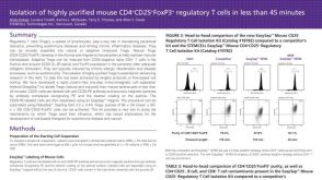 Isolation of Highly Purified Mouse CD4+CD25+Foxp3+ Regulatory T Cells in Less
 文献
文献 BrochureProducts for Human Pluripotent Stem Cells
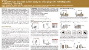 科学海报A Novel 96-well Plate Cell Culture Assay for Lineage-Specific Hematopoietic Cell Toxicity Screening
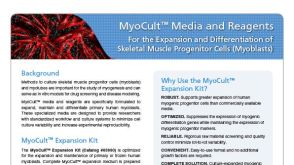 BrochureMyoCult™ Media and Reagents
 沪公网安备31010102008431号