Akopian V et al. (APR 2010)
In vitro cellular & developmental biology. Animal 46 3-4 247--258
Comparison of defined culture systems for feeder cell free propagation of human embryonic stem cells.
There are many reports of defined culture systems for the propagation of human embryonic stem cells in the absence of feeder cell support,but no previous study has undertaken a multi-laboratory comparison of these diverse methodologies. In this study,five separate laboratories,each with experience in human embryonic stem cell culture,used a panel of ten embryonic stem cell lines (including WA09 as an index cell line common to all laboratories) to assess eight cell culture methods,with propagation in the presence of Knockout Serum Replacer,FGF-2,and mouse embryonic fibroblast feeder cell layers serving as a positive control. The cultures were assessed for up to ten passages for attachment,death,and differentiated morphology by phase contrast microscopy,for growth by serial cell counts,and for maintenance of stem cell surface marker expression by flow cytometry. Of the eight culture systems,only the control and those based on two commercial media,mTeSR1 and STEMPRO,supported maintenance of most cell lines for ten passages. Cultures grown in the remaining media failed before this point due to lack of attachment,cell death,or overt cell differentiation. Possible explanations for relative success of the commercial formulations in this study,and the lack of success with other formulations from academic groups compared to previously published results,include: the complex combination of growth factors present in the commercial preparations; improved development,manufacture,and quality control in the commercial products; differences in epigenetic adaptation to culture in vitro between different ES cell lines grown in different laboratories.
View Publication
产品类型:
产品号#:
85850
85857
产品名:
mTeSR™1
mTeSR™1
文献
Conklin JF et al. ( 2012)
Nature communications 3 May 1244
The RB family is required for the self-renewal and survival of human embryonic stem cells.
The mechanisms ensuring the long-term self-renewal of human embryonic stem cells are still only partly understood,limiting their use in cellular therapies. Here we found that increased activity of the RB cell cycle inhibitor in human embryonic stem cells induces cell cycle arrest,differentiation and cell death. Conversely,inactivation of the entire RB family (RB,p107 and p130) in human embryonic stem cells triggers G2/M arrest and cell death through functional activation of the p53 pathway and the cell cycle inhibitor p21. Differences in E2F target gene activation upon loss of RB family function between human embryonic stem cells,mouse embryonic stem cells and human fibroblasts underscore key differences in the cell cycle regulatory networks of human embryonic stem cells. Finally,loss of RB family function promotes genomic instability in both human and mouse embryonic stem cells,uncoupling cell cycle defects from chromosomal instability. These experiments indicate that a homeostatic level of RB activity is essential for the self-renewal and the survival of human embryonic stem cells.
View Publication
产品类型:
产品号#:
85850
85857
产品名:
mTeSR™1
mTeSR™1
文献
Massumi M et al. ( 2016)
PloS one 11 10 e0164457
An Abbreviated Protocol for In Vitro Generation of Functional Human Embryonic Stem Cell-Derived Beta-Like Cells.
The ability to yield glucose-responsive pancreatic beta-cells from human pluripotent stem cells in vitro will facilitate the development of the cell replacement therapies for the treatment of Type 1 Diabetes. Here,through the sequential in vitro targeting of selected signaling pathways,we have developed an abbreviated five-stage protocol (25-30 days) to generate human Embryonic Stem Cell-Derived Beta-like Cells (ES-DBCs). We showed that Geltrex,as an extracellular matrix,could support the generation of ES-DBCs more efficiently than that of the previously described culture systems. The activation of FGF and Retinoic Acid along with the inhibition of BMP,SHH and TGF-beta led to the generation of 75% NKX6.1+/NGN3+ Endocrine Progenitors. The inhibition of Notch and tyrosine kinase receptor AXL,and the treatment with Exendin-4 and T3 in the final stage resulted in 35% mono-hormonal insulin positive cells,1% insulin and glucagon positive cells and 30% insulin and NKX6.1 co-expressing cells. Functionally,ES-DBCs were responsive to high glucose in static incubation and perifusion studies,and could secrete insulin in response to successive glucose stimulations. Mitochondrial metabolic flux analyses using Seahorse demonstrated that the ES-DBCs could efficiently metabolize glucose and generate intracellular signals to trigger insulin secretion. In conclusion,targeting selected signaling pathways for 25-30 days was sufficient to generate ES-DBCs in vitro. The ability of ES-DBCs to secrete insulin in response to glucose renders them a promising model for the in vitro screening of drugs,small molecules or genes that may have potential to influence beta-cell function.
View Publication
Haploinsufficiency for ribosomal protein genes causes selective activation of p53 in human erythroid progenitor cells.
Haploinsufficiency for ribosomal protein genes has been implicated in the pathophysiology of Diamond-Blackfan anemia (DBA) and the 5q-syndrome,a subtype of myelodysplastic syndrome. The p53 pathway is activated by ribosome dysfunction,but the molecular basis for selective impairment of the erythroid lineage in disorders of ribosome function has not been determined. We found that p53 accumulates selectively in the erythroid lineage in primary human hematopoietic progenitor cells after expression of shRNAs targeting RPS14,the ribosomal protein gene deleted in the 5q-syndrome,or RPS19,the most commonly mutated gene in DBA. Induction of p53 led to lineage-specific accumulation of p21 and consequent cell cycle arrest in erythroid progenitor cells. Pharmacologic inhibition of p53 rescued the erythroid defect,whereas nutlin-3,a compound that activates p53 through inhibition of HDM2,selectively impaired erythropoiesis. In bone marrow biopsies from patients with DBA or del(5q) myelodysplastic syndrome,we found an accumulation of nuclear p53 staining in erythroid progenitor cells that was not present in control samples. Our findings indicate that the erythroid lineage has a low threshold for the induction of p53,providing a basis for the failure of erythropoiesis in the 5q-syndrome,DBA,and perhaps other bone marrow failure syndromes.
View Publication
产品类型:
产品号#:
03334
03434
03444
产品名:
MethoCult™M3334
MethoCult™GF M3434
MethoCult™GF M3434
文献
Escobedo-Lucea C et al. (MAR 2012)
Stem Cell Reviews and Reports 8 1 170--183
Development of a human extracellular matrix for applications related with stem cells and tissue engineering.
Pettinato G et al. (NOV 2014)
PLoS ONE 9 11 e100742
ROCK inhibitor is not required for embryoid body formation from singularized human embryonic stem cells
We report a technology to form human embryoid bodies (hEBs) from singularized human embryonic stem cells (hESCs) without the use of the p160 rho-associated coiled-coil kinase inhibitor (ROCKi) or centrifugation (spin). hEB formation was tested under four conditions: +ROCKi/+spin,+ROCKi/-spin,-ROCKi/+spin,and -ROCKi/-spin. Cell suspensions of BG01V/hOG and H9 hESC lines were pipetted into non-adherent hydrogel substrates containing defined microwell arrays. hEBs of consistent size and spherical geometry can be formed in each of the four conditions,including the -ROCKi/-spin condition. The hEBs formed under the -ROCKi/-spin condition differentiated to develop the three embryonic germ layers and tissues derived from each of the germ layers. This simplified hEB production technique offers homogeneity in hEB size and shape to support synchronous differentiation,elimination of the ROCKi xeno-factor and rate-limiting centrifugation treatment,and low-cost scalability,which will directly support automated,large-scale production of hEBs and hESC-derived cells needed for clinical,research,or therapeutic applications.
View Publication
Cell-surface marker signatures for the Isolation of neural stem cells, glia and neurons derived from human pluripotent stem cells
BACKGROUND: Neural induction of human pluripotent stem cells often yields heterogeneous cell populations that can hamper quantitative and comparative analyses. There is a need for improved differentiation and enrichment procedures that generate highly pure populations of neural stem cells (NSC),glia and neurons. One way to address this problem is to identify cell-surface signatures that enable the isolation of these cell types from heterogeneous cell populations by fluorescence activated cell sorting (FACS). METHODOLOGY/PRINCIPAL FINDINGS: We performed an unbiased FACS- and image-based immunophenotyping analysis using 190 antibodies to cell surface markers on naïve human embryonic stem cells (hESC) and cell derivatives from neural differentiation cultures. From this analysis we identified prospective cell surface signatures for the isolation of NSC,glia and neurons. We isolated a population of NSC that was CD184(+)/CD271(-)/CD44(-)/CD24(+) from neural induction cultures of hESC and human induced pluripotent stem cells (hiPSC). Sorted NSC could be propagated for many passages and could differentiate to mixed cultures of neurons and glia in vitro and in vivo. A population of neurons that was CD184(-)/CD44(-)/CD15(LOW)/CD24(+) and a population of glia that was CD184(+)/CD44(+) were subsequently purified from cultures of differentiating NSC. Purified neurons were viable,expressed mature and subtype-specific neuronal markers,and could fire action potentials. Purified glia were mitotic and could mature to GFAP-expressing astrocytes in vitro and in vivo. CONCLUSIONS/SIGNIFICANCE: These findings illustrate the utility of immunophenotyping screens for the identification of cell surface signatures of neural cells derived from human pluripotent stem cells. These signatures can be used for isolating highly pure populations of viable NSC,glia and neurons by FACS. The methods described here will enable downstream studies that require consistent and defined neural cell populations.
View Publication
产品类型:
产品号#:
85850
85857
产品名:
mTeSR™1
mTeSR™1
文献
Wang Y et al. (DEC 2012)
Circulation research 111 12 1494--1503
Genome editing of human embryonic stem cells and induced pluripotent stem cells with zinc finger nucleases for cellular imaging
RATIONALE: Molecular imaging has proven to be a vital tool in the characterization of stem cell behavior in vivo. However,the integration of reporter genes has typically relied on random integration,a method that is associated with unwanted insertional mutagenesis and positional effects on transgene expression.backslashnbackslashnOBJECTIVE: To address this barrier,we used genome editing with zinc finger nuclease (ZFN) technology to integrate reporter genes into a safe harbor gene locus (PPP1R12C,also known as AAVS1) in the genome of human embryonic stem cells and human induced pluripotent stem cells for molecular imaging.backslashnbackslashnMETHODS AND RESULTS: We used ZFN technology to integrate a construct containing monomeric red fluorescent protein,firefly luciferase,and herpes simplex virus thymidine kinase reporter genes driven by a constitutive ubiquitin promoter into a safe harbor locus for fluorescence imaging,bioluminescence imaging,and positron emission tomography imaging,respectively. High efficiency of ZFN-mediated targeted integration was achieved in both human embryonic stem cells and induced pluripotent stem cells. ZFN-edited cells maintained both pluripotency and long-term reporter gene expression. Functionally,we successfully tracked the survival of ZFN-edited human embryonic stem cells and their differentiated cardiomyocytes and endothelial cells in murine models,demonstrating the use of ZFN-edited cells for preclinical studies in regenerative medicine.backslashnbackslashnCONCLUSION: Our study demonstrates a novel application of ZFN technology to the targeted genetic engineering of human pluripotent stem cells and their progeny for molecular imaging in vitro and in vivo.
View Publication
产品类型:
产品号#:
85850
85857
产品名:
mTeSR™1
mTeSR™1
文献
Pearce DJ and Bonnet D (SEP 2007)
Experimental hematology 35 9 1437--46
The combined use of Hoechst efflux ability and aldehyde dehydrogenase activity to identify murine and human hematopoietic stem cells.
OBJECTIVE: In murine hematopoietic tissue,direct repopulation experiments have demonstrated that the side population (SP) represents a remarkable enrichment of hematopoietic stem cells. Human SP has been phenotyped as negative for lineage antigens as well as CD34. However,in the 9 years since the original publication,no long-term hematopoietic reconstitution has been reported for the adult human SP/CD34(-) subset. Elevated levels of aldehyde dehydrogenase (ALDH) have been demonstrated in murine and human progenitor cells when compared to other hematopoietic cells. METHODS: Here,we report the phenotype of human cord blood SP cells. We established the technique of simultaneous phenotyping,Hoechst exclusion,and ALDH labeling on murine tissues. We then performed the simultaneous analysis of phenotype,SP,and ALDH activity on human cord blood and bone marrow cells. Finally,we analyzed the phenotype and functional potential of human cord blood ALDH(+) cells to determine whether Lin(-)/CD34(-) cells are identified via this technique. RESULTS: We demonstrate that human Lin(-)/CD34(-)/ALDH(+) cells are capable of long-term repopulation. Although the SP technique identifies cells that overlap with the ALDH(+) cell population,this is restricted to the CD34(+) cell subset. CONCLUSION: Hoechst exclusion ability does not seem to be the method of choice for the isolation of human hematopoietic stem cells.
View Publication

 EasySep™小鼠TIL(CD45)正选试剂盒


 文献
文献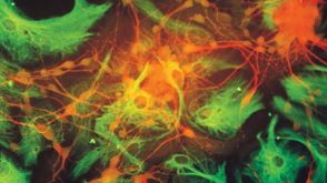 41:19
Standardized Media and Reagents for Neural Stem Cell Research with NeuroCult™
41:19
Standardized Media and Reagents for Neural Stem Cell Research with NeuroCult™ 科学海报A Robust, Xeno- and Feeder-Free Culture System for Expansion of Human Peripheral Blood NK Cells
 沪公网安备31010102008431号