Three-Dimensional Neuroepithelial Culture from Human Embryonic Stem Cells and Its Use for Quantitative Conversion to Retinal Pigment Epithelium
A goal in human embryonic stem cell (hESC) research is the faithful differentiation to given cell types such as neural lineages. During embryonic development,a basement membrane surrounds the neural plate that forms a tight,apico-basolaterally polarized epithelium before closing to form a neural tube with a single lumen. Here we show that the three-dimensional epithelial cyst culture of hESCs in Matrigel combined with neural induction results in a quantitative conversion into neuroepithelial cysts containing a single lumen. Cells attain a defined neuroepithelial identity by 5 days. The neuroepithelial cysts naturally generate retinal epithelium,in part due to IGF-1/insulin signaling. We demonstrate the utility of this epithelial culture approach by achieving a quantitative production of retinal pigment epithelial (RPE) cells from hESCs within 30 days. Direct transplantation of this RPE into a rat model of retinal degeneration without any selection or expansion of the cells results in the formation of a donor-derived RPE monolayer that rescues photoreceptor cells. The cyst method for neuroepithelial differentiation of pluripotent stem cells is not only of importance for RPE generation but will also be relevant to the production of other neuronal cell types and for reconstituting complex patterning events from three-dimensional neuroepithelia.
View Publication
产品类型:
产品号#:
05854
05855
07913
85850
85857
产品名:
mFreSR™
mFreSR™
Dispase(5 U/mL)
mTeSR™1
mTeSR™1
文献
Kim J et al. (NOV 2013)
Stem Cell Research 11 3 978--989
Alginate microcapsule as a 3D platform for the efficient differentiation of human embryonic stem cells to dopamine neurons
Human embryonic stem cells (hESCs) are emerging as an attractive alternative source for cell replacement therapy since the cells can be expanded in culture indefinitely and differentiated into any cell types in the body. In order to optimize cell-to-cell interaction,cell proliferation and differentiation into specific lineages as well as tissue organization,it is important to provide a microenvironment for the hESCs which mimics the stem cell niche. One approach is to provide a three-dimensional (3D) environment such as encapsulation. We present an approach to culture and differentiate hESCs into midbrain dopamine (mdDA) neurons in a 3D microenvironment using alginate microcapsules for the first time. A detailed gene and protein expression analysis during neuronal differentiation showed an increased gene and protein expression of various specific DA neuronal markers,particularly tyrosine hydroxylase (TH) by textgreater100 folds after 2weeks and at least 50% higher expression after 4weeks respectively,compared to cells differentiated under conventional two-dimensional (2D) platform. The encapsulated TH+ cells co-expressed mdDA neuronal markers,forkhead box protein A-2 (FOXA2) and pituitary homeobox-3 (PITX3) after 4weeks and secreted approximately 60pg/ml/106 cells higher DA level when induced. We propose that the 3D platform facilitated an early onset of DA neuronal generation compared to that with conventional 2D system which also secretes more DA under potassium-induction. It is a very useful model to study the proliferation and directed differentiation of hESCs to various lineages,particularly to mdDA neurons. This 3D system also allows the separation of feeder cells from hESCs during the process of differentiation and also has potential for immune-isolation during transplantation studies. ?? 2013 Elsevier B.V.
View Publication
Mirabelli P et al. (JAN 2008)
BMC physiology 8 1 13
Extended flow cytometry characterization of normal bone marrow progenitor cells by simultaneous detection of aldehyde dehydrogenase and early hematopoietic antigens: implication for erythroid differentiation studies.
BACKGROUND: Aldehyde dehydrogenase (ALDH) is a cytosolic enzyme highly expressed in hematopoietic precursors from cord blood and granulocyte-colony stimulating factor mobilized peripheral blood,as well as in bone marrow from patients with acute myeloblastic leukemia. As regards human normal bone marrow,detailed characterization of ALDH+ cells has been addressed by one single study (Gentry et al,2007). The goal of our work was to provide new information about the dissection of normal bone marrow progenitor cells based upon the simultaneous detection by flow cytometry of ALDH and early hematopoietic antigens,with particular attention to the expression of ALDH on erythroid precursors. To this aim,we used three kinds of approach: i) multidimensional analytical flow cytometry,detecting ALDH and early hematopoietic antigens in normal bone marrow; ii) fluorescence activated cell sorting of distinct subpopulations of progenitor cells,followed by in vitro induction of erythroid differentiation; iii) detection of ALDH+ cellular subsets in bone marrow from pure red cell aplasia patients. RESULTS: In normal bone marrow,we identified three populations of cells,namely ALDH+CD34+,ALDH-CD34+ and ALDH+CD34- (median percentages were 0.52,0.53 and 0.57,respectively). As compared to ALDH-CD34+ cells,ALDH+CD34+ cells expressed the phenotypic profile of primitive hematopoietic progenitor cells,with brighter expression of CD117 and CD133,accompanied by lower display of CD38 and CD45RA. Of interest,ALDH+CD34- population disclosed a straightforward erythroid commitment,on the basis of three orders of evidences. First of all,ALDH+CD34- cells showed a CD71bright,CD105+,CD45- phenotype. Secondly,induction of differentiation experiments evidenced a clear-cut expression of glycophorin A (CD235a). Finally,ALDH+CD34- precursors were not detectable in patients with pure red cell aplasia (PRCA). CONCLUSION: Our study,comparing surface antigen expression of ALDH+/CD34+,ALDH-/CD34+ and ALDH+/CD34- progenitor cell subsets in human bone marrow,clearly indicated that ALDH+CD34- cells are mainly committed towards erythropoiesis. To the best of our knowledge this finding is new and could be useful for basic studies about normal erythropoietic differentiation as well as for enabling the employment of ALDH as a red cell marker in polychromatic flow cytometry characterization of bone marrow from patients with aplastic anemia and myelodysplasia.
View Publication
A Novel Role for miR-1305 in Regulation of Pluripotency-Differentiation Balance, Cell Cycle, and Apoptosis in Human Pluripotent Stem Cells
Human embryonic stem cells (hESCs) and human induced pluripotent stem cells (hiPSCs) are defined as pluripotent in view of their self-renewal ability and potential to differentiate to cells of all three germ layers. Recent studies have indicated that microRNAs (miRNAs) play an important role in the maintenance of pluripotency and cell cycle regulation. We used a microarray based approach to identify miRNAs that were enriched in hESCs when compared to differentiated cells and at the same time showed significant expression changes between different phases of cell cycle. We identified 34 candidate miRNAs and performed functional studies on one of these,miR-1305,which showed the highest expression change during cell cycle transition. Overexpression of miR-1305 induced differentiation of pluripotent stem cells,increased cell apoptosis and sped up G1/S transition,while its downregulation facilitated the maintenance of pluripotency and increased cell survival. Using target prediction software and luciferase based reporter assays we identified POLR3G as a downstream target by which miR-1305 regulates the fine balance between maintenance of pluripotency and onset of differentiation. Overexpression of POLR3G rescued pluripotent stem cell differentiation induced by miR-1305 overexpression. In contrast,knock-down of POLR3G expression abolished the miR-1305-knockdown mediated enhancement of pluripotency,thus validating its role as miR-1305 target in human pluripotent stem cells. Together our data point to an important role for miR-1305 as a novel regulator of pluripotency,cell survival and cell cycle and uncovers new mechanisms and networks by which these processes are intertwined in human pluripotent stem cells. This article is protected by copyright. All rights reserved.
View Publication
产品类型:
产品号#:
85850
85857
产品名:
mTeSR™1
mTeSR™1
文献
Jones C et al. (MAY 2004)
Cancer research 64 9 3037--45
Expression profiling of purified normal human luminal and myoepithelial breast cells: identification of novel prognostic markers for breast cancer.
The normal duct-lobular system of the breast is lined by two epithelial cell types,inner luminal secretory cells and outer contractile myoepithelial cells. We have generated comprehensive expression profiles of the two normal cell types,using immunomagnetic cell separation and gene expression microarray analysis. The cell-type specificity was confirmed at the protein level by immunohistochemistry in normal breast tissue. New prognostic markers for survival were identified when the luminal- and myoepithelial-specific molecules were evaluated on breast tumor tissue microarrays. Nuclear expression of luminal epithelial marker galectin 3 correlated with a shorter overall survival in these patients,and the expression of SPARC (osteonectin),a myoepithelial marker,was an independent marker of poor prognosis in breast cancers as a whole. These data provide a framework for the interpretation of breast cancer molecular profiling experiments,the identification of potential new diagnostic markers,and development of novel indicators of prognosis.
View Publication
产品类型:
产品号#:
产品名:
文献
Mousa SA et al. (MAR 2010)
Cancer Letters 289 2 208--216
Stress resistant human embryonic stem cells as a potential source for the identification of novel cancer stem cell markers
Cancer stem cells are known for their inherent resistance to therapy. Here we investigated whether normal stem cells with acquired resistance to stress can be used to identify novel markers of cancer stem cells. For this,we generated a human embryonic stem cell line resistant to Trichostatin A and analyzed changes in its gene expression. The resistant cells over-expressed various genes associated with tumor aggressiveness,many of which are also expressed in the CD133+ glioma cancer stem cells. These findings suggest that stress-resistant stem cells generated in vitro may be useful for the discovery of novel markers of cancer stem cells.
View Publication
产品类型:
产品号#:
85850
85857
产品名:
mTeSR™1
mTeSR™1
文献
Agrawal B et al. (SEP 1998)
Cancer research 58 18 4079--81
Expression of MUC1 mucin on activated human T cells: implications for a role of MUC1 in normal immune regulation.
MUC1 mucin is expressed by normal and malignant epithelial cells and is thought to function through cell-cell interactions and transmembrane signal transduction events. Secreted cancer-associated MUC1 is immunosuppressive and inhibits human T-cell proliferation. We report here that newly synthesized MUC1 is expressed on the surface of mitogen-activated human T cells and is also found in soluble form in the supernatants from cultures of mitogen-activated human T cells. After removal of the mitogenic stimulus from the T-cell cultures,MUC1 expression is downregulated. The addition of anti-MUC1 monoclonal antibody to mitogen-activated cultures partially inhibits the T-cell proliferative response. These data suggest that MUC1 serves an immunodulatory function for human T lymphocytes.
View Publication
产品类型:
产品号#:
产品名:
文献
Sebaa M et al. (JAN 2015)
Journal of Biomedical Materials Research - Part A 103 1 25--37
The effects of poly(3,4-ethylenedioxythiophene) coating on magnesium degradation and cytocompatibility with human embryonic stem cells for potential neural applications
Magnesium (Mg) is a promising conductive metallic biomaterial due to its desirable mechanical properties for load bearing and biodegradability in human body. Controlling the rapid degradation of Mg in physiological environment continues to be the key challenge toward clinical translation. In this study,we investigated the effects of conductive poly(3,4-ethylenedioxythiophene) (PEDOT) coating on the degradation behavior of Mg substrates and their cytocompatibility. Human embryonic stem cells (hESCs) were used as the in vitro model system to study cellular responses to Mg degradation because they are sensitive and can potentially differentiate into many cell types of interest (e.g.,neurons) for regenerative medicine. The PEDOT was deposited on Mg substrates using electrochemical deposition. The greater number of cyclic voltammetry (CV) cycles yielded thicker PEDOT coatings on Mg substrates. Specifically,the coatings produced by 2,5,and 10 CV cycles (denoted as 2×-PEDOT-Mg,5×-PEDOT-Mg,and 10×-PEDOT-Mg) had an average thickness of 31,63,and 78 µm,respectively. Compared with non-coated Mg samples,all PEDOT coated Mg samples showed slower degradation rates,as indicated by Tafel test results and Mg ion concentrations in the post-culture media. The 5×-PEDOT-Mg showed the best coating adhesion and slowest Mg degradation among the tested samples. Moreover,hESCs survived for the longest period when cultured with the 5×-PEDOT-Mg samples compared with the non-coated Mg and 2×-PEDOT-Mg. Overall,the results of this study showed promise in using PEDOT coating on biodegradable Mg-based implants for potential neural recording,stimulation and tissue engineering applications,thus encouraging further research.
View Publication

 EasySep™小鼠TIL(CD45)正选试剂盒


 文献
文献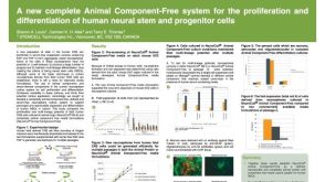 A New Complete Animal Component-Free System for the Proliferation and Differentiation of Human Neural Stem and Progenitor Cells
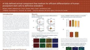 A Fully Defined Animal Component Free Medium for Efficient Differentiation of Human Pluripotent Stem Cells to Definitive Endoderm
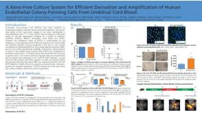 科学海报A Xeno-Free Culture System for Efficient Derivation and Amplification of Human Endothelial Colony-Forming Cells From Umbilical Cord Blood

 沪公网安备31010102008431号