Liberski AR et al. (JUL 2013)
Journal of Proteome Research 12 7 3233--3245
Adaptation of a Commonly Used, Chemically Defined Medium for Human Embryonic Stem Cells to Stable Isotope Labeling with Amino Acids in Cell Culture
Metabolic labeling with stable isotopes is a prominent technique for comparative quantitative proteomics,and stable isotope labeling with amino acids in cell culture (SILAC) is the most commonly used approach. SILAC is,however,traditionally limited to simple tissue culture regimens and only rarely employed in the context of complex culturing conditions as those required for human embryonic stem cells (hESCs). Classic hESC culture is based on the use of mouse embryonic fibroblasts (MEFs) as a feeder layer,and as a result,possible xenogeneic contamination,contribution of unlabeled amino acids by the feeders,interlaboratory variability of MEF preparation,and the overall complexity of the culture system are all of concern in conjunction with SILAC. We demonstrate a feeder-free SILAC culture system based on a customized version of a commonly used,chemically defined hESC medium developed by Ludwig et al. and commercially available as mTeSR1 [mTeSR1 is a trade mark of WiCell (Madison,WI) licensed to STEMCELL Technologies (Vancouver,Canada)]. This medium,together with adjustments to the culturing protocol,facilitates reproducible labeling that is easily scalable to the protein amounts required by proteomic work flows. It greatly enhances the usability of quantitative proteomics as a tool for the study of mechanisms underlying hESCs differentiation and self-renewal. Associated data have been deposited to the ProteomeXchange with the identifier PXD000151.
View Publication
产品类型:
产品号#:
07923
85850
85857
产品名:
Dispase (1 U/mL)
mTeSR™1
mTeSR™1
文献
Wang JC et al. (JUN 1997)
Blood 89 11 3919--24
Primitive human hematopoietic cells are enriched in cord blood compared with adult bone marrow or mobilized peripheral blood as measured by the quantitative in vivo SCID-repopulating cell assay.
We have previously reported the development of in vivo functional assays for primitive human hematopoietic cells based on their ability to repopulate the bone marrow (BM) of severe combined immunodeficient (SCID) and nonobese diabetic/SCID (NOD/SCID) mice following intravenous transplantation. Accumulated data from gene marking and cell purification experiments indicate that the engrafting cells (defined as SCID-repopulating cells or SRC) are biologically distinct from and more primitive than most cells that can be assayed in vitro. Here we demonstrate through limiting dilution analysis that the NOD/SCID xenotransplant model provides a quantitative assay for SRC. Using this assay,the frequency of SRC in cord blood (CB) was found to be 1 in 9.3 x 10(5) cells. This was significantly higher than the frequency of 1 SRC in 3.0 x 10(6) adult BM cells or 1 in 6.0 x 10(6) mobilized peripheral blood (PB) cells from normal donors. Mice transplanted with limiting numbers of SRC were engrafted with both lymphoid and multilineage myeloid human cells. This functional assay is currently the only available method for quantitative analysis of human hematopoietic cells with repopulating capacity. Both CB and mobilized PB are increasingly being used as alternative sources of hematopoietic stem cells in allogeneic transplantation. Thus,the findings reported here will have important clinical as well as biologic implications.
View Publication
产品类型:
产品号#:
28600
产品名:
L-Calc™有限稀释软件
文献
Kwok CTD et al. (MAR 2016)
Stem Cell Research 16 3 651--661
The Forkhead box transcription factor FOXM1 is required for the maintenance of cell proliferation and protection against oxidative stress in human embryonic stem cells
Human embryonic stem cells (hESCs) exhibit unique cell cycle structure,self-renewal and pluripotency. The Forkhead box transcription factor M1 (FOXM1) is critically required for the maintenance of pluripotency in mouse embryonic stem cells and mouse embryonal carcinoma cells,but its role in hESCs remains unclear. Here,we show that FOXM1 expression was enriched in undifferentiated hESCs and was regulated in a cell cycle-dependent manner with peak levels detected at the G2/M phase. Expression of FOXM1 did not correlate with OCT4 and NANOG during in vitro differentiation of hESCs. Importantly,knockdown of FOXM1 expression led to aberrant cell cycle distribution with impairment in mitotic progression but showed no profound effect on the undifferentiated state. Interestingly,FOXM1 depletion sensitized hESCs to oxidative stress. Moreover,genome-wide analysis of FOXM1 targets by ChIP-seq identified genes important for M phase including CCNB1 and CDK1,which were subsequently confirmed by ChIP and RNA interference analyses. Further peak set comparison against a differentiating hESC line and a cancer cell line revealed a substantial difference in the genomic binding profile of FOXM1 in hESCs. Taken together,our findings provide the first evidence to support FOXM1 as an important regulator of cell cycle progression and defense against oxidative stress in hESCs.
View Publication
产品类型:
产品号#:
05110
85850
85857
产品名:
STEMdiff™权威内胚层检测试剂盒
mTeSR™1
mTeSR™1
文献
Salvagiotto G et al. (JAN 2011)
PLoS ONE 6 3 e17829
A defined, feeder-free, serum-free system to generate In Vitro hematopoietic progenitors and differentiated blood cells from hESCs and hiPSCs
Human ESC and iPSC are an attractive source of cells of high quantity and purity to be used to elucidate early human development processes,for drug discovery,and in clinical cell therapy applications. To efficiently differentiate pluripotent cells into a pure population of hematopoietic progenitors we have developed a new 2-dimensional,defined and highly efficient protocol that avoids the use of feeder cells,serum or embryoid body formation. Here we showed that a single matrix protein in combination with growth factors and a hypoxic environment is sufficient to generate from pluripotent cells hematopoietic progenitors capable of differentiating further in mature cell types of different lineages of the blood system. We tested the differentiation method using hESCs and 9 iPSC lines generated from different tissues. These data indicate the robustness of the protocol providing a valuable tool for the generation of clinical-grade hematopoietic cells from pluripotent cells.
View Publication
产品类型:
产品号#:
05850
05857
05870
05875
85850
85857
85870
85875
产品名:
mTeSR™1
mTeSR™1
文献
Wognum AW et al. ( )
Archives of medical research 34 6 461--75
Identification and isolation of hematopoietic stem cells.
Hematopoietic stem cells (HSCs) are defined by their ability to repopulate all of the hematopoietic lineages in vivo and sustain the production of these cells for the life span of the individual. In the absence of reliable direct markers for HSCs,their identification and enumeration depends on functional long-term,multilineage,in vivo repopulation assays. The extremely low frequency of HSCs in any tissue and the absence of a specific HSC phenotype have made their purification and characterization a highly challenging goal. HSCs and primitive hematopoietic cells can be distinguished from mature blood cells by their lack of lineage-specific markers and presence of certain other cell-surface antigens,such as CD133 (for human cells) and c-kit and Sca-1 (for murine cells). Functional analyses of purified subpopulations of primitive hematopoietic cells have led to the development of several procedures for isolating cell populations that are highly enriched in cells with in vivo stem cell activity. Simplified methods for obtaining these cells at high yield have been important to the practical exploitation of such advances. This article reviews recent progress in identifying human and mouse HSCs and current techniques for their purification.
View Publication
Enhanced cytotoxicity of an anti-transferrin receptor IgG3-avidin fusion protein in combination with gambogic acid against human malignant hematopoietic cells: functional relevance of iron, the receptor, and reactive oxygen species.
The human transferrin receptor (hTfR) is a target for cancer immunotherapy due to its overexpression on the surface of cancer cells. We previously developed an antibody-avidin fusion protein that targets hTfR (anti-hTfR IgG3-Av) and exhibits intrinsic cytotoxicity against certain malignant cells. Gambogic acid (GA),a drug that also binds hTfR,induces cytotoxicity in several malignant cell lines. We now report that anti-hTfR IgG3-Av and GA induce cytotoxicity in a new broader panel of hematopoietic malignant cell lines. Our results show that the effect of anti-hTfR IgG3-Av is iron-dependent whereas that of GA is iron-independent in all cells tested. In addition,we observed that GA exerts a TfR-independent cytotoxicity. We also found that GA increases the generation of reactive oxygen species that may play a role in the cytotoxicity induced by this drug. Additive cytotoxicity was observed by simultaneous combination treatment with these drugs and synergy by using anti-hTfR IgG3-Av as a chemosensitizing agent. In addition,we found a concentration of GA that is toxic to malignant hematopoietic cells but not to human hematopoietic progenitor cells. Our results suggest that these two compounds may be effective,alone or in combination,for the treatment of human hematopoietic malignancies.
View Publication
Zandstra PW et al. (APR 1997)
Proceedings of the National Academy of Sciences of the United States of America 94 9 4698--703
Cytokine manipulation of primitive human hematopoietic cell self-renewal.
Previous studies have shown that primitive human hematopoietic cells detectable as long-term culture-initiating cells (LTC-ICs) and colony-forming cells (CFCs) can be amplified when CD34(+) CD38(-) marrow cells are cultured for 10 days in serum-free medium containing flt3 ligand (FL),Steel factor (SF),interleukin (IL)-3,IL-6,and granulocyte colony-stimulating factor. We now show that the generation of these two cell types in such cultures is differentially affected at the single cell level by changes in the concentrations of these cytokines. Thus,maximal expansion of LTC-ICs (60-fold) was obtained in the presence of 30 times more FL,SF,IL-3,IL-6,and granulocyte colony-stimulating factor than could concomitantly stimulate the near-maximal (280-fold) amplification of CFCs. Furthermore,the reduced ability of suboptimal cytokine concentrations to support the production of LTC-ICs could be ascribed to a differential response of the stimulated cells since this was not accompanied by a change in the number of input CD34(+) CD38(-) cells that proliferated. Reduced LTC-IC amplification in the absence of a significant effect on CFC generation also occurred when the concentrations of FL and SF were decreased but the concentration of IL-3 was high (as compared with cultures containing high levels of all three cytokines). To our knowledge,these findings provide the first evidence suggesting that extrinsically acting cytokines can alter the self-renewal behavior of primary human hematopoietic stem cells independent of effects on their viability or proliferation.
View Publication
产品类型:
产品号#:
05150
产品名:
MyeloCult™H5100
文献
Gerrits A et al. (APR 2010)
Blood 115 13 2610--8
Cellular barcoding tool for clonal analysis in the hematopoietic system.
Clonal analysis is important for many areas of hematopoietic stem cell research,including in vitro cell expansion,gene therapy,and cancer progression and treatment. A common approach to measure clonality of retrovirally transduced cells is to perform integration site analysis using Southern blotting or polymerase chain reaction-based methods. Although these methods are useful in principle,they generally provide a low-resolution,biased,and incomplete assessment of clonality. To overcome those limitations,we labeled retroviral vectors with random sequence tags or barcodes." On integration�
View Publication

 EasySep™小鼠TIL(CD45)正选试剂盒


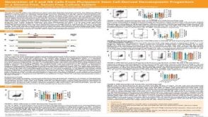 科学海报Generation of T and NK Cells From Pluripotent Stem Cell-Derived Hematopoietic Progenitors in a Stroma-Free, Serum-Free Culture System
 文献
文献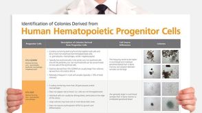 Identification of Colonies Derived from Human Hematopoietic Progenitors

 沪公网安备31010102008431号