Roubal I et al. ( 2016)
Methods in molecular biology (Clifton,N.J.) 1341 345--357
Derivation of Neural Precursor Cells from Human Embryonic Stem Cells for DNA Methylomic Analysis.
Embryonic stem cells are self-renewing pluripotent cells with competency to differentiate into all three-germ lineages. Many studies have demonstrated the importance of genetic and epigenetic molecular mechanisms in the maintenance of self-renewal and pluripotency. Stem cells are under unique molecular and cellular regulations different from somatic cells. Proper regulation should be ensured to maintain their unique self-renewal and undifferentiated characteristics. Understanding key mechanisms in stem cell biology will be important for the successful application of stem cells for regenerative therapeutic medicine. More importantly practical use of stem cells will require our knowledge on how to properly direct and differentiate stem cells into the necessary type of cells. Embryonic stem cells and adult stem cells have been used as study models to unveil molecular and cellular mechanisms in various signaling pathways. They are especially beneficial to developmental studies where in vivo molecular/cellular study models are not available. We have derived neural stem cells from human embryonic stem cells as a model to study the effect of teratogen in neural development. We have tested commercial neural differentiation system and successfully derived neural precursor cells exhibiting key molecular features of neural stem cells,which will be useful for experimental application.
View Publication
产品类型:
产品号#:
85850
85857
产品名:
mTeSR™1
mTeSR™1
文献
Rapti K et al. (FEB 2015)
Molecular Therapy — Methods & Clinical Development 2 May 2014 14067
Effectiveness of gene delivery systems for pluripotent and differentiated cells.
Human embryonic stem cells (hESC) and induced pluripotent stem cells (hiPSC) assert a great future for the cardiovascular diseases,both to study them and to explore therapies. However,a comprehensive assessment of the viral vectors used to modify these cells is lacking. In this study,we aimed to compare the transduction efficiency of recombinant adeno-associated vectors (AAV),adenoviruses and lentiviral vectors in hESC,hiPSC,and the derived cardiomyocytes. In undifferentiated cells,adenoviral and lentiviral vectors were superior,whereas in differentiated cells AAV surpassed at least lentiviral vectors. We also tested four AAV serotypes,1,2,6,and 9,of which 2 and 6 were superior in their transduction efficiency. Interestingly,we observed that AAVs severely diminished the viability of undifferentiated cells,an effect mediated by induction of cell cycle arrest genes and apoptosis. Furthermore,we show that the transduction efficiency of the different viral vectors correlates with the abundance of their respective receptors. Finally,adenoviral delivery of the calcium-transporting ATPase SERCA2a to hESC and hiPSC-derived cardiomyocytes successfully resulted in faster calcium reuptake. In conclusion,adenoviral vectors prove to be efficient for both differentiated and undifferentiated lines,whereas lentiviral vectors are more applicable to undifferentiated cells and AAVs to differentiated cells.
View Publication
Pax7 is required for the specification of myogenic satellite cells.
The paired box transcription factor Pax7 was isolated by representational difference analysis as a gene specifically expressed in cultured satellite cell-derived myoblasts. In situ hybridization revealed that Pax7 was also expressed in satellite cells residing in adult muscle. Cell culture and electron microscopic analysis revealed a complete absence of satellite cells in Pax7(-/-) skeletal muscle. Surprisingly,fluorescence-activated cell sorting analysis indicated that the proportion of muscle-derived stem cells was unaffected. Importantly,stem cells from Pax7(-/-) muscle displayed almost a 10-fold increase in their ability to form hematopoietic colonies. These results demonstrate that satellite cells and muscle-derived stem cells represent distinct cell populations. Together these studies suggest that induction of Pax7 in muscle-derived stem cells induces satellite cell specification by restricting alternate developmental programs.
View Publication
Bagó et al. (FEB 2017)
Science Translational Medicine 9 375 eaah6510
Tumor-homing cytotoxic human induced neural stem cells for cancer therapy
Engineered neural stem cells (NSCs) are a promising approach to treating glioblastoma (GBM). The ideal NSC drug carrier for clinical use should be easily isolated and autologous to avoid immune rejection. We transdifferentiated (TD) human fibroblasts into tumor-homing early-stage induced NSCs (h-iNSC(TE)),engineered them to express optical reporters and different therapeutic gene products,and assessed the tumor-homing migration and therapeutic efficacy of cytotoxic h-iNSC(TE) in patient-derived GBM models of surgical and nonsurgical disease. Molecular and functional analysis revealed that our single-factor SOX2 TD strategy converted human skin fibroblasts into h-iNSC(TE) that were nestin(+) and expressed pathways associated with tumor-homing migration in 4 days. Time-lapse motion analysis showed that h-iNSC(TE) rapidly migrated to human GBM cells and penetrated human GBM spheroids,a process inhibited by blockade of CXCR4. Serial imaging showed that h-iNSC(TE) delivery of the proapoptotic agent tumor necrosis factor-α-related apoptosis-inducing ligand (TRAIL) reduced the size of solid human GBM xenografts 250-fold in 3 weeks and prolonged median survival from 22 to 49 days. Additionally,h-iNSC(TE) thymidine kinase/ganciclovir enzyme/prodrug therapy (h-iNSC(TE)-TK) reduced the size of patient-derived GBM xenografts 20-fold and extended survival from 32 to 62 days. Mimicking clinical NSC therapy,h-iNSC(TE)-TK therapy delivered into the postoperative surgical resection cavity delayed the regrowth of residual GBMs threefold and prolonged survival from 46 to 60 days. These results suggest that TD of human skin into h-iNSC(TE) is a platform for creating tumor-homing cytotoxic cell therapies for cancer,where the potential to avoid carrier rejection could maximize treatment durability in human trials.
View Publication
产品类型:
产品号#:
05835
05839
08581
08582
产品名:
STEMdiff™ 神经诱导培养基
STEMdiff™ 神经诱导培养基
STEMdiff™SMADi神经诱导试剂盒
STEMdiff™SMADi神经诱导试剂盒,2套
文献
Mujtaba T et al. (OCT 1999)
Developmental biology 214 1 113--27
Lineage-restricted neural precursors can be isolated from both the mouse neural tube and cultured ES cells.
We have previously identified multipotent neuroepithelial (NEP) stem cells and lineage-restricted,self-renewing precursor cells termed NRPs (neuron-restricted precursors) and GRPs (glial-restricted precursors) present in the developing rat spinal cord (A. Kalyani,K. Hobson,and M. S. Rao,1997,Dev. Biol. 186,202-223; M. S. Rao and M. Mayer-Proschel,1997,Dev. Biol. 188,48-63; M. Mayer-Proschel,A. J. Kalyani,T. Mujtaba,and M. S. Rao,1997,Neuron 19,773-785). We now show that cells identical to rat NEPs,NRPs,and GRPs are present in mouse neural tubes and that immunoselection against cell surface markers E-NCAM and A2B5 can be used to isolate NRPs and GRPs,respectively. Restricted precursors similar to NRPs and GRPs can also be isolated from mouse embryonic stem cells (ES cells). ES cell-derived NRPs are E-NCAM immunoreactive,undergo self-renewal in defined medium,and differentiate into multiple neuronal phenotypes in mass culture. ES cells also generate A2B5-immunoreactive cells that are similar to E9 NEP-cell-derived GRPs and can differentiate into oligodendrocytes and astrocytes. Thus,lineage restricted precursors can be generated in vitro from cultured ES cells and these restricted precursors resemble those derived from mouse neural tubes. These results demonstrate the utility of using ES cells as a source of late embryonic precursor cells.
View Publication
产品类型:
产品号#:
产品名:
文献
Maciejewski JP et al. (SEP 1996)
Blood 88 6 1983--91
A severe and consistent deficit in marrow and circulating primitive hematopoietic cells (long-term culture-initiating cells) in acquired aplastic anemia.
We examined the stem cell compartment of patients with acquired aplastic anemia (AA) using the long-term culture-initiating cell assay (LTC-IC),in parallel with measurements of CD34+ cells and mature hematopoietic progenitors. Secondary colonies from cells surviving 5 weeks of long-term bone marrow culture (LTBMC) were determined for the peripheral blood (PB) of 68 AA patients and 13 normal controls and for BM of 49 AA patients and 14 controls; because of low cell numbers,formal limiting dilution analysis could only be performed in 10 patients. The relationship of cell input in LTBMC and the output of secondary colonies was linear,allowing quantification of LTC-IC number from bulk cultures. Secondary colony formation was markedly abnormal in severe AA. In contrast to 7.8 colony-forming cells (CFC)/10(5) mononuclear cells in normal BM and 0.14 CFC/10(5) normal PB mononuclear cells,patients with severe disease showed 0.024 CFC/10(5) in BM and 0.0068 CFC/10(5) in PB. Under limiting dilution conditions,patients' cells also showed markedly lower colony-forming ability. In contrast to 4.3 +/- 1 colonies/normal LTC-IC,we obtained only 1.27 +/- 0.09 and 2.0 +/- 0.35 colonies from BM of acute and recovered cases,respectively. These values were used to extrapolate LTC-IC numbers from secondary colony formation in suspension cultures. In PB,calculated LTC-IC were decreased 7.4-fold in new and relapsed severe AA and 2.8-fold in recovered AA. In BM,LTC-IC were decreased 10-fold in new and relapsed AA and sixfold in recovered cases. Compared with measurements obtained on presentation,LTC-IC were lower in post-treatment samples from patients who had failed to recover after intensive immunosuppression and relatively higher in cases at relapse. In recovered patients,LTC-IC number increased but remained below the normal range in 20 of 25. In patients studied serially for 3 to 12 months after treatment,LTC-IC numbers remained stable but low. LTC-IC number correlated with concurrently determined CD34+ cell number and primary hematopoietic colony formation. These results indicate that stem cell numbers,as quantitated by the LTC-IC assay,are markedly diminished in number in all severe AA. Additionally,the function of the stem cell or the stem cell compartment in AA is also abnormal,as inferred from the low clonogenic potential in secondary colony assays. Early hematologic improvement in some patients occurs without increasing numbers of LTC-IC,and a minority of recovered cases show apparent repopulation of the LTC-IC compartment years after treatment.
View Publication
Gene expression profiling and localization of Hoechst-effluxing CD45- and CD45+ cells in the embryonic mouse lung.
Hoechst-effluxing cells (side population cells) are a rare subset of cells found in adult tissues that are highly enriched for stem and progenitor cell activity. To identify potential stem and progenitor cells during lung development,we generated gene expression profiles for CD45- and CD45+ side population cells in the embryonic day 17.5 lung. We found that side population cells comprise 1% of total embryonic day 17.5 lung cells (55% CD45+,45% CD45-). Gene profiling data demonstrated an overrepresentation of endothelial genes within the CD45- side population. We used expression of several distinct genes to identify two types of CD45- side population cells: 1) von Willebrand factor+/smooth muscle actin+ cells that reside in the muscular layer of select large vessels and 2) von Willebrand factor+/intercellular adhesion molecule+ cells that reside within the endothelial layer of select small vessels. Gene profiling of the CD45+ side population indicated an overrepresentation of genes associated with myeloid cell differentiation. Consistent with this,culturing CD45+ side population cells was associated with induction of mature dendritic markers (CD86). The microarray results suggested that expression of myeloperoxidase and proteinase-3 might be used to identify CD45+ side population cells. By immunohistochemistry,we found that myeloperoxidase+/proteinase-3+ cells represent a small subset of total CD45+ cells in the embryonic day 17.5 lung and that they reside in the mesenchyme and perivascular regions. This is the first detailed information regarding the phenotype and localization of side population cells in a developing organ.
View Publication
Murdoch B et al. (MAR 2003)
Proceedings of the National Academy of Sciences of the United States of America 100 6 3422--7
Wnt-5A augments repopulating capacity and primitive hematopoietic development of human blood stem cells in vivo.
Human hematopoietic stem cells are defined by their ability to repopulate multiple hematopoietic lineages in the bone marrow of transplanted recipients and therefore are functionally distinct from hematopoietic progenitors detected in vitro. Although factors capable of regulating progenitors are well established,in vivo regulators of hematopoietic repopulating function are unknown. By using a member of the vertebrate Wnt family,Wnt-5A,the proliferation and differentiation of progenitors cocultured on stromal cells transduced with Wnt-5A or treated with Wnt-5A conditioned medium (CM) was unaffected. However,i.p. injection of Wnt-5A CM into mice engrafted with human repopulating cells increased multilineage reconstitution by textgreater3-fold compared with controls. Furthermore,in vivo treatment of human repopulating cells with Wnt-5A CM produced a greater proportion of phenotypically primitive hematopoietic progeny that could be isolated and shown to possess enhanced progenitor function independent of continued Wnt-5A treatment. Our study demonstrates that Wnt-5A augments primitive hematopoietic development in vivo and represents an in vivo regulator of hematopoietic stem cell function in the human. Based on these findings,we suggest a potential role for activation of Wnt signaling in managing patients exhibiting poor hematopoietic recovery shortly after stem cell transplantation.
View Publication
产品类型:
产品号#:
05150
产品名:
MyeloCult™H5100
文献
Lemoli RM et al. (SEP 2004)
Blood 104 6 1662--70
Extracellular nucleotides are potent stimulators of human hematopoietic stem cells in vitro and in vivo.
Although extracellular nucleotides support a wide range of biologic responses of mature blood cells,little is known about their effect on blood cell progenitor cells. In this study,we assessed whether receptors for extracellular nucleotides (P2 receptors [P2Rs]) are expressed on human hematopoietic stem cells (HSCs),and whether activation by their natural ligands,adenosine triphosphate (ATP) and uridine triphosphate (UTP),induces HSC proliferation in vitro and in vivo. Our results demonstrated that CD34(+) HSCs express functional P2XRs and P2YRs of several subtypes. Furthermore,stimulation of CD34(+) cells with extracellular nucleotides caused a fast release of Ca(2+) from intracellular stores and an increase in ion fluxes across the plasma membrane. Functionally,ATP and,to a higher extent,UTP acted as potent early acting growth factors for HSCs,in vitro,because they strongly enhanced the stimulatory activity of several cytokines on clonogenic CD34(+) and lineage-negative CD34(-) progenitors and expanded more primitive CD34(+)-derived long-term culture-initiating cells. Furthermore,xenogenic transplantation studies showed that short-term preincubation with UTP significantly expanded the number of marrow-repopulating HSCs in nonobese diabetic/severe combined immunodeficiency mice. Our data suggest that extracellular nucleotides may provide a novel and powerful tool to modulate HSC functions.
View Publication

 EasySep™小鼠TIL(CD45)正选试剂盒


 文献
文献 3:39
Why Use Semi-Solid Medium for Cloning Hybridomas and CHO Cells
3:39
Why Use Semi-Solid Medium for Cloning Hybridomas and CHO Cells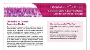 BrochurePneumaCult™-Ex Plus: Generate More Airway Epithelial Cells for Extended Passages
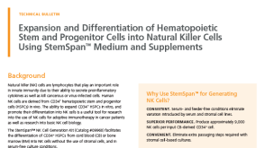 技术窍门Expansion and Differentiation of Hematopoietic Stem and Progenitor Cells into Natural Killer (NK) Cells Using StemSpan™ Medium and Supplements
 沪公网安备31010102008431号