Ma X et al. ( 2012)
Journal of biomedicine & biotechnology 2012 741416
Development of new technologies for stem cell research.
Since the 1960s,the stem cells have been extensively studied including embryonic stem cells,neural stem cells,bone marrow hematopoietic stem cells,and mesenchymal stem cells. In the recent years,several stem cells have been initially used in the treatment of diseases,such as in bone marrow transplant. At the same time,isolation and culture experimental technologies for stem cell research have been widely developed in recent years. In addition,molecular imaging technologies including optical molecular imaging,positron emission tomography,single-photon emission computed tomography,and computed tomography have been developed rapidly in recent the 10 years and have also been used in the research on disease mechanism and evaluation of treatment of disease related with stem cells. This paper will focus on recent typical isolation,culture,and observation techniques of stem cells followed by a concise introduction. Finally,the current challenges and the future applications of the new technologies in stem cells are given according to the understanding of the authors,and the paper is then concluded.
View Publication
产品类型:
产品号#:
85850
85857
产品名:
mTeSR™1
mTeSR™1
Stern P et al. (SEP 2008)
Proceedings of the National Academy of Sciences of the United States of America 105 37 13895--900
A system for Cre-regulated RNA interference in vivo.
We report a system for Cre-regulated expression of RNA interference in vivo. Expression cassettes comprise selectable and FACS-sortable markers in tandem with additional marker genes and shRNAs in the antisense orientation. The cassettes are flanked by tandem LoxP sites arranged so that Cre expression inverts the marker-shRNA construct,allowing its regulated expression (and,at the same time,deletes the original selection/marker genes). The cassettes can be incorporated into retroviral or lentiviral vectors and delivered to cells in culture or used to generate transgenic mice. We describe cassettes incorporating various combinations of reporter genes,miRNA-based RNAi (including two shRNA constructs at once),and oncogenes and demonstrate the delivery of effective RNA interference in cells in culture,efficient transduction into hematopoietic stem cells with cell-type-specific knockdown in their progeny,and rapid generation of regulated shRNA knockdown in transgenic mice. These vector systems allow regulated combinatorial manipulation (both overexpression and loss of function) of gene expression in multiple systems in vitro and in vivo.
View Publication
产品类型:
产品号#:
09600
09650
产品名:
StemSpan™ SFEM
StemSpan™ SFEM
Lee SB et al. (JAN 2016)
Nature 529 7585 172--7
An ID2-dependent mechanism for VHL inactivation in cancer.
Mechanisms that maintain cancer stem cells are crucial to tumour progression. The ID2 protein supports cancer hallmarks including the cancer stem cell state. HIFα transcription factors,most notably HIF2α (also known as EPAS1),are expressed in and required for maintenance of cancer stem cells (CSCs). However,the pathways that are engaged by ID2 or drive HIF2α accumulation in CSCs have remained unclear. Here we report that DYRK1A and DYRK1B kinases phosphorylate ID2 on threonine 27 (Thr27). Hypoxia downregulates this phosphorylation via inactivation of DYRK1A and DYRK1B. The activity of these kinases is stimulated in normoxia by the oxygen-sensing prolyl hydroxylase PHD1 (also known as EGLN2). ID2 binds to the VHL ubiquitin ligase complex,displaces VHL-associated Cullin 2,and impairs HIF2α ubiquitylation and degradation. Phosphorylation of Thr27 of ID2 by DYRK1 blocks ID2-VHL interaction and preserves HIF2α ubiquitylation. In glioblastoma,ID2 positively modulates HIF2α activity. Conversely,elevated expression of DYRK1 phosphorylates Thr27 of ID2,leading to HIF2α destabilization,loss of glioma stemness,inhibition of tumour growth,and a more favourable outcome for patients with glioblastoma.
View Publication
产品类型:
产品号#:
05700
05701
05702
产品名:
NeuroCult™ 基础培养基(小鼠&大鼠)
NeuroCult™ 扩增添加物 (小鼠&大鼠)
NeuroCult™ 扩增试剂盒 (小鼠&大鼠)
Rahman M et al. (SEP 2013)
Future Oncology 9 9 1389--1396
Controlling tumor invasion: bevacizumab and BMP4 for glioblastoma
AIM Bevacizumab has been reported to result in increased tumor invasion when used to treat malignant glioma. We hypothesized that BMP4 would prevent diffuse tumor infiltration induced by bevacizumab for malignant glioma in a xenograft model. METHODS Human glioblastoma (GBM) tumor cells were implanted in the striatum of immunocompromised mice. The animals were treated with bevacizumab and BMP4. Tumor growth and invasion were measured. RESULTS The bevacizumab-treated mice had increased survival compared with control animals (p = 0.02). BMP4 alone did not result in improved survival (p = 1.0). The bevacizumab (p = 0.006) and bevacizumab plus BMP4 (p = 0.006) groups demonstrated significantly decreased total tumor size compared with control. Tumor invasion was significantly decreased in the bevacizumab (p = 0.005),BMP4 (p = 0.04) alone and bevacizumab plus BMP4 (p = 0.002) groups compared with control. No synergistic effect between bevacizumab and BMP4 was observed. CONCLUSION Bevacizumab treatment did not result in diffuse infiltration of human GBM in a mouse xenograft model. BMP4 did have an independent favorable effect on GBM that was not synergistic with bevacizumab treatment.
View Publication
Sharei A et al. (FEB 2013)
Proceedings of the National Academy of Sciences 110 6 2082--2087
A vector-free microfluidic platform for intracellular delivery
Intracellular delivery of macromolecules is a challenge in research and therapeutic applications. Existing vector-based and physical methods have limitations,including their reliance on exogenous materials or electrical fields,which can lead to toxicity or off-target effects. We describe a microfluidic approach to delivery in which cells are mechanically deformed as they pass through a constriction 30–80% smaller than the cell diameter. The resulting controlled application of compression and shear forces results in the formation of transient holes that enable the diffusion of material from the surrounding buffer into the cytosol. The method has demonstrated the ability to deliver a range of material,such as carbon nanotubes,proteins,and siRNA,to 11 cell types,including embryonic stem cells and immune cells. When used for the delivery of transcription factors,the microfluidic devices produced a 10-fold improvement in colony formation relative to electroporation and cell-penetrating peptides. Indeed,its ability to deliver structurally diverse materials and its applicability to difficult-to-transfect primary cells indicate that this method could potentially enable many research and clinical applications.
View Publication
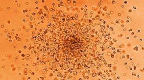
Retinoic acid enhances the generation of hematopoietic progenitors from human embryonic stem cell-derived hemato-vascular precursors.
Current induction schemes directing hematopoietic differentiation of human embryonic stem cells (hESCs) are not well defined to mimic the sequential stages of hematopoietic development in vivo. Here,we report a 3-stage method to direct differentiation of hESCs toward hematopoietic progenitors in chemically defined mediums. In the first 2 stages,we efficiently generated T-positive primitive streak/mesendoderm cells and kinase domain receptor-positive (KDR(+)) platelet-derived growth factor receptor α-negative (PDGFRα(-)) hemato-vascular precursors sequentially. In the third stage,we found that cells in a spontaneous differentiation condition mainly formed erythroid colonies. Addition of all-trans retinoic acid (RA) greatly enhanced generation of hematopoietic progenitors in this stage while suppressing erythroid development. The RA-treated cells highly expressed definitive hematopoietic genes,formed large numbers of multilineage and myeloid colonies,and gave rise to greater than 45% CD45(+) hematopoietic cells. When hematopoietic progenitors were selected with CD34 and C-Kit,greater than 95% CD45(+) hematopoietic cells could be generated. In addition,we found that endogenous RA signaling at the second stage was required for vascular endothelial growth factor/basic fibroblast growth factor-induced hemato-vascular specification,whereas exogenously applied RA efficiently induced KDR(-)PDGFRα(+) paraxial mesoderm cells. Our study suggests that RA signaling plays diverse roles in human mesoderm and hematopoietic development.
View Publication
产品类型:
产品号#:
04436
09600
09650
产品名:
MethoCult™ SF H4436
StemSpan™ SFEM
StemSpan™ SFEM
(May 2024)
Cell Communication and Signaling : CCS 22 1
Megakaryocytic IGF1 coordinates activation and ferroptosis to safeguard hematopoietic stem cell regeneration after radiation injury
BackgroundHematopoietic stem cell (HSC) regeneration underlies hematopoietic recovery from myelosuppression,which is a life-threatening side effect of cytotoxicity. HSC niche is profoundly disrupted after myelosuppressive injury,while if and how the niche is reshaped and regulates HSC regeneration are poorly understood.MethodsA mouse model of radiation injury-induced myelosuppression was built by exposing mice to a sublethal dose of ionizing radiation. The dynamic changes in the number,distribution and functionality of HSCs and megakaryocytes were determined by flow cytometry,immunofluorescence,colony assay and bone marrow transplantation,in combination with transcriptomic analysis. The communication between HSCs and megakaryocytes was determined using a coculture system and adoptive transfer. The signaling mechanism was investigated both in vivo and in vitro,and was consolidated using megakaryocyte-specific knockout mice and transgenic mice.ResultsMegakaryocytes become a predominant component of HSC niche and localize closer to HSCs after radiation injury. Meanwhile,transient insulin-like growth factor 1 (IGF1) hypersecretion is predominantly provoked in megakaryocytes after radiation injury,whereas HSCs regenerate paralleling megakaryocytic IGF1 hypersecretion. Mechanistically,HSCs are particularly susceptible to megakaryocytic IGF1 hypersecretion,and mTOR downstream of IGF1 signaling not only promotes activation including proliferation and mitochondrial oxidative metabolism of HSCs,but also inhibits ferritinophagy to restrict HSC ferroptosis. Consequently,the delicate coordination between proliferation,mitochondrial oxidative metabolism and ferroptosis ensures functional HSC expansion after radiation injury. Importantly,punctual IGF1 administration simultaneously promotes HSC regeneration and hematopoietic recovery after radiation injury,representing a superior therapeutic approach for myelosuppression.ConclusionsOur study identifies megakaryocytes as a last line of defense against myelosuppressive injury and megakaryocytic IGF1 as a novel niche signal safeguarding HSC regeneration.Supplementary InformationThe online version contains supplementary material available at 10.1186/s12964-024-01651-5.
View Publication

 EasySep™小鼠TIL(CD45)正选试剂盒


 42:14
线上讲座Scaling-Up hPSC-Derived Cardiomyocytes for Preclinical and Therapeutic Applications发布日期: 02/21/2025
42:14
线上讲座Scaling-Up hPSC-Derived Cardiomyocytes for Preclinical and Therapeutic Applications发布日期: 02/21/2025


 沪公网安备31010102008431号