Dichlberger A et al. (DEC 2011)
Journal of lipid research 52 12 2198--208
Lipid body formation during maturation of human mast cells.
Lipid droplets,also called lipid bodies (LB) in inflammatory cells,are important cytoplasmic organelles. However,little is known about the molecular characteristics and functions of LBs in human mast cells (MC). Here,we have analyzed the genesis and components of LBs during differentiation of human peripheral blood-derived CD34(+) progenitors into connective tissue-type MCs. In our serum-free culture system,the maturing MCs,derived from 18 different donors,invariably developed triacylglycerol (TG)-rich LBs. Not known heretofore,the MCs transcribe the genes for perilipins (PLIN)1-4,but not PLIN5,and PLIN2 and PLIN3 display different degrees of LB association. Upon MC activation and ensuing degranulation,the LBs were not cosecreted with the cytoplasmic secretory granules. Exogenous arachidonic acid (AA) enhanced LB genesis in Triacsin C-sensitive fashion,and it was found to be preferentially incorporated into the TGs of LBs. The large TG-associated pool of AA in LBs likely is a major precursor for eicosanoid production by MCs. In summary,we demonstrate that cultured human MCs derived from CD34(+) progenitors in peripheral blood provide a new tool to study regulatory mechanisms involving LB functions,with particular emphasis on AA metabolism,eicosanoid biosynthesis,and subsequent release of proinflammatory lipid mediators from these cells.
View Publication
产品类型:
产品号#:
09500
产品名:
BIT 9500血清替代物
F. W. Pagliuca et al. (oct 2014)
Cell 159 2 428--39
Generation of functional human pancreatic $\beta$ cells in vitro.
The generation of insulin-producing pancreatic $\beta$ cells from stem cells in vitro would provide an unprecedented cell source for drug discovery and cell transplantation therapy in diabetes. However,insulin-producing cells previously generated from human pluripotent stem cells (hPSC) lack many functional characteristics of bona fide $\beta$ cells. Here,we report a scalable differentiation protocol that can generate hundreds of millions of glucose-responsive $\beta$ cells from hPSC in vitro. These stem-cell-derived $\beta$ cells (SC-$\beta$) express markers found in mature $\beta$ cells,flux Ca(2+) in response to glucose,package insulin into secretory granules,and secrete quantities of insulin comparable to adult $\beta$ cells in response to multiple sequential glucose challenges in vitro. Furthermore,these cells secrete human insulin into the serum of mice shortly after transplantation in a glucose-regulated manner,and transplantation of these cells ameliorates hyperglycemia in diabetic mice.
View Publication
产品类型:
产品号#:
100-0548
100-0549
产品名:
3,3',5-三碘- l -甲状腺原氨酸(钠盐水合物)
3,3',5-三碘- l -甲状腺原氨酸(钠盐水合物)
Y. Tokumoto et al. (jan 2022)
Clinical and experimental immunology 207 1 95--103
Induction of memory-like CD8+ T cells and CD4+ T cells from human naive T cells in culture.
Memory T cells are crucial players in vertebrate adaptive immunity but their development is incompletely understood. Here,we describe a method to produce human memory-like T cells from naive human T cells in culture. Using commercially available human T-cell differentiation kits,both purified naive CD8+ T cells and purified naive CD4+ T cells were activated via T-cell receptor signaling and appropriate cytokines for several days in culture. All the T-cell activators were then removed from the medium and the cultures were continued in hypoxic condition (1% O2 atmosphere) for several more days; during this period,most of the cells died,but some survived in a quiescent state for a month. The survivors had small round cell bodies,expressed differentiation markers characteristic of memory T cells and restarted proliferation when the T-cell activators were added back. We could also induce memory-like T cells from naive human T cells without hypoxia,if we froze the activated T cells or prepared the naive T cells from chilled filter buffy coats.
View Publication
Hertsenberg AJ and Funderburgh JL ( 2015)
1341 285--294
Generation of corneal keratocytes from human embryonic stem cells
Human Embryonic Stem Cells (hESC) offer an important resource as a limitless supply of any differentiated cell type of the human body. Keratocytes,cells from the corneal stroma,may have the potential for restoration of vision in cell therapy and biomedical engineering applications,but these specialized cells are not readily expanded in vitro. Here we describe a two-part method to produce keratocytes from the H1 hESC cell line. The hESC cells,maintained and expanded in feeder-free culture medium are first differentiated to neural crest cells using the stromal-derived inducing activity (SDIA) of the PA6 mouse embryonic fibroblast cell line. The resulting neural crest cells are selected by their expression of cell-surface CD271 and subsequently cultured as 3D pellets in a defined differentiation medium to induce a keratocyte phenotype.
View Publication
产品类型:
产品号#:
85850
85857
产品名:
mTeSR™1
mTeSR™1
Gué et al. (JUN 2017)
Diabetes 66 6 1470--1478
Functional Human Beige Adipocytes From Induced Pluripotent Stem Cells.
Activation of thermogenic beige adipocytes has recently emerged as a promising therapeutic target in obesity and diabetes. Relevant human models for beige adipocyte differentiation are essential to implement such therapeutic strategies. We report a straightforward and efficient protocol to generate functional human beige adipocytes from human induced pluripotent stem cells (hiPSCs). Without overexpression of exogenous adipogenic genes,our method recapitulates an adipogenic developmental pathway through successive mesodermal and adipogenic progenitor stages. hiPSC-derived adipocytes are insulin sensitive and display beige-specific markers and functional properties,including upregulation of thermogenic genes,increased mitochondrial content,and increased oxygen consumption upon activation with cAMP analogs. Engraftment of hiPSC-derived adipocytes in mice produces well-organized and vascularized adipose tissue,capable of β-adrenergic-responsive glucose uptake. Our model of human beige adipocyte development provides a new and scalable tool for disease modeling and therapeutic screening.
View Publication
产品类型:
产品号#:
85850
85857
产品名:
mTeSR™1
mTeSR™1
Gore A et al. (MAR 2011)
Nature 471 7336 63--7
Somatic coding mutations in human induced pluripotent stem cells.
Defined transcription factors can induce epigenetic reprogramming of adult mammalian cells into induced pluripotent stem cells. Although DNA factors are integrated during some reprogramming methods,it is unknown whether the genome remains unchanged at the single nucleotide level. Here we show that 22 human induced pluripotent stem (hiPS) cell lines reprogrammed using five different methods each contained an average of five protein-coding point mutations in the regions sampled (an estimated six protein-coding point mutations per exome). The majority of these mutations were non-synonymous,nonsense or splice variants,and were enriched in genes mutated or having causative effects in cancers. At least half of these reprogramming-associated mutations pre-existed in fibroblast progenitors at low frequencies,whereas the rest occurred during or after reprogramming. Thus,hiPS cells acquire genetic modifications in addition to epigenetic modifications. Extensive genetic screening should become a standard procedure to ensure hiPS cell safety before clinical use.
View Publication
产品类型:
产品号#:
85850
85857
产品名:
mTeSR™1
mTeSR™1
Guan X et al. (MAY 2012)
Stem Cell Research 8 3 410--5
Derivation of human embryonic stem cells with NEMO deficiency.
Deficiency of the nuclear factor-kappa-B essential modulator (NEMO) is a rare X-linked disorder that presents in boys as hypohydrotic ectodermal dysplasia with immunodeficiency due to defective nuclear factor-κB activation. Here we report on the generation of 2 human embryonic stem cell lines from discarded in vitro fertilization (IVF) embryos ascertained via preimplantation genetic diagnosis. We have derived two human embryonic stem cell lines that carry a T458G hypomorphic mutation in exon 4 of the NEMO (or IKBKG) gene. One of the lines is diploid male; the other is diploid female but has clonally inactivated the X-chromosome that harbors the wild-type IKBKG gene. We show that both lines are pluripotent,have the capacity to differentiate into hematopoietic progenitors,and have defective inhibitor of nuclear factor kappa-B kinase activity. These NEMO deficiency hES cell lines provide an unlimited source for differentiated cell types and may serve as a unique tool to study NEMO deficiency and potentially lead to the development of new therapies for this disease.
View Publication
产品类型:
产品号#:
85850
85857
产品名:
mTeSR™1
mTeSR™1
Gilbert AE et al. (JAN 2011)
PloS one 6 4 e19330
Monitoring the systemic human memory B cell compartment of melanoma patients for anti-tumor IgG antibodies.
Melanoma,a potentially lethal skin cancer,is widely thought to be immunogenic in nature. While there has been much focus on T cell-mediated immune responses,limited knowledge exists on the role of mature B cells. We describe an approach,including a cell-based ELISA,to evaluate mature IgG antibody responses to melanoma from human peripheral blood B cells. We observed a significant increase in antibody responses from melanoma patients (n = 10) to primary and metastatic melanoma cells compared to healthy volunteers (n = 10) (Ptextless0.0001). Interestingly,we detected a significant reduction in antibody responses to melanoma with advancing disease stage in our patient cohort (n = 21) (Ptextless0.0001). Overall,28% of melanoma patient-derived B cell cultures (n = 1,800) compared to 2% of cultures from healthy controls (n = 600) produced antibodies that recognized melanoma cells. Lastly,a patient-derived melanoma-specific monoclonal antibody was selected for further study. This antibody effectively killed melanoma cells in vitro via antibody-mediated cellular cytotoxicity. These data demonstrate the presence of a mature systemic B cell response in melanoma patients,which is reduced with disease progression,adding to previous reports of tumor-reactive antibodies in patient sera,and suggesting the merit of future work to elucidate the clinical relevance of activating humoral immune responses to cancer.
View Publication
产品类型:
产品号#:
15024
15064
产品名:
RosetteSep™ 人B细胞富集抗体混合物
RosetteSep™人B细胞富集抗体混合物
Lu HF et al. (MAR 2012)
Biomaterials 33 8 2419--30
A 3D microfibrous scaffold for long-term human pluripotent stem cell self-renewal under chemically defined conditions.
Realizing the potential of human pluripotent stem cell (hPSC)-based therapy requires the development of defined scalable culture systems with efficient expansion,differentiation and isolation protocols. We report an engineered 3D microfiber system that efficiently supports long-term hPSCs self-renewal under chemically defined conditions. The unique feature of this system lies in the application of a 3D ECM-like environment in which cells are embedded,that affords: (i) uniform high cell loading density in individual cell-laden constructs (∼10 7 cells/ml); (ii) quick recovery of encapsulated cells (textless10min at 37°C) with excellent preservation of cell viability and 3D multicellular structure; (iii) direct cryopreservation of the encapsulated cells in situ in the microfibers with textgreater17-fold higher cell viability compared to those cultured on Matrigel surface; (iv) long-term hPSC propagation under chemically defined conditions. Four hPSC lines propagated in the microfibrous scaffold for 10 consecutive passages were capable of maintaining an undifferentiated phenotype as demonstrated by the expression of stem cell markers and stable karyotype invitro and the ability to form derivatives of the three germ layers both invitro and invivo. Our 3D microfibrous system has the potential for large-scale cultivation of transplantable hESCs and derivatives for clinical applications. textcopyright 2011 Elsevier Ltd.
View Publication

 EasySep™小鼠TIL(CD45)正选试剂盒


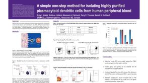 科学海报Isolation of Plasmacytoid Dendritic Cells from Human Peripheral Blood
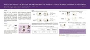 科学海报Pre-Enrichment of Dendritic Cells from Human Peripheral Blood Samples

 沪公网安备31010102008431号