Prasain N et al. (NOV 2014)
Nature biotechnology 32 11 1151--1157
Differentiation of human pluripotent stem cells to cells similar to cord-blood endothelial colony-forming cells.
The ability to differentiate human pluripotent stem cells into endothelial cells with properties of cord-blood endothelial colony-forming cells (CB-ECFCs) may enable the derivation of clinically relevant numbers of highly proliferative blood vessel-forming cells to restore endothelial function in patients with vascular disease. We describe a protocol to convert human induced pluripotent stem cells (hiPSCs) or embryonic stem cells (hESCs) into cells similar to CB-ECFCs at an efficiency of textgreater10(8) ECFCs produced from each starting pluripotent stem cell. The CB-ECFC-like cells display a stable endothelial phenotype with high clonal proliferative potential and the capacity to form human vessels in mice and to repair the ischemic mouse retina and limb,and they lack teratoma formation potential. We identify Neuropilin-1 (NRP-1)-mediated activation of KDR signaling through VEGF165 as a critical mechanism for the emergence and maintenance of CB-ECFC-like cells.
View Publication
产品类型:
产品号#:
85850
85857
产品名:
mTeSR™1
mTeSR™1
E. Vokali et al. (jan 2020)
Nature communications 11 1 538
Lymphatic endothelial cells prime na\ive CD8+ T cells into memory cells under steady-state conditions."
Lymphatic endothelial cells (LECs) chemoattract na{\{i}}ve T cells and promote their survival in the lymph nodes and can cross-present antigens to na{\"{i}}ve CD8+ T cells to drive their proliferation despite lacking key costimulatory molecules. However the functional consequence of LEC priming of CD8+ T cells is unknown. Here we show that while many proliferating LEC-educated T cells enter early apoptosis the remainders comprise a long-lived memory subset with transcriptional metabolic and phenotypic features of central memory and stem cell-like memory T cells. In vivo these memory cells preferentially home to lymph nodes and display rapid proliferation and effector differentiation following memory recall and can protect mice against a subsequent bacterial infection. These findings introduce a new immunomodulatory role for LECs in directly generating a memory-like subset of quiescent yet antigen-experienced CD8+ T cells that are long-lived and can rapidly differentiate into effector cells upon inflammatory antigenic challenge."""
View Publication
产品类型:
产品号#:
19853
19853RF
产品名:
EasySep™小鼠CD8+ T细胞分选试剂盒
RoboSep™ 小鼠CD8+ T细胞分选试剂盒
Hudson J et al. (JUN 2012)
Stem cells and development 21 9 1513--23
Primitive cardiac cells from human embryonic stem cells.
Pluripotent stem cell-derived cardiomyocytes are currently being investigated for in vitro human heart models and as potential therapeutics for heart failure. In this study,we have developed a differentiation protocol that minimizes the need for specific human embryonic stem cell (hESC) line optimization. We first reduced the heterogeneity that exists within the starting population of bulk cultured hESCs by using cells adapted to single-cell passaging in a 2-dimensional (2D) culture format. Compared with bulk cultures,single-cell cultures comprised larger fractions of TG30(hi)/OCT4(hi) cells,corresponding to an increased expression of pluripotency markers OCT4 and NANOG,and reduced expression of early lineage-specific markers. A 2D temporal differentiation protocol was then developed,aimed at reducing the inherent heterogeneity and variability of embryoid body-based protocols,with induction of primitive streak cells using bone morphogenetic protein 4 and activin A,followed by cardiogenesis via inhibition of Wnt signaling using the small molecules IWP-4 or IWR-1. IWP-4 treatment resulted in a large percentage of cells expressing low amounts of cardiac myosin heavy chain and expression of early cardiac progenitor markers ISL1 and NKX2-5,thus indicating the production of large numbers of immature cardiomyocytes (˜65,000/cm(2) or ˜1.5 per input hESC). This protocol was shown to be effective in HES3,H9,and,to a lesser,extent,MEL1 hESC lines. In addition,we observed that IWR-1 induced predominantly atrial myosin light chain (MLC2a) expression,whereas IWP-4 induced expression of both atrial (MLC2a) and ventricular (MLC2v) forms. The intrinsic flexibility and scalability of this 2D protocol mean that the output population of primitive cardiomyocytes will be particularly accessible and useful for the investigation of molecular mechanisms driving terminal cardiomyocyte differentiation,and potentially for the future treatment of heart failure.
View Publication
产品类型:
产品号#:
72552
72554
85850
85857
产品名:
IWP-4
IWP-4
mTeSR™1
mTeSR™1
Kovarova M and Koller B (APR 2012)
Current protocols in immunology / edited by John E. Coligan ... [et al.] Chapter 22 Unit 22F.10.1--16
Differentiation of mast cells from embryonic stem cells.
In this unit,we describe a simple coculture-free method for obtaining mast cells from mouse and human embryonic stem (ES) cells. Much of our knowledge regarding the mechanisms by which mast cells are activated comes from studies of mouse bone marrow-derived mast cells. Studies of human mast cells have been hampered by the limited sources from which they can be cultured,the difficulty in introducing specific genetic changes into these cells,and differences between established cultures that reflect the unique genetic makeup of the tissue donor. Derivation of mast cells from embryonic stem cells addresses these limitations. ES-derived mast cells can be generated in numbers sufficient for studies of the pathways involved in mast cell effector functions. These ES cell-derived mast cells respond to antigens and other stimuli by releasing histamine,cytokines,lipids,and other bioactive mediators. The derivation of human mast cells from ES cells carrying mutations introduced by homologous recombination should provide a novel means of testing the function of genes in both the development and the effector functions of mast cells.
View Publication
产品类型:
产品号#:
85850
85857
产品名:
mTeSR™1
mTeSR™1
Maeda M et al. (JAN 2006)
The Journal of biological chemistry 281 1 59--68
Src activation is not necessary for transforming growth factor (TGF)-beta-mediated epithelial to mesenchymal transitions (EMT) in mammary epithelial cells. PP1 directly inhibits TGF-beta receptors I and II.
Epithelial to mesenchymal transitions (EMTs) are key events during embryonic development and cancer progression. It has been proposed that Src plays a major role in some EMT models,as shown by the overexpression of viral Src (v-Src) in epithelial cells. It is clear that Src family kinases can regulate the integrity of both adherens junctions and focal adhesions; however,their significance in EMT,especially in the physiological context,remains to be elucidated. Here we showed that Src is activated in transforming growth factor-beta1 (TGF-beta1)-mediated EMT in mammary epithelial cells and that the Src family kinase inhibitor,PP1,prevents EMT. However,neither a more specific Src family kinase inhibitor,SU6656,nor a dominant-negative Src inhibited TGF-beta1-mediated EMT,leading us to speculate that Src activation is not an essential component of TGF-beta1-mediated EMT. Unexpectedly,PP1 prevented Smad2/3 activation by TGF-beta1,whereas SU6656 did not. Most interestingly,an in vitro kinase assay showed that PP1 strongly inhibited the TGF-beta receptor type I,and to a lesser extent,the TGF-beta receptor type II. Taken together,our data indicated that PP1 interferes with TGF-beta1-mediated EMT not by inhibiting Src family kinases but by inhibiting the Smad pathway via a direct inhibition of TGF-beta receptor kinase activity.
View Publication
产品类型:
产品号#:
73112
73114
产品名:
PP1
PP1
挂图
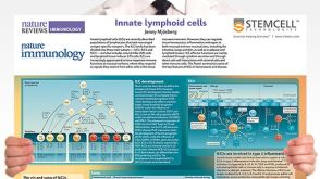
Innate Lymphoid Cells
Overview of innate lymphoid cells (ILCs) development, classification, plasticity and functional diversity
挂图
Neural Stem Cells
Overview of the types of NSCs and their potential use as therapeutic agents for disease
挂图
Regulatory T Cells
Overview of the development, phenotype and functions of regulatory T cells
Lei IL et al. (JAN 2015)
Journal of visualized experiments : JoVE January 52047. doi: 10.3791/52047.
Derivation of cardiac progenitor cells from embryonic stem cells.
Cardiac progenitor cells (CPCs) have the capacity to differentiate into cardiomyocytes,smooth muscle cells (SMC),and endothelial cells and hold great promise in cell therapy against heart disease. Among various methods to isolate CPCs,differentiation of embryonic stem cell (ESC) into CPCs attracts great attention in the field since ESCs can provide unlimited cell source. As a result,numerous strategies have been developed to derive CPCs from ESCs. In this protocol,differentiation and purification of embryonic CPCs from both mouse and human ESCs is described. Due to the difficulty of using cell surface markers to isolate embryonic CPCs,ESCs are engineered with fluorescent reporters activated by CPC-specific cre recombinase expression. Thus,CPCs can be enriched by fluorescence-activated cell sorting (FACS). This protocol illustrates procedures to form embryoid bodies (EBs) from ESCs for CPC specification and enrichment. The isolated CPCs can be subsequently cultured for cardiac lineage differentiation and other biological assays. This protocol is optimized for robust and efficient derivation of CPCs from both mouse and human ESCs.
View Publication
产品类型:
产品号#:
85850
85857
产品名:
mTeSR™1
mTeSR™1
B. A. Jonas et al. ( 2016)
PloS one 11 7 e0159189
Alkylator-Induced and Patient-Derived Xenograft Mouse Models of Therapy-Related Myeloid Neoplasms Model Clinical Disease and Suggest the Presence of Multiple Cell Subpopulations with Leukemia Stem Cell Activity.
Acute myeloid leukemia (AML) is a heterogeneous group of aggressive bone marrow cancers arising from transformed hematopoietic stem and progenitor cells (HSPC). Therapy-related AML and MDS (t-AML/MDS) comprise a subset of AML cases occurring after exposure to alkylating chemotherapy and/or radiation and are associated with a very poor prognosis. Less is known about the pathogenesis and disease-initiating/leukemia stem cell (LSC) subpopulations of t-AML/MDS compared to their de novo counterparts. Here,we report the development of mouse models of t-AML/MDS. First,we modeled alkylator-induced t-AML/MDS by exposing wild type adult mice to N-ethyl-N-nitrosurea (ENU),resulting in several models of AML and MDS that have clinical and pathologic characteristics consistent with human t-AML/MDS including cytopenia,myelodysplasia,and shortened overall survival. These models were limited by their inability to transplant clinically aggressive disease. Second,we established three patient-derived xenograft models of human t-AML. These models led to rapidly fatal disease in recipient immunodeficient xenografted mice. LSC activity was identified in multiple HSPC subpopulations suggesting there is no canonical LSC immunophenotype in human t-AML. Overall,we report several new t-AML/MDS mouse models that could potentially be used to further define disease pathogenesis and test novel therapeutics.
View Publication

 EasySep™小鼠TIL(CD45)正选试剂盒


 科学海报Using Human Pluripotent Stem Cell-Derived Microglia As Models For Neurological Disease Research
 挂图Innate Lymphoid Cells Overview of innate lymphoid cells (ILCs) development, classification, plasticity and functional diversity
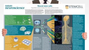 挂图Neural Stem Cells Overview of the types of NSCs and their potential use as therapeutic agents for disease
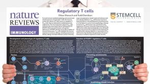 挂图Regulatory T Cells Overview of the development, phenotype and functions of regulatory T cells
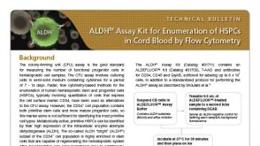 技术公告ALDHbr Assay Kit for Enumeration of HSPCs in Cord Blood by Flow Cytometry
 沪公网安备31010102008431号