Mesenchymal stem cells can be differentiated into endothelial cells in vitro.
Human bone marrow-derived mesenchymal stem cells (MSCs) have the potential to differentiate into mesenchymal tissues like osteocytes,chondrocytes,and adipocytes in vivo and in vitro. The aim of this study was to investigate the in vitro differentiation of MSCs into cells of the endothelial lineage. MSCs were generated out of mononuclear bone marrow cells from healthy donors separated by density gradient centrifugation. Cells were characterized by flow cytometry using a panel of monoclonal antibodies and were tested for their potential to differentiate along different mesenchymal lineages. Isolated MSCs were positive for the markers CD105,CD73,CD166,CD90,and CD44 and negative for typical hematopoietic and endothelial markers. They were able to differentiate into adipocytes and osteocytes after cultivation in respective media. Differentiation into endothelial-like cells was induced by cultivation of confluent cells in the presence of 2% fetal calf serum and 50 ng/ml vascular endothelial growth factor. Laser scanning cytometry analysis of the confluent cells in situ showed a strong increase of expression of endothelial-specific markers like KDR and FLT-1,and immunofluorescence analysis showed typical expression of the von Willebrand factor. The functional behavior of the differentiated cells was tested with an in vitro angiogenesis test kit where cells formed characteristic capillary-like structures. We could show the differentiation of expanded adult human MSCs into cells with phenotypic and functional features of endothelial cells. These predifferentiated cells provide new options for engineering of artificial tissues based on autologous MSCs and vascularized engineered tissues.
View Publication
产品类型:
产品号#:
05401
产品名:
MesenCult™ MSC基础培养基 (人)
Feng Y et al. (SEP 2010)
Progress in biophysics and molecular biology 103 1 148--56
Unique biomechanical interactions between myeloma cells and bone marrow stroma cells.
We observed that BMSCs (bone marrow stromal cells) from myeloma patients (myeloma BMSCs) were significantly stiffer than control BMSCs using a cytocompression device. The stiffness of myeloma BMSCs and control BMSCs was further increased upon priming by myeloma cells. Additionally,myeloma cells became stiffer when primed by myeloma BMSCs. The focal adhesion kinase activity of myeloma cells was increased when cells were on stiffer collagen gels and on myeloma BMSCs. This change in myeloma stiffness is associated with increased colony formation of myeloma cells and FAK activation when co-cultured with stiffer myeloma BMSCs or stiffer collagen. Additionally,stem cells of RPMI8226 cells became stiffer after priming by myeloma BMSCs,with concomitant increases of stem cell colony formation. These results suggest the presence of a mechanotransduction loop between myeloma cells and myeloma BMSCs to increase the stiffness of both types of cells via FAK activation. The increase of stiffness may in turn support the growth of myeloma cells and myeloma stem cells.
View Publication
产品类型:
产品号#:
05401
05402
05411
产品名:
MesenCult™ MSC基础培养基 (人)
MesenCult™ MSC 刺激补充剂(人)
MesenCult™ 增殖试剂盒(人)
Lippmann ES et al. (AUG 2012)
Nature biotechnology 30 8 783--791
Derivation of blood-brain barrier endothelial cells from human pluripotent stem cells.
The blood-brain barrier (BBB) is crucial to the health of the brain and is often compromised in neurological disease. Moreover,because of its barrier properties,this endothelial interface restricts uptake of neurotherapeutics. Thus,a renewable source of human BBB endothelium could spur brain research and pharmaceutical development. Here we show that endothelial cells derived from human pluripotent stem cells (hPSCs) acquire BBB properties when co-differentiated with neural cells that provide relevant cues,including those involved in Wnt/β-catenin signaling. The resulting endothelial cells have many BBB attributes,including well-organized tight junctions,appropriate expression of nutrient transporters and polarized efflux transporter activity. Notably,they respond to astrocytes,acquiring substantial barrier properties as measured by transendothelial electrical resistance (1,450 ± 140 Ω cm2),and they possess molecular permeability that correlates well with in vivo rodent blood-brain transfer coefficients.
View Publication
A fast, automated, polynomial-based cosmic ray spike-removal method for the high-throughput processing of Raman spectra.
Raman spectra often contain undesirable,randomly positioned,intense,narrow-bandwidth,positive,unidirectional spectral features generated when cosmic rays strike charge-coupled device cameras. These must be removed prior to analysis,but doing so manually is not feasible for large data sets. We developed a quick,simple,effective,semi-automated procedure to remove cosmic ray spikes from spectral data sets that contain large numbers of relatively homogenous spectra. Although some inhomogeneous spectral data sets can be accommodated—it requires replacing excessively modified spectra with the originals and removing their spikes with a median filter instead—caution is advised when processing such data sets. In addition,the technique is suitable for interpolating missing spectra or replacing aberrant spectra with good spectral estimates. The method is applied to baseline-flattened spectra and relies on fitting a third-order (or higher) polynomial through all the spectra at every wavenumber. Pixel intensities in excess of a threshold of 3× the noise standard deviation above the fit are reduced to the threshold level. Because only two parameters (with readily specified default values) might require further adjustment,the method is easily implemented for semi-automated processing of large spectral sets.
View Publication
产品类型:
产品号#:
85850
85857
产品名:
mTeSR™1
mTeSR™1
Puri MC and Bernstein A (OCT 2003)
Proceedings of the National Academy of Sciences of the United States of America 100 22 12753--8
Requirement for the TIE family of receptor tyrosine kinases in adult but not fetal hematopoiesis.
In mammals,the continuous production of hematopoietic cells (HCs) is sustained by a small number of hematopoietic stem cells (HSCs) residing in the bone marrow. Early HSC activity arises in the aorta-gonad mesonephros region,within cells localized to the ventral floor of the major blood vessels,suggesting that the first HSCs may be derived from cells capable of giving rise to the hematopoietic system and to the endothelial cells of the vasculature. TIE1 (TIE) and TIE2 (TEK) are related receptor tyrosine kinases with an embryonic expression pattern in endothelial cells,their precursors,and HCs,suggestive of a role in the divergence and function of both lineages. Indeed,gene targeting approaches have shown that TIE1,TIE2,and ligands for TIE2,the angiopoietins,are essential for vascular development and maintenance. To explore possible roles for these receptors in HCs,we have examined the ability of embryonic cells lacking both TIE1 and TIE2 to contribute to developmental and adult hematopoiesis by generating chimeric animals between normal embryonic cells and cells lacking these receptors. We show here that TIE receptors are not required for differentiation and proliferation of definitive hematopoietic lineages in the embryo and fetus; surprisingly,however,these receptors are specifically required during postnatal bone marrow hematopoiesis.
View Publication
产品类型:
产品号#:
03434
03444
产品名:
MethoCult™GF M3434
MethoCult™GF M3434
Park I-K et al. (MAR 2009)
Blood 113 11 2470--7
The Axl/Gas6 pathway is required for optimal cytokine signaling during human natural killer cell development.
Interleukin-15 (IL-15) is essential for natural killer (NK) cell differentiation. In this study,we assessed whether the receptor tyrosine kinase Axl and its ligand,Gas6,are involved in IL-15-mediated human NK differentiation from CD34(+) hematopoietic progenitor cells (HPCs). Blocking the Axl-Gas6 interaction with a soluble Axl fusion protein (Axl-Fc) or the vitamin K inhibitor warfarin significantly diminished the absolute number and percentage of CD3(-)CD56(+) NK cells derived from human CD34(+) HPCs cultured in the presence of IL-15,probably resulting in part from reduced phosphorylation of STAT5. In addition,CD3(-)CD56(+) NK cells derived from culture of CD34(+) HPCs with IL-15 and Axl-Fc had a significantly diminished capacity to express interferon-gamma or its master regulator,T-BET. Culture of CD34(+) HPCs in the presence of c-Kit ligand and Axl-Fc resulted in a significant decrease in the frequency of NK precursor cells responding to IL-15,probably the result of reduced c-Kit phosphorylation. Collectively,our data suggest that the Axl/Gas6 pathway contributes to normal human NK-cell development,at least in part via its regulatory effects on both the IL-15 and c-Kit signaling pathways in CD34(+) HPCs,and to functional NK-cell maturation via an effect on the master regulatory transcription factor T-BET.
View Publication
产品类型:
产品号#:
15026
15066
产品名:
RosetteSep™ 人造血祖细胞富集抗体混合物
RosetteSep™人造血祖细胞富集抗体混合物
Jimeno A et al. (FEB 2009)
Molecular cancer therapeutics 8 2 310--4
A direct pancreatic cancer xenograft model as a platform for cancer stem cell therapeutic development.
There is an enormous gap between the antiproliferative and in vivo antitumor efficacy of gemcitabine in cell line-based models and its clinical efficacy. This may be due to insensitiveness of the precursor,cancer stem cell (CSC) compartment to cytotoxic agents. The hedgehog pathway is associated with CSC signaling and control. We used a direct xenograft model of pancreatic cancer and a two-stage approach was used to test the hypotheses that targeting CSC could increase the efficacy of gemcitabine. Tumors from a gemcitabine-sensitive xenograft were treated with gemcitabine first,and randomized,after tumor regression to continuing treatment with gemcitabine,a hedgehog inhibitor alone or in combination with gemcitabine. We tested markers described as associated with CSC such as CD24,CD44,ALDH,nestin,and the hedgehog pathway. After induction with gemcitabine,treated tumor showed an enrichment in CSC markers such as ALDH and CD24. Subsequently,a release from gemcitabine prompted a repopulation of proliferating cells and a decrease in such markers to equilibrate from pretreatment levels. Combined treatment with gemcitabine and cyclopamine induced tumor regression and decrease in CSC markers and hedgehog signaling. Cytoplasmic CD24 and ALDH were inversely and strongly associated with growth and were expressed in a minority of cells that we propose constitute the CSC compartment. Hedgehog inhibitors as part of a dual compartment therapeutic approach were able to further reduce tumor growth and decreased both static and dynamic markers of CSC. Direct tumor xenografts are a valid platform to test multicompartment therapeutic approaches in pancreatic cancer.
View Publication
产品类型:
产品号#:
01700
01702
01705
产品名:
ALDEFLUOR™ 试剂盒
ALDEFLUOR™测定缓冲液
ALDEFLUOR™ DEAB试剂
Hansen SK et al. (AUG 2016)
Stem cell research 17 2 306--317
Induced pluripotent stem cell - derived neurons for the study of spinocerebellar ataxia type 3.
The neurodegenerative disease spinocerebellar ataxia type 3 (SCA3) is caused by a CAG-repeat expansion in the ATXN3 gene. In this study,induced pluripotent stem cell (iPSC) lines were established from two SCA3 patients. Dermal fibroblasts were reprogrammed using an integration-free method and the resulting SCA3 iPSCs were differentiated into neurons. These neuronal lines harbored the disease causing mutation,expressed comparable levels of several neuronal markers and responded to the neurotransmitters,glutamate/glycine,GABA and acetylcholine. Additionally,all neuronal cultures formed networks displaying synchronized spontaneous calcium oscillations within 28days of maturation,and expressed the mature neuronal markers NeuN and Synapsin 1 implying a relatively advanced state of maturity,although not comparable to that of the adult human brain. Interestingly,we were not able to recapitulate the glutamate-induced ataxin-3 aggregation shown in a previously published iPSC-derived SCA3 model. In conclusion,we have generated a panel of SCA3 patient iPSCs and a robust protocol to derive neurons of relatively advanced maturity,which could potentially be valuable for the study of SCA3 disease mechanisms.
View Publication
产品类型:
产品号#:
85850
85857
产品名:
mTeSR™1
mTeSR™1
Lehmann JM et al. (JUN 1995)
The Journal of biological chemistry 270 22 12953--6
An antidiabetic thiazolidinedione is a high affinity ligand for peroxisome proliferator-activated receptor gamma (PPAR gamma).
Thiazolidinedione derivatives are antidiabetic agents that increase the insulin sensitivity of target tissues in animal models of non-insulin-dependent diabetes mellitus. In vitro,thiazolidinediones promote adipocyte differentiation of preadipocyte and mesenchymal stem cell lines; however,the molecular basis for this adipogenic effect has remained unclear. Here,we report that thiazolidinediones are potent and selective activators of peroxisome proliferator-activated receptor gamma (PPAR gamma),a member of the nuclear receptor superfamily recently shown to function in adipogenesis. The most potent of these agents,BRL49653,binds to PPAR gamma with a Kd of approximately 40 nM. Treatment of pluripotent C3H10T1/2 stem cells with BRL49653 results in efficient differentiation to adipocytes. These data are the first demonstration of a high affinity PPAR ligand and provide strong evidence that PPAR gamma is a molecular target for the adipogenic effects of thiazolidinediones. Furthermore,these data raise the intriguing possibility that PPAR gamma is a target for the therapeutic actions of this class of compounds.
View Publication
产品类型:
产品号#:
72622
72624
产品名:
Rosiglitazone
罗格列酮(Rosiglitazone)
Park SY et al. (FEB 2010)
Clinical cancer research : an official journal of the American Association for Cancer Research 16 3 876--87
Heterogeneity for stem cell-related markers according to tumor subtype and histologic stage in breast cancer.
PURPOSE: To evaluate the expression of stem cell-related markers at the cellular level in human breast tumors of different subtypes and histologic stage. EXPERIMENTAL DESIGN: We performed immunohistochemical analyses of 12 proteins [CD44,CD24,ALDH1,vimentin,osteonectin,EPCR,caveolin 1,connexin 43,cytokeratin 18 (CK18),MUC1,claudin 7,and GATA3] selected based on their differential expression in breast cancer cells with more differentiated and stem cell-like characteristics in 47 cases of invasive ductal carcinoma (IDC) only,135 cases of IDC with ductal carcinoma in situ (DCIS),35 cases of DCIS with microinvasion,and 58 cases of pure DCIS. We also analyzed 73 IDCs with adjacent DCIS to determine the differences in the expression of markers by histology within individual tumors. CD44+/CD24- and CD24-/CD24+ cells were detected using double immunohistochemistry. RESULTS: CD44 and EPCR expression was different among the four histologic groups and was lower in invasive compared with in situ tumors,especially in luminal A subtype. The expression of vimentin,osteonectin,connexin 43,ALDH1,CK18,GATA3,and MUC1 differed by tumor subtype in some histologic groups. ALDH1-positive cells were more frequent in basal-like and HER2+ than in luminal tumors. CD44+/CD24- cells were detected in 69% of all tumors with 100% of the basal-like and 52% of HER2+ tumors having some of these cells. CONCLUSIONS: Our findings suggest that in breast cancer,the frequency of tumor cells positive for stem cell-like and more differentiated cell markers varies according to tumor subtype and histologic stage.
View Publication

 EasySep™小鼠TIL(CD45)正选试剂盒


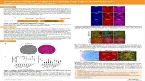 科学海报Efficient Generation of Human Pluripotent Stem Cells to Neural Crest Cells
 科学海报Efficient Generation of Lung Progenitor Cells From Human Pluripotent Stem Cells
 沪公网安备31010102008431号