Sato K et al. (JAN 2006)
The Journal of experimental medicine 203 1 239--50
TRAIL-expressing T cells induce apoptosis of vascular smooth muscle cells in the atherosclerotic plaque.
Acute coronary syndromes (ACS) are precipitated by a rupture of the atherosclerotic plaque,often at the site of T cell and macrophage infiltration. Here,we show that plaque-infiltrating CD4 T cells effectively kill vascular smooth muscle cells (VSMC). VSMCs sensitive to T cell-mediated killing express the death receptor DR5 (TNF-related apoptosis-inducing ligand [TRAIL] receptor 2),and anti-TRAIL and anti-DR5 antibodies block T cell-mediated apoptosis. CD4 T cells that express TRAIL upon stimulation are expanded in patients with ACS and more effectively induce VSMC apoptosis. Adoptive transfer of plaque-derived CD4 T cells into immunodeficient mice that are engrafted with human atherosclerotic plaque results in apoptosis of VSMCs,which was prevented by coadministration of anti-TRAIL antibody. These data identify that the death pathway is triggered by TRAIL-producing CD4 T cells as a direct mechanism of VSMC apoptosis,a process which may lead to plaque destabilization.
View Publication
产品类型:
产品号#:
15022
15062
产品名:
RosetteSep™人CD4+ T细胞富集抗体混合物
RosetteSep™人CD4+ T细胞富集抗体混合物
Wang R et al. (DEC 2015)
BMC cancer 16 1 56
Fusion with stem cell makes the hepatocellular carcinoma cells similar to liver tumor-initiating cells.
BACKGROUND Cell fusion is a fast and highly efficient technique for cells to acquire new properties. The fusion of somatic cells with stem cells can reprogram somatic cells to a pluripotent state. Our research on the fusion of stem cells and cancer cells demonstrates that the fused cells can exhibit stemness and cancer cell-like characteristics. Thus,tumor-initiating cell-like cells are generated. METHODS We employed laser-induced single-cell fusion technique to fuse the hepatocellular carcinoma cells and human embryonic stem cells (hESC). Real-time RT-PCR,flow cytometry and in vivo tumorigenicity assay were adopted to identify the gene expression difference. RESULTS We successfully produced a fused cell line that coalesces the gene expression information of hepatocellular carcinoma cells and stem cells. Experimental results showed that the fused cells expressed cancer and stemness markers as well as exhibited increased resistance to drug treatment and enhanced tumorigenesis. CONCLUSIONS Fusion with stem cells transforms liver cancer cells into tumor initiating-like cells. Results indicate that fusion between cancer cell and stem cell may generate tumor initiating-like cells.
View Publication
产品类型:
产品号#:
85850
85857
产品名:
mTeSR™1
mTeSR™1
Kim J-HHH et al. (MAR 2016)
ACS nano 10 3 3342--3355
Nanotopography Promotes Pancreatic Differentiation of Human Embryonic Stem Cells and Induced Pluripotent Stem Cells.
Although previous studies suggest that nanotopographical features influence properties and behaviors of stem cells,only a few studies have attempted to derive clinically useful somatic cells from human pluripotent stem cells using nanopatterned surfaces. In the present study,we report that polystyrene nanopore-patterned surfaces significantly promote the pancreatic differentiation of human embryonic and induced pluripotent stem cells. We compared different diameters of nanopores and showed that 200 nm nanopore-patterned surfaces highly upregulated the expression of PDX1,a critical transcription factor for pancreatic development,leading to an approximately 3-fold increase in the percentage of differentiating PDX1(+) pancreatic progenitors compared with control flat surfaces. Furthermore,in the presence of biochemical factors,200 nm nanopore-patterned surfaces profoundly enhanced the derivation of pancreatic endocrine cells producing insulin,glucagon,or somatostatin. We also demonstrate that nanopore-patterned surface-induced upregulation of PDX1 is associated with downregulation of TAZ,suggesting the potential role of TAZ in nanopore-patterned surface-mediated mechanotransduction. Our study suggests that appropriate cytokine treatments combined with nanotopographical stimulation could be a powerful tool for deriving a high purity of desired cells from human pluripotent stem cells.
View Publication
产品类型:
产品号#:
85850
85857
产品名:
mTeSR™1
mTeSR™1
Duelen R et al. ( 2017)
Stem cells international 2017 4651238
Activin A Modulates CRIPTO-1/HNF4α(+) Cells to Guide Cardiac Differentiation from Human Embryonic Stem Cells.
The use of human pluripotent stem cells in basic and translational cardiac research requires efficient differentiation protocols towards cardiomyocytes. In vitro differentiation yields heterogeneous populations of ventricular-,atrial-,and nodal-like cells hindering their potential applications in regenerative therapies. We described the effect of the growth factor Activin A during early human embryonic stem cell fate determination in cardiac differentiation. Addition of high levels of Activin A during embryoid body cardiac differentiation augmented the generation of endoderm derivatives,which in turn promoted cardiomyocyte differentiation. Moreover,a dose-dependent increase in the coreceptor expression of the TGF-β superfamily member CRIPTO-1 was observed in response to Activin A. We hypothesized that interactions between cells derived from meso- and endodermal lineages in embryoid bodies contributed to improved cell maturation in early stages of cardiac differentiation,improving the beating frequency and the percentage of contracting embryoid bodies. Activin A did not seem to affect the properties of cardiomyocytes at later stages of differentiation,measuring action potentials,and intracellular Ca(2+) dynamics. These findings are relevant for improving our understanding on human heart development,and the proposed protocol could be further explored to obtain cardiomyocytes with functional phenotypes,similar to those observed in adult cardiac myocytes.
View Publication
Ahmad N et al. (APR 2000)
Archives of biochemistry and biophysics 376 2 338--46
Green tea polyphenol epigallocatechin-3-gallate differentially modulates nuclear factor kappaB in cancer cells versus normal cells.
Green tea has shown remarkable anti-inflammatory and cancer chemopreventive effects in many animal tumor bioassays,cell culture systems,and epidemiological studies. Many of these biological effects of green tea are mediated by epigallocatechin 3-gallate (EGCG),the major polyphenol present therein. We have earlier shown that EGCG treatment results in apoptosis of several cancer cells,but not of normal cells (J. Natl. Cancer Inst. 89,1881-1886 (1997)). The mechanism of this differential response of EGCG is not known. In this study,we investigated the involvement of NF-kappaB during these differential responses of EGCG. EGCG treatment resulted in a dose-dependent (i) inhibition of cell growth,(ii) G0/G1-phase arrest of the cell cycle,and (iii) induction of apoptosis in human epidermoid carcinoma (A431) cells,but not in normal human epidermal keratinocytes (NHEK). Electromobility shift assay revealed that EGCG (10-80 microM) treatment results in lowering of NF-kappaB levels in both the cytoplasm and nucleus in a dose-dependent manner in both A431 cells and NHEK,albeit at different concentrations. EGCG treatment was found to result in a dose-based differential inhibition of TNF-alpha- and LPS-mediated activation of NF-kappaB in these cells. The inhibition of NF-kappaB constitutive expression and activation in NHEK was observed only at high concentrations. The immunoblot analysis also demonstrated a similar pattern of inhibition of the constitutive expression as well as activation of NF-kappaB/p65 nuclear protein. This inhibition of TNF-alpha-caused NF-kappaB activation was mediated via the phosphorylative degradation of its inhibitory protein IkappaBalpha. Taken together,EGCG was found to impart differential dose-based NF-kappaB inhibitory response in cancer cells vs normal cells; i.e.,EGCG-mediated inhibition of NF-kappaB constitutive expression and activation was found to occur at much higher dose of EGCG in NHEK as compared to A431 cells. This study suggests that EGCG-caused cell cycle deregulation and apoptosis of cancer cells may be mediated through NF-kappaB inhibition.
View Publication
产品类型:
产品号#:
73644
产品名:
(-)-Epigallocatechin Gallate
Richardson T et al. (DEC 2013)
Tissue Engineering: Part A 20 23-24 Epub ahead of print
Alginate encapsulation of human embryonic stem cells to enhance directed differentiation to pancreatic islet-like cells
The pluripotent property of hESCs makes them attractive for treatment of degenerative diseases such as diabetes. We have developed a stage-wise directed differentiation protocol to produce alginate-encapsulated islet-like cells derived from hESCs,which can be directly implanted for diabetes therapy. The advantage of alginate encapsulation lies in its capability to immunoisolate,along with the added possibility of scalable culture. We have evaluated the possibility of encapsulating hESCs at different stages of differentiation. Encapsulation of predifferentiated cells resulted in insufficient cellular yield and differentiation. On the other hand,encapsulation of undifferentiated hESCs followed by differentiation induction upon encapsulation,resulted in the highest viability and differentiation. More striking was that alginate encapsulation resulted in a much stronger differentiation compared to parallel 2D cultures,resulting in 20-fold increase in c-peptide protein synthesis. To elucidate the mechanism contributing to encapsulation-mediated enhancement in hESC maturation,investigation of the signaling pathways revealed interesting insight. While the phospho-protein levels of all the tested signaling molecules were lower under encapsulation,the ratio of pSMAD/pAKT was significantly higher,indicating a more efficient signal transduction under encapsulation. These results clearly demonstrate that alginate encapsulation of hESCs and differentiation to islet-cells types provides a potentially translatable treatment option for type1 diabetes.
View Publication
产品类型:
产品号#:
85850
85857
产品名:
mTeSR™1
mTeSR™1
Ji H et al. (JAN 2015)
The Journal of allergy and clinical immunology 135 1 236--244
Dynamic transcriptional and epigenomic reprogramming from pediatric nasal epithelial cells to induced pluripotent stem cells
BACKGROUND Induced pluripotent stem cells (iPSCs) hold tremendous potential,both as a biological tool to uncover the pathophysiology of disease by creating relevant human cell models and as a source of cells for cell-based therapeutic applications. Studying the reprogramming process will also provide significant insight into tissue development. OBJECTIVE We sought to characterize the derivation of iPSC lines from nasal epithelial cells (NECs) isolated from nasal mucosa samples of children,a highly relevant and easily accessible tissue for pediatric populations. METHODS We performed detailed comparative analysis on the transcriptomes and methylomes of NECs,iPSCs derived from NECs (NEC-iPSCs),and embryonic stem cells (ESCs). RESULTS NEC-iPSCs express pluripotent cell markers,can differentiate into all 3 germ layers in vivo and in vitro,and have a transcriptome and methylome remarkably similar to those of ESCs. However,residual DNA methylation marks exist,which are differentially methylated between NEC-iPSCs and ESCs. A subset of these methylation markers related to epithelium development and asthma and specific to NEC-iPSCs persisted after several passages in vitro,suggesting the retention of an epigenetic memory of their tissue of origin. Our analysis also identified novel candidate genes with dynamic gene expression and DNA methylation changes during reprogramming,which are indicative of possible roles in airway epithelium development. CONCLUSION NECs are an excellent tissue source to generate iPSCs in pediatric asthmatic patients,and detailed characterization of the resulting iPSC lines would help us better understand the reprogramming process and retention of epigenetic memory.
View Publication
产品类型:
产品号#:
85850
85857
产品名:
mTeSR™1
mTeSR™1
De Giorgi U et al. (MAY 2011)
Cancer biology & therapy 11 9 812--5
Mesenchymal stem cells expressing GD2 and CD271 correlate with breast cancer-initiating cells in bone marrow.
Purpose: The bone marrow microenvironment is considered a critical component in the dissemination and fate of cancer cells in the metastatic process. We explored the possible correlation between bone marrow mesenchymal stem cells (BM-MSC) and disseminated breast cancer-initiating cells (BCIC) in primary breast cancer patients. Experimental design: Bone marrow mononuclear cells (BM-MNC) were collected at the time of primary surgery in 12 breast cancer patients. BM-MNC was immunophenotyped and BCIC was defined as epithelial cells (CD326+CD45-) with a stem-like" phenotype (CD44+CD24low/-�
View Publication

 EasySep™小鼠TIL(CD45)正选试剂盒


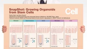 挂图SnapShot: Growing Organoids from Stem Cells Key culture conditions and organoid-forming cells from a variety of epithelial tissues

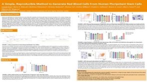 科学海报A Simple, Reproducible Method to Generate Red Blood Cells From Human Pluripotent Stem Cells

 沪公网安备31010102008431号