Choi SA et al. (NOV 2012)
Cancer Letters 324 2 221--230
A distinct subpopulation within CD133 positive brain tumor cells shares characteristics with endothelial progenitor cells
The cell surface marker CD133 has been proposed as a brain tumor stem cell marker. However,there have been substantial controversies regarding the necessity and role of CD133 in tumorigenesis. This study aimed to characterize CD133(+) cells in brain tumors. Human brain tumor specimens and whole blood were collected from the same patients (N=12). We carried out dual FACS staining for CD133/CD34 and functional tumorigenesis and angiogenesis analyses of CD133(+) cells from different origins. We also investigated the in vivo tumorigenic potential and histological characteristics of four distinct groups on the basis of expression of CD133/CD34 markers (CD133(+),CD133(+)/CD34(+),CD133(+)/CD34(-),and CD133(-)). CD133(+) brain tumor cells coexpressed significantly higher positivity for CD34 (70.7±5.2% in CD133(+) vs. 12.3±4.2% in CD133(-) cells,P<0.001). CD133(+) brain tumor cells formed neurosphere-like spheroids and differentiated into multiple nervous system lineages unlike CD133(+) blood cells. They showed biological characteristics of endothelial cells,including vWF expression,LDL uptake and tube formation in vitro,unlike CD133(-) brain tumors cells. Pathologic analysis of brains implanted with CD133(+) cells showed large,markedly hypervascular tumors with well-demarcated boundary. CD133(+)/CD34(-) cells produced smaller but highly infiltrative tumors. Notably,pure angiogenic cell fractions (CD133(+)/CD34(+)) and CD133(-) tumor cells did not generate tumors in vivo. Our data suggest the presence of a distinct subpopulation of CD133(+) cells isolated from human brain tumors,with characteristics of endothelial progenitor cells (EPCs).
View Publication
产品类型:
产品号#:
05750
05752
产品名:
NeuroCult™ NS-A 基础培养基(人)
NeuroCult™ NS-A 分化试剂盒 (人)
Slukvin II et al. (MAR 2006)
Journal of immunology (Baltimore,Md. : 1950) 176 5 2924--32
Directed differentiation of human embryonic stem cells into functional dendritic cells through the myeloid pathway.
We have established a system for directed differentiation of human embryonic stem (hES) cells into myeloid dendritic cells (DCs). As a first step,we induced hemopoietic differentiation by coculture of hES cells with OP9 stromal cells,and then,expanded myeloid cells with GM-CSF using a feeder-free culture system. Myeloid cells had a CD4+CD11b+CD11c+CD16+CD123(low)HLA-DR- phenotype,expressed myeloperoxidase,and included a population of M-CSFR+ monocyte-lineage committed cells. Further culture of myeloid cells in serum-free medium with GM-CSF and IL-4 generated cells that had typical dendritic morphology; expressed high levels of MHC class I and II molecules,CD1a,CD11c,CD80,CD86,DC-SIGN,and CD40; and were capable of Ag processing,triggering naive T cells in MLR,and presenting Ags to specific T cell clones through the MHC class I pathway. Incubation of DCs with A23187 calcium ionophore for 48 h induced an expression of mature DC markers CD83 and fascin. The combination of GM-CSF with IL-4 provided the best conditions for DC differentiation. DCs obtained with GM-CSF and TNF-alpha coexpressed a high level of CD14,and had low stimulatory capacity in MLR. These data clearly demonstrate that hES cells can be used as a novel and unique source of hemopoietic and DC precursors as well as DCs at different stages of maturation to address essential questions of DC development and biology. In addition,because ES cells can be expanded without limit,they can be seen as a potential scalable source of cells for DC vaccines or DC-mediated induction of immune tolerance.
View Publication
产品类型:
产品号#:
09600
09650
产品名:
StemSpan™ SFEM
StemSpan™ SFEM
Dottori M et al. (MAY 2008)
Stem cells (Dayton,Ohio) 26 5 1146--54
Lysophosphatidic acid inhibits neuronal differentiation of neural stem/progenitor cells derived from human embryonic stem cells.
Lysophospholipids are signaling molecules that play broad and major roles within the nervous system during both early development and neural injury. We used neural differentiation of human embryonic stem cells (hESC) as an in vitro model to examine the specific effects of lysophosphatidic acid (LPA) at various stages of neural development,from neural induction to mature neurons and glia. We report that LPA inhibits neurosphere formation and the differentiation of neural stem cells (NSC) toward neurons,without modifying NSC proliferation,apoptosis,or astrocytic differentiation. LPA acts through the activation of the Rho/ROCK and the phosphatidylinositol 3-kinase/Akt pathways to inhibit neuronal differentiation. This study is the first demonstration of a role for LPA signaling in neuronal differentiation of hESC. As LPA concentrations increase during inflammation,the inhibition of neuronal differentiation by LPA might contribute to the low level of neurogenesis observed following neurotrauma.
View Publication
产品类型:
产品号#:
72694
产品名:
1-Oleoyl Lysophosphatidic Acid (Sodium Salt)
E. Menares et al. (sep 2019)
Nature communications 10 1 4401
Tissue-resident memory CD8+ T cells amplify anti-tumor immunity by triggering antigen spreading through dendritic cells.
Tissue-resident memory CD8+ T (Trm) cells mediate potent local innate and adaptive immune responses and play a central role against solid tumors. However,whether Trm cells cross-talk with dendritic cells (DCs) to support anti-tumor immunity remains unclear. Here we show that antigen-specific activation of skin Trm cells leads to maturation and migration to draining lymph nodes of cross-presenting dermal DCs. Tumor rejection mediated by Trm cells triggers the spread of cytotoxic CD8+ T cell responses against tumor-derived neo- and self-antigens via dermal DCs. These responses suppress the growth of intradermal tumors and disseminated melanoma lacking the Trm cell-targeted epitope. Moreover,analysis of RNA sequencing data from human melanoma tumors reveals that enrichment of a Trm cell gene signature associates with DC activation and improved survival. This work unveils the ability of Trm cells to amplify the breath of cytotoxic CD8+ T cell responses through DCs,thereby strengthening anti-tumor immunity.
View Publication
产品类型:
产品号#:
09605
09655
产品名:
StemSpan™ SFEM II
StemSpan™ SFEM II
(Feb 2025)
Journal for Immunotherapy of Cancer 13 2
Frizzled-7-targeting antibody-derived bifunctional protein retargets NK cells against triple-negative breast cancer cells via MICA-NKG2D axis
AbstractBackgroundHypoxia is associated with the evasion of triple-negative breast cancer (TNBC) from immune surveillance. Hypoxia increases the subpopulation of putative TNBC stem-like cells (TNBCSCs) through activating Wnt/β-Catenin signaling. The shedding of MHC class I-related chain A (MICA) is particularly noteworthy in cancer stem cells (CSCs),promoting the resistance of CSCs to natural killer (NK) cell cytotoxicity. To reestablish MICA/NKG2D-mediated immunosurveillance,we proposed the design of a fusion protein (SHH002-hu1-MICA) which consists of Frizzled-7 (Fzd7)-targeting antibody and MICA,serving as an engager retargeting NK cells against TNBCs,especially TNBCSCs.MethodsOpal multicolor immunohistochemistry staining was used to validate the expression of membrane MICA (mMICA) and existence of NK cells in TNBC tumors; flow cytometry (FCM) assay was used to detect the expression of Fzd7/mMICA on TNBCs. Biolayer interferometry (BLI) and surface plasmon resonance (SPR) assays were executed to assess the affinity of SHH002-hu1-MICA towards rhFzd7/rhNKG2D; near-infrared imaging assay was used to evaluate the targeting capability. A cytotoxicity assay was conducted to assess the effects of SHH002-hu1-MICA on NK cell-mediated killing of TNBCs,and FCM assay to analyze the effects of SHH002-hu1-MICA on the degranulation of NK cells. Finally,TNBC cell-line-derived xenografts were established to evaluate the anti-tumor activities of SHH002-hu1-MICA in vivo.ResultsThe expression of mMICA is significantly downregulated in hypoxic TNBCs and TNBCSCs,leading to the evasion of immune surveillance exerted by NK cells. The expression of Fzd7 is significantly upregulated in TNBCSCs and exhibits a negative correlation with the expression of mMICA and infiltration level of NK cells. On accurate assembly,SHH002-hu1-MICA shows a strong affinity for rhFzd7/rhNKG2D,specifically targets TNBC tumor tissues,and disrupts Wnt/β-Catenin signaling. SHH002-hu1-MICA significantly enhances the cytotoxicity of NK cells against hypoxic TNBCs and TNBCSCs by inducing the degranulation of NK cells and promotes the infiltration of NK cells in CD44high regions within TNBC xenograft tumors,exhibiting superior anti-tumor activities than SHH002-hu1.ConclusionsSHH002-hu1-MICA maintains the targeting property of SHH002-hu1,successfully activates and retargets NK cells against TNBCs,especially TNBCSCs,exhibiting superior antitumor activities than SHH002-hu1. SHH002-hu1-MICA represents a promising new engager for NK cell-based immunotherapy for TNBC.
View Publication
产品类型:
产品号#:
19055
19055RF
产品名:
EasySep™人NK细胞富集试剂盒
RoboSep™ 人NK细胞富集试剂盒含滤芯吸头
(Apr 2025)
NPJ Vaccines 10
Emulsion adjuvant-induced uric acid release modulates optimal immunogenicity by targeting dendritic cells and B cells
Squalene-based emulsion (SE) adjuvants like MF59 and AS03 are used in protein subunit vaccines against influenza virus (e.g.,Fluad,Pandemrix,Arepanrix) and SARS-CoV-2 (e.g.,Covifenz,SKYCovione). We demonstrate the critical role of uric acid (UA),a damage-associated molecular pattern (DAMP),in triggering immunogenicity by SE adjuvants. In mice,SE adjuvants elevated DAMP levels in draining lymph nodes. Strikingly,inhibition of UA synthesis reduced vaccine-induced innate immunity,subsequently impairing optimal antibody and T cell responses. In vivo treatment with UA crystals elicited partial adjuvant effects. In vitro stimulation with UA crystals augmented the activation of dendritic cells (DCs) and B cells and altered multiple pathways in these cells,including inflammation and antigen presentation in DCs and cell proliferation in B cells. In an influenza vaccine model,UA contributed to protection against influenza viral infection. These results demonstrate the importance of DAMPs,specifically the versatile role of UA in the immunogenicity of SE adjuvants,by regulating DCs and B cells.
View Publication
产品类型:
产品号#:
18000
产品名:
EasySep™磁极
Vasir B et al. (FEB 2005)
Journal of immunology (Baltimore,Md. : 1950) 174 4 2376--86
Dendritic cells induce MUC1 expression and polarization on human T cells by an IL-7-dependent mechanism.
The MUC1 transmembrane mucin is expressed on the surface of activated human T cells; however,the physiologic signals responsible for the regulation of MUC1 in T cells are not known. The present studies demonstrate that IL-7,but not IL-2 or IL-4,markedly induces MUC1 expression on CD3+ T cells. MUC1 was also up-regulated by IL-15,but to a lesser extent than that found with IL-7. The results show that IL-7 up-regulates MUC1 on CD4+,CD8+,CD25+,CD69+,naive CD45RA+,and memory CD45RO+ T cells. In concert with induction of MUC1 expression by IL-7,activated dendritic cells (DC) that produce IL-7 up-regulate MUC1 on allogeneic CD3+ T cells. DC also induce MUC1 expression on autologous CD3+ T cells in the presence of recall Ag. Moreover,DC-induced MUC1 expression on T cells is blocked by a neutralizing anti-IL-7 Ab. The results also demonstrate that DC induce polarization of MUC1 on T cells at sites opposing the DC-T cell synapse. These findings indicate that DC-mediated activation of Ag-specific T cells is associated with induction and polarization of MUC1 expression by an IL-7-dependent mechanism.
View Publication
产品类型:
产品号#:
15271HLA
产品名:
RosetteSep™ HLA 淋系细胞富集试剂盒
A. Haase et al. ( 2017)
Stem cell research 21 71--73
Generation of non-transgenic iPS cells from human cord blood CD34+ cells under animal component-free conditions.
Recently,many hurdles and limitations for production of clinically applicable iPSC derivatives have been overcome. Transgene-free iPSCs can be efficiently derived from easily accessible cell sources such as blood. Here we describe the generation of transgene-free hiPS cells from cord blood derived CD34+ cells,reprogrammed using CytoTune™ Sendai reprogramming vectors. CD34+ cell isolation,cultivation,reprogramming and establishment of resulting hiPSC lines were performed under the exclusive usage of animal-derived component-free (ADCF) materials and components.
View Publication
Tang C et al. (SEP 2011)
Nature biotechnology 29 9 829--34
An antibody against SSEA-5 glycan on human pluripotent stem cells enables removal of teratoma-forming cells.
An important risk in the clinical application of human pluripotent stem cells (hPSCs),including human embryonic and induced pluripotent stem cells (hESCs and hiPSCs),is teratoma formation by residual undifferentiated cells. We raised a monoclonal antibody against hESCs,designated anti-stage-specific embryonic antigen (SSEA)-5,which binds a previously unidentified antigen highly and specifically expressed on hPSCs--the H type-1 glycan. Separation based on SSEA-5 expression through fluorescence-activated cell sorting (FACS) greatly reduced teratoma-formation potential of heterogeneously differentiated cultures. To ensure complete removal of teratoma-forming cells,we identified additional pluripotency surface markers (PSMs) exhibiting a large dynamic expression range during differentiation: CD9,CD30,CD50,CD90 and CD200. Immunohistochemistry studies of human fetal tissues and bioinformatics analysis of a microarray database revealed that concurrent expression of these markers is both common and specific to hPSCs. Immunodepletion with antibodies against SSEA-5 and two additional PSMs completely removed teratoma-formation potential from incompletely differentiated hESC cultures.
View Publication
产品类型:
产品号#:
85850
85857
产品名:
mTeSR™1
mTeSR™1
Luo LZ et al. (JAN 2012)
PLoS ONE 7 3 e30541
DNA repair in human pluripotent stem cells is distinct from that in non-pluripotent human cells.
The potential for human disease treatment using human pluripotent stem cells,including embryonic stem cells and induced pluripotent stem cells (iPSCs),also carries the risk of added genomic instability. Genomic instability is most often linked to DNA repair deficiencies,which indicates that screening/characterization of possible repair deficiencies in pluripotent human stem cells should be a necessary step prior to their clinical and research use. In this study,a comparison of DNA repair pathways in pluripotent cells,as compared to those in non-pluripotent cells,demonstrated that DNA repair capacities of pluripotent cell lines were more heterogeneous than those of differentiated lines examined and were generally greater. Although pluripotent cells had high DNA repair capacities for nucleotide excision repair,we show that ultraviolet radiation at low fluxes induced an apoptotic response in these cells,while differentiated cells lacked response to this stimulus,and note that pluripotent cells had a similar apoptotic response to alkylating agent damage. This sensitivity of pluripotent cells to damage is notable since viable pluripotent cells exhibit less ultraviolet light-induced DNA damage than do differentiated cells that receive the same flux. In addition,the importance of screening pluripotent cells for DNA repair defects was highlighted by an iPSC line that demonstrated a normal spectral karyotype,but showed both microsatellite instability and reduced DNA repair capacities in three out of four DNA repair pathways examined. Together,these results demonstrate a need to evaluate DNA repair capacities in pluripotent cell lines,in order to characterize their genomic stability,prior to their pre-clinical and clinical use.
View Publication
产品类型:
产品号#:
85850
85857
产品名:
mTeSR™1
mTeSR™1
P. J. Gokhale and P. W. Andrews ( 2013)
NeuroReport
Characterization of human pluripotent stem cells
Characterization of pluripotent stem cells is required for the registration of stem cell lines and allows for an impartial and objective comparison of the results obtained when generating multiple lines. It is therefore crucial to establish specific,fast and reliable protocols to detect the hallmarks of pluripotency. Such protocols should include immunocytochemistry (takes 2 d),identification of the three germ layers in in vitro-derived embryoid bodies by immunocytochemistry (immunodetection takes 3 d) and detection of differentiation markers in in vivo-generated teratomas by immunohistochemistry (differentiation marker detection takes 4 d). Standardization of the immunodetection protocols used ensures minimum variations owing to the source,the animal species,the endogenous fluorescence or the inability to collect large amounts of cells,thereby yielding results as fast as possible without loss of quality. This protocol provides a description of all the immunodetection procedures necessary to characterize mouse and human stem cell lines in different circumstances.
View Publication

 EasySep™小鼠TIL(CD45)正选试剂盒


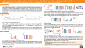 科学海报A Reproducible and Simple Method to Generate Red Blood Cells From Human Pluripotent Stem Cells
 沪公网安备31010102008431号