Hidalgo LG et al. (MAR 2008)
American journal of transplantation : official journal of the American Society of Transplantation and the American Society of Transplant Surgeons 8 3 627--36
The transcriptome of human cytotoxic T cells: similarities and disparities among allostimulated CD4(+) CTL, CD8(+) CTL and NK cells.
Transcripts expressed in cytotoxic T lymphocytes (CTL) have mechanistic and diagnostic importance in transplantation. We used microarrays to select CTL-associated transcripts (CATs) expressed in human CD4(+) CTL,CD8(+) CTL and NK cells,excluding transcripts expressed in B cells,monocytes and kidney. This generated three transcript sets: CD4(+)-associated,CD8(+)-associated and NK-associated. Surprisingly,many CATs were expressed in effector memory cells e.g. granzyme B/GZMB,interferon-gamma/IFNG. Transcript expression was very similar between CD4(+) and CD8(+) CTL. There were no transcripts highly selective for CD4(+) CTL or CD8(+) CTL: for example,cytotoxic molecule transcripts (perforin,granzymes,granulysin) were shared between CD8(+) CTL and CD4(+) CTL although expression remained higher in CD8(+) CTL. Transcripts that differentiated between CD8(+) CTL and CD4(+) CTL were primarily those shared between CD8(+) CTL and NK cells (e.g. NK receptors KLRC1,KLRC3,KLRD1,KLRK1). No transcripts could differentiate CD4(+) CTL from CD8(+) CTL but NK cell-associated transcripts could differentiate NK cells from CTL. This study serves as a foundation for the interpretation of CATs in rejecting allografts and highlights the extensive sharing of CATs among CD4(+) CTL,CD8(+) CTL and effector memory T cells.
View Publication
产品类型:
产品号#:
19052
19052RF
19053
19053RF
19054
19054RF
19055
19055RF
产品名:
EasySep™人CD4+ T细胞富集试剂盒
RoboSep™ 人CD4+ T细胞富集试剂盒含滤芯吸头
EasySep™人CD8+ T细胞富集试剂盒
RoboSep™ 人CD8+ T细胞富集试剂盒含滤芯吸头
EasySep™人B细胞富集试剂盒
RoboSep™ 人B细胞富集试剂盒含滤芯吸头
EasySep™人NK细胞富集试剂盒
RoboSep™ 人NK细胞富集试剂盒含滤芯吸头
Lee SJ et al. (DEC 2014)
Stem Cells and Development 23 23 2831--2840
Adult Stem Cells from the Hyaluronic Acid-Rich Node and Duct System Differentiate into Neuronal Cells and Repair Brain Injury
The existence of a hyaluronic acid-rich node and duct system (HAR-NDS) within the lymphatic and blood vessels was demonstrated previously. The HAR-NDS was enriched with small (3.0-5.0 μm in diameter),adult stem cells with properties similar to those of the very small embryonic-like stem cells (VSELs). Sca-1(+)Lin(-)CD45(-) cells were enriched approximately 100-fold in the intravascular HAR-NDS compared with the bone marrow. We named these adult stem cells node and duct stem cells (NDSCs)." NDSCs formed colonies on C2C12 feeder layers were positive for fetal alkaline phosphatase and could be subcultured on the feeder layers. NDSCs were Oct4(+)Nanog(+)SSEA-1(+)Sox2(+) while VSELs were Oct4(+)Nanog(+)SSEA-1(+)Sox2(-). NDSCs had higher sphere-forming efficiency and proliferative potential than VSELs and they were found to differentiate into neuronal cells in vitro. Injection of NDSCs into mice partially repaired ischemic brain damage. Thus we report the discovery of potential adult stem cells that may be involved in tissue regeneration. The intravascular HAR-NDS may serve as a route that delivers these stem cells to their target tissues.
View Publication
产品类型:
产品号#:
05700
产品名:
NeuroCult™ 基础培养基(小鼠&大鼠)
Kang H et al. (DEC 2015)
Molecular therapy. Nucleic acids 4 October e268
CCR5 Disruption in Induced Pluripotent Stem Cells Using CRISPR/Cas9 Provides Selective Resistance of Immune Cells to CCR5-tropic HIV-1 Virus.
The chemokine (C-C motif) receptor 5 (CCR5) serves as an HIV-1 co-receptor and is essential for cell infection with CCR5-tropic viruses. Loss of functional receptor protects against HIV infection. Here,we report the successful targeting of CCR5 in GFP-marked human induced pluripotent stem cells (iPSCs) using CRISPR/Cas9 with single and dual guide RNAs (gRNAs). Following CRISPER/Cas9-mediated gene editing using a single gRNA,12.5% of cell colonies demonstrated CCR5 editing,of which 22.2% showed biallelic editing as determined by a Surveyor nuclease assay and direct sequencing. The use of dual gRNAs significantly increased the efficacy of CCR5 editing to 27% with a biallelic gene alteration frequency of 41%. To ensure the homogeneity of gene editing within cells,we used single cell sorting to establish clonal iPSC lines. Single cell-derived iPSC lines with homozygous CCR5 mutations displayed the typical characteristics of pluripotent stem cells and differentiated efficiently into hematopoietic cells,including macrophages. Although macrophages from both wild-type and CCR5-edited iPSCs supported CXCR4-tropic virus replication,macrophages from CCR5-edited iPSCs were uniquely resistant to CCR5-tropic virus challenge. This study demonstrates the feasibility of applying iPSC technology for the study of the role of CCR5 in HIV infection in vitro,and generation of HIV-resistant cells for potential therapeutic applications.
View Publication
产品类型:
产品号#:
85850
85857
产品名:
mTeSR™1
mTeSR™1
Crispí et al. (OCT 2009)
Journal of immunology (Baltimore,Md. : 1950) 183 7 4675--81
Human TCR-alpha beta+ CD4- CD8- T cells can derive from CD8+ T cells and display an inflammatory effector phenotype.
The origin and function of human double negative (DN) TCR-alphabeta+ T cells is unknown. They are thought to contribute to the pathogenesis of systemic lupus erythematosus because they expand and accumulate in inflamed organs. In this study,we provide evidence that human TCR-alphabeta+ CD4- CD8- DN T cells can derive from activated CD8+ T cells. Freshly isolated TCR-alphabeta+ DN T cells display a distinct gene expression and cytokine production profile. DN cells isolated from peripheral blood as well as DN cells derived in vitro from CD8+ T cells produce a defined array of proinflammatory mediators that includes IL-1beta,IL-17,IFN-gamma,CXCL3,and CXCL2. These results indicate that,upon activation,CD8+ T cells have the capacity to acquire a distinct phenotype that grants them inflammatory capacity.
View Publication
产品类型:
产品号#:
15021
15061
产品名:
RosetteSep™人T细胞富集抗体混合物
RosetteSep™人T细胞富集抗体混合物
Wang X et al. (DEC 2010)
Blood 116 26 5972--82
Sequential treatment of CD34+ cells from patients with primary myelofibrosis with chromatin-modifying agents eliminate JAK2V617F-positive NOD/SCID marrow repopulating cells.
Because primary myelofibrosis (PMF) originates at the level of the pluripotent hematopoietic stem cell (HSC),we examined the effects of various therapeutic agents on the in vitro and in vivo behavior of PMF CD34(+) cells. Treatment of PMF CD34(+) cells with chromatin-modifying agents (CMAs) but not hydroxyurea,Janus kinase 2 (JAK2) inhibitors,or low doses of interferon-α led to the generation of greater numbers of CD34(+) chemokine (C-X-C motif) receptor (CXCR)4(+) cells,which were capable of migrating in response to chemokine (C-X-C motif) ligand (CXCL)12 and resulted in a reduction in the proportion of hematopoietic progenitor cells (HPCs) that were JAK2V617F(+). Furthermore,sequential treatment of PMF CD34(+) cells but not normal CD34(+) cells with decitabine (5-aza-2'-deoxycytidine [5azaD]),followed by suberoylanilide hydroxamic acid (SAHA; 5azaD/SAHA),or trichostatin A (5azaD/TSA) resulted in a higher degree of apoptosis. Two to 6 months after the transplantation of CMAs treated JAK2V617F(+) PMF CD34(+) cells into nonobese diabetic/severe combined immunodeficient (SCID)/IL-2Rγ(null) mice,the percentage of JAK2V617F/JAK2(total) in human CD45(+) marrow cells was dramatically reduced. These findings suggest that both PMF HPCs,short-term and long-term SCID repopulating cells (SRCs),are JAK2V617F(+) and that JAK2V617F(+) HPCs and SRCs can be eliminated by sequential treatment with CMAs. Sequential treatment with CMAs,therefore,represents a possible effective means of treating PMF at the level of the malignant SRC.
View Publication
产品类型:
产品号#:
04230
产品名:
MethoCult™H4230
Hu K et al. (APR 2011)
Blood 117 14 e109--19
Efficient generation of transgene-free induced pluripotent stem cells from normal and neoplastic bone marrow and cord blood mononuclear cells.
Reprogramming blood cells to induced pluripotent stem cells (iPSCs) provides a novel tool for modeling blood diseases in vitro. However,the well-known limitations of current reprogramming technologies include low efficiency,slow kinetics,and transgene integration and residual expression. In the present study,we have demonstrated that iPSCs free of transgene and vector sequences could be generated from human BM and CB mononuclear cells using non-integrating episomal vectors. The reprogramming described here is up to 100 times more efficient,occurs 1-3 weeks faster compared with the reprogramming of fibroblasts,and does not require isolation of progenitors or multiple rounds of transfection. Blood-derived iPSC lines lacked rearrangements of IGH and TCR,indicating that their origin is non-B- or non-T-lymphoid cells. When cocultured on OP9,blood-derived iPSCs could be differentiated back to the blood cells,albeit with lower efficiency compared to fibroblast-derived iPSCs. We also generated transgene-free iPSCs from the BM of a patient with chronic myeloid leukemia (CML). CML iPSCs showed a unique complex chromosomal translocation identified in marrow sample while displaying typical embryonic stem cell phenotype and pluripotent differentiation potential. This approach provides an opportunity to explore banked normal and diseased CB and BM samples without the limitations associated with virus-based methods.
View Publication
产品类型:
产品号#:
09600
09650
72252
72254
100-0247
产品名:
StemSpan™ SFEM
StemSpan™ SFEM
Thiazovivin
Thiazovivin
Thiazovivin
Schwarz A et al. (MAY 1995)
The Journal of biological chemistry 270 18 10990--8
A regulatory role for sphingolipids in neuronal growth. Inhibition of sphingolipid synthesis and degradation have opposite effects on axonal branching.
Sphingolipids,particularly gangliosides,are enriched in neuronal membranes where they have been implicated as mediators of various regulatory events. We recently provided evidence that sphingolipid synthesis is necessary to maintain neuronal growth by demonstrating that in hippocampal neurons,inhibition of ceramide synthesis by Fumonisin B1 (FB1) disrupted axonal outgrowth (Harel,R. and Futerman,A. H. (1993) J. Biol. Chem. 268,14476-14481). We now analyze further the relationship between neuronal growth and sphingolipid metabolism by examining the effect of an inhibitor of glucosylceramide synthesis,D-threo-1-phenyl-2-decanoylamino-3-morpholino-1- propanol (PDMP) and by examining the effects of both FB1 and PDMP at various stages of neuronal development. No effects of FB1 or PDMP were observed during the first 2 days in culture,but by day 3 axonal morphology was significantly altered,irrespective of the time of addition of the inhibitors to the cultures. Cells incubated with FB1 or PDMP had a shorter axon plexus and less axonal branches. FB1 appeared to cause a retraction of axonal branches between days 2 and 3,although long term incubation had no apparent effect on neuronal morphology or on the segregation of axonal or dendritic proteins. In contrast,incubation of neurons with conduritol B-epoxide,an inhibitor of glucosylceramide degradation,caused an increase in the number of axonal branches and a corresponding increase in the length of the axon plexus. A direct correlation was observed between the number of axonal branch points per cell and the extent of inhibition of either sphingolipid synthesis or degradation. These results suggest that sphingolipids play an important role in the formation or stabilization of axonal branches.
View Publication
产品类型:
产品号#:
73682
73684
产品名:
Fumonisin B1
Fumonisin B1
Smith GH (JAN 1996)
Breast cancer research and treatment 39 1 21--31
Experimental mammary epithelial morphogenesis in an in vivo model: evidence for distinct cellular progenitors of the ductal and lobular phenotype.
An in vivo transplantation system has been used to evaluate the developmental capacities of specific mouse mammary epithelial cell populations. Specifically,mouse mammary epithelial cells with distinctly limited developmental potentials have been identified using this procedure. Two distinct epithelial cell progenitors have been identified by experiments designed to determine whether basal lobular and ductal phenotypes could develop independently under conditions imposed by a limiting dilution. The prediction that these separate epithelial progenitors must exist was based upon the results from transplantation experiments carried out in epithelium-divested mammary fat pads of syngeneic mice with mammary epithelium from two different transgenic mouse models. The results presented here demonstrate the following points: 1) lobular,i.e. secretory,progenitor cells are present as distinct entities among the mammary epithelial cells found in immature virgin female mice; 2) similarly,ductal epithelial progenitors are present within the same population; 3) lobular progenitors are present in greater numbers,although both cell populations are extremely small; 4) as expected,some inocula produce outgrowths with simultaneous development of both lobular and ductal phenotypes--it is not known whether this indicates cooperative interaction between the two epithelial progenitors or signals the presence of a third progenitor type capable of producing both ductular and lobular committed daughters; 5) these findings have important consequences in the design of experiments aimed at testing the effects of known and putative mammary oncogenes and tumor suppressor genes,using techniques which include cellular transformation in vitro followed by in vivo cultivation and evaluation.
View Publication
产品类型:
产品号#:
01700
01705
05601
05610
05620
01702
产品名:
ALDEFLUOR™ 试剂盒
ALDEFLUOR™ DEAB试剂
EpiCult™-B 人培养基
EpiCult™-B 小鼠培养基试剂盒
MammoCult™人培养基试剂盒
ALDEFLUOR™测定缓冲液
Farnie G et al. (APR 2007)
Journal of the National Cancer Institute 99 8 616--27
Novel cell culture technique for primary ductal carcinoma in situ: role of Notch and epidermal growth factor receptor signaling pathways.
BACKGROUND The epidermal growth factor receptor (EGFR) and Notch signaling pathways have been implicated in self-renewal of normal breast stem cells. We investigated the involvement of these signaling pathways in ductal carcinoma in situ (DCIS) of the breast. METHODS Samples of normal breast tissue (n = 15),pure DCIS tissue of varying grades (n = 35),and DCIS tissue surrounding an invasive cancer (n = 7) were used for nonadherent (i.e.,mammosphere) culture. Mammosphere cultures were treated at day 0 with gefitinib (an EGFR inhibitor),DAPT (N-[N-(3,5-difluorophenacetyl-L-alanyl)]-S-phenylglycine t-butyl ester) (a gamma-secretase inhibitor),or Notch 4-neutralizing antibody. Mammosphere-forming efficiency (MFE) was calculated by dividing the number of mammospheres of 60 microm or more formed by the number of single cells seeded and is expressed as a percentage. The Notch 1 intracellular domain (NICD) was detected immunohistochemically in paraffin-embedded DCIS tissue from 50 patients with at least 60 months of follow-up. All statistical tests were two-sided. RESULTS DCIS had a greater MFE than normal breast tissue (1.5% versus 0.5%,difference = 1%,95% confidence interval [CI] = 0.62% to 1.25%,Ptextless.001). High-grade DCIS had a greater MFE than low-grade DCIS (1.6% versus 1.09%,difference = 0.51%,95% CI = 0.07% to 0.94%,P = .01). The MFE of high-grade DCIS treated with gefitinib in the absence of exogenous EGF was lower than that of high-grade DCIS treated with mammosphere medium lacking gefitinib and exogenous EGF (0.56% versus 1.36%,difference 0.8%,95% CI = 0.33% to 1.4%,P = .004). Increased Notch signaling as detected by NICD staining was associated with recurrence at 5 years (P = .012). DCIS MFE was reduced when Notch signaling was inhibited using either DAPT (0.89% versus 0.51%,difference = 0.38%,95% CI = 0.2% to 0.6%,Ptextless.001) or a Notch 4-neutralizing antibody (0.97% versus 0.2%,difference = 0.77%,95% CI = 0.52% to 1.0%,Ptextless.001). CONCLUSION We describe a novel primary culture technique for DCIS. Inhibition of the EGFR or Notch signaling pathways reduced DCIS MFE.
View Publication
产品类型:
产品号#:
05620
72082
73162
产品名:
MammoCult™人培养基试剂盒
DAPT
吉非替尼
Mao Y et al. (APR 1999)
Chemistry & biology 6 4 251--263
Molecular characterization and analysis of the biosynthetic gene cluster for the antitumor antibiotic mitomycin C from Streptomyces lavendulae NRRL 2564.
BACKGROUND: The mitomycins are natural products that contain a variety of functional groups,including aminobenzoquinone- and aziridine-ring systems. Mitomycin C (MC) was the first recognized bioreductive alkylating agent,and has been widely used clinically for antitumor therapy. Precursor-feeding studies showed that MC is derived from 3-amino-5-hydroxybenzoic acid (AHBA),D-glucosamine,L-methionine and carbamoyl phosphate. A genetically linked AHBA biosynthetic gene and MC resistance genes were identified previously in the MC producer Streptomyces lavendulae NRRL 2564. We set out to identify other genes involved in MC biosynthesis. RESULTS: A cluster of 47 genes spanning 55 kilobases of S. lavendulae DNA governs MC biosynthesis. Fourteen of 22 disruption mutants did not express or overexpressed MC. Seven gene products probably assemble the AHBA intermediate through a variant of the shikimate pathway. The gene encoding the first presumed enzyme in AHBA biosynthesis is not,however,linked within the MC cluster. Candidate genes for mitosane nucleus formation and functionalization were identified. A putative MC translocase was identified that comprises a novel drug-binding and export system,which confers cellular self-protection on S. lavendulae. Two regulatory genes were also identified. CONCLUSIONS: The overall architecture of the MC biosynthetic gene cluster in S. lavendulae has been determined. Targeted manipulation of a putative MC pathway regulator led to a substantial increase in drug production. The cloned genes should help elucidate the molecular basis for creation of the mitosane ring system,as well efforts to engineer the biosynthesis of novel natural products.
View Publication
产品类型:
产品号#:
100-1048
73274
产品名:
丝裂霉素C
丝裂霉素C
Jin Q et al. (SEP 2011)
Virology 417 2 449--56
Role for the conserved N-terminal cysteines in the anti-chemokine activities by the chemokine-like protein MC148R1 encoded by Molluscum contagiosum virus.
Molluscum contagiosum poxvirus (MCV) type 1 and type 2 encode two chemokine-like proteins MC148R1 and MC148R2. It is believed that MC148R proteins function by blocking the inflammatory response. However,the mechanism of the proposed biological activities of MC148R proteins and the role of the additional C-terminal cysteines that do not exist in other chemokines are not understood. Here,we demonstrated in two different assay systems that His-tagged MC148R1 displaces the interaction between CXCL12α and CXCR4. The N-terminal cysteines but not the additional C-terminal cysteines modulate this displacement. His-tagged MC148R1 blocked both CXCL12α-mediated and MIP-1α-mediated chemotaxis. In contrast,MC148R2 blocked MIP-1α-mediated but not CXCL12α-mediated chemotaxis. Immunoprecipitation by antibodies to MC148R1 or CXCL12α followed by immunoblotting and detection by antibodies to the other protein demonstrated physical interaction of His-tagged CXCL12α and His-tagged MC148R1. Interaction with chemokines might mask the receptor interaction site resulting in decreased binding and impairment of the biological activities.
View Publication

 EasySep™小鼠TIL(CD45)正选试剂盒


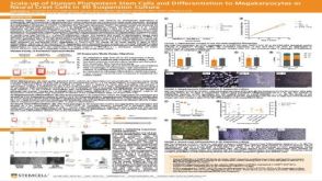 科学海报Scale-up of Human Pluripotent Stem Cells and Differentiation to Megakaryocytes or Neural Crest Cells in 3D Suspension Culture

 沪公网安备31010102008431号