Nagaoka M et al. (JAN 2010)
BMC developmental biology 10 60
Culture of human pluripotent stem cells using completely defined conditions on a recombinant E-cadherin substratum.
BACKGROUND: To maintain pluripotency of human embryonic stem (huES) cells in feeder-free culture it has been necessary to provide a Matrigel substratum,which is a complex of poorly defined extracellular matrices and growth factors derived from mouse Engelbreth-Holm-Swarm sarcoma cells. Culture of stem cells under ill-defined conditions can inhibit the effectiveness of maintaining cells in a pluripotent state and reduce reproducibility of differentiation protocols. Moreover recent batches of Matrigel have been found to be contaminated with the single stranded RNA virus,Lactate Dehydrogenase Elevating Virus (LDEV),raising concerns regarding the safety of using stem cells that have been cultured on Matrigel in a therapeutic setting. To circumvent such concerns,we attempted to identify a recombinant matrix that could be used as an alternative to Matrigel for the culture of human pluripotent stem cells. huES and human induced pluripotent stem (hiPS) cells were grown on plates coated with a fusion protein consisting of E-cadherin and the IgG Fc domain using mTeSR1 medium.backslashnbackslashnRESULTS: Cells grown under these conditions maintained similar morphology and growth rate to those grown on Matrigel and retained all pluripotent stem cell features,including an ability to differentiate into multiple cell lineages in teratoma assays. We,therefore,present a culture system that maintains the pluripotency of huES and hiPS cells under completely defined conditions.backslashnbackslashnCONCLUSIONS: We propose that this system should facilitate growth of stem cells using good manufacturing practices (GMP),which will be necessary for the clinical use of pluripotent stem cells and their derivatives.
View Publication
产品类型:
产品号#:
85850
85857
产品名:
mTeSR™1
mTeSR™1
Dorosko SM and Connor RI (OCT 2010)
Journal of virology 84 20 10533--42
Primary human mammary epithelial cells endocytose HIV-1 and facilitate viral infection of CD4+ T lymphocytes.
The contribution of mammary epithelial cells (MEC) to human immunodeficiency virus type 1 (HIV-1) in breast milk remains largely unknown. While breast milk contains CD4(+) cells throughout the breast-feeding period,it is not known whether MEC directly support HIV-1 infection or facilitate infection of CD4(+) cells in the breast compartment. This study evaluated primary human MEC for direct infection with HIV-1 and for indirect transfer of infection to CD4(+) target cells. Primary human MEC were isolated and assessed for expression of HIV-1 receptors. MEC were exposed to CCR5-,CXCR4- and dual-tropic strains of HIV-1 and evaluated for viral reverse transcription and integration and productive viral infection. MEC were also tested for the ability to transfer HIV to CD4(+) target cells and to activate resting CD4(+) T cells. Our results demonstrate that MEC express HIV-1 receptor proteins CD4,CCR5,CXCR4,and galactosyl ceramide (GalCer). While no evidence for direct infection of MEC was found,HIV-1 virions were observed in MEC endosomal compartments. Coculture of HIV-exposed MEC resulted in productive infection of activated CD4(+) T cells. In addition,MEC secretions increased HIV-1 replication and proliferation of infected target cells. Overall,our results indicate that MEC are capable of endosomal uptake of HIV-1 and can facilitate virus infection and replication in CD4(+) target cells. These findings suggest that MEC may serve as a viral reservoir for HIV-1 and may enhance infection of CD4(+) T lymphocytes in vivo.
View Publication
产品类型:
产品号#:
19052
19052RF
产品名:
EasySep™人CD4+ T细胞富集试剂盒
RoboSep™ 人CD4+ T细胞富集试剂盒含滤芯吸头
Avitabile D et al. (MAY 2011)
American journal of physiology. Heart and circulatory physiology 300 5 H1875--84
Human cord blood CD34+ progenitor cells acquire functional cardiac properties through a cell fusion process.
The efficacy of cardiac repair by stem cell administration relies on a successful functional integration of injected cells into the host myocardium. Safety concerns have been raised about the possibility that stem cells may induce foci of arrhythmia in the ischemic myocardium. In a previous work (36),we showed that human cord blood CD34(+) cells,when cocultured on neonatal mouse cardiomyocytes,exhibit excitation-contraction coupling features similar to those of cardiomyocytes,even though no human genes were upregulated. The aims of the present work are to investigate whether human CD34(+) cells,isolated after 1 wk of coculture with neonatal ventricular myocytes,possess molecular and functional properties of cardiomyocytes and to discriminate,using a reporter gene system,whether cardiac differentiation derives from a (trans)differentiation or a cell fusion process. Umbilical cord blood CD34(+) cells were isolated by a magnetic cell sorting method,transduced with a lentiviral vector carrying the enhanced green fluorescent protein (EGFP) gene,and seeded onto primary cultures of spontaneously beating rat neonatal cardiomyocytes. Cocultured EGFP(+)/CD34(+)-derived cells were analyzed for their electrophysiological features at different time points. After 1 wk in coculture,EGFP(+) cells,in contact with cardiomyocytes,were spontaneously contracting and had a maximum diastolic potential (MDP) of -53.1 mV,while those that remained isolated from the surrounding myocytes did not contract and had a depolarized resting potential of -11.4 mV. Cells were then resuspended and cultured at low density to identify EGFP(+) progenitor cell derivatives. Under these conditions,we observed single EGFP(+) beating cells that had acquired an hyperpolarization-activated current typical of neonatal cardiomyocytes (EGFP(+) cells,-2.24 ± 0.89 pA/pF; myocytes,-1.99 ± 0.63 pA/pF,at -125 mV). To discriminate between cell autonomous differentiation and fusion,EGFP(+)/CD34(+) cells were cocultured with cardiac myocytes infected with a red fluorescence protein-lentiviral vector; under these conditions we found that 100% of EGFP(+) cells were also red fluorescent protein positive,suggesting cell fusion as the mechanism by which cardiac functional features are acquired.
View Publication
产品类型:
产品号#:
09600
09650
产品名:
StemSpan™ SFEM
StemSpan™ SFEM
S. Yang et al. ( 2019)
Stem cells international 2019 1351860
Promoting Osteogenic Differentiation of Human Adipose-Derived Stem Cells by Altering the Expression of Exosomal miRNA.
Human adipose-derived stem cells (ADSCs) can release exosomes; however,their specific functions remain elusive. In this study,we verified that exosomes derived from osteogenically differentiated ADSCs can promote osteogenic differentiation of ADSCs. Furthermore,in order to investigate the importance of exosomal microRNAs (miRNAs) in osteogenic differentiation of ADSCs,we used microarray assays to analyze the expression profiles of exosomal miRNAs derived from undifferentiated as well as osteogenically differentiated ADSCs; 201 miRNAs were upregulated and 33 miRNAs were downregulated between the two types of exosomes. Additionally,bioinformatic analyses,which included gene ontology analyses,pathway analysis,and miRNA-mRNA-network investigations,were performed. The results of these analyses revealed that the differentially expressed exosomal miRNAs participate in multiple biological processes,such as gene expression,synthesis of biomolecules,cell development,differentiation,and signal transduction,among others. Moreover,we found that these differentially expressed exosomal miRNAs connect osteogenic differentiation to processes such as axon guidance,MAPK signaling,and Wnt signaling. To the best of our knowledge,this is the first study to identify and characterize exosomal miRNAs derived from osteogenically differentiated ADSCs. This study confirms that alterations in the expression of exosomal miRNAs can promote osteogenic differentiation of ADSCs,which also provides the foundation for further research on the regulatory functions of exosomal miRNAs in the context of ADSC osteogenesis.
View Publication
产品类型:
产品号#:
05412
05455
产品名:
MesenCult™ 脂肪分化试剂盒 (人)
MesenCult™-ACF软骨细胞分化试剂盒
N. S. Bharadwaj et al. (Apr 2024)
iScience 27 5
Human CD4 + memory phenotype T cells use mitochondrial metabolism to generate sensitive IFN-γ responses
The transition of naive T lymphocytes into antigenically activated effector cells is associated with a metabolic shift from oxidative phosphorylation to aerobic glycolysis. This shift facilitates production of the key anti-tumor cytokine interferon (IFN)-γ; however,an associated loss of mitochondrial efficiency in effector T cells ultimately limits anti-tumor immunity. Memory phenotype (MP) T cells are a newly recognized subset that arises through homeostatic activation signals following hematopoietic transplantation. We show here that human CD4 + MP cell differentiation is associated with increased glycolytic and oxidative metabolic activity,but MP cells retain less compromised mitochondria compared to effector CD4 + T cells,and their IFN-γ response is less dependent on glucose and more reliant on glutamine. MP cells also produced IFN-γ more efficiently in response to weak T cell receptor (TCR) agonism than effectors and mediated stronger responses to transformed B cells. MP cells may thus be particularly well suited to carry out sustained immunosurveillance against neoplastic cells. Subject areas: immunity,cell biology
View Publication
产品类型:
产品号#:
100-0784
10971
10991
产品名:
ImmunoCult™ 人CD3/CD28 T细胞激活剂
ImmunoCult™ 人CD3/CD28 T细胞激活剂
ImmunoCult™ 人CD3/CD28 T细胞激活剂
M. Peter et al. (May 2024)
iScience 27 6
Limitations of fluorescent timer protein maturation kinetics to isolate transcriptionally synchronized human neural progenitor cells
Differentiation of human pluripotent stem cells (hPSCs) into subtype-specific neurons holds substantial potential for disease modeling in vitro . For successful differentiation,a detailed understanding of the transcriptional networks regulating cell fate decisions is critical. The heterochronic nature of neurodevelopment,during which distinct cells in the brain and during in vitro differentiation acquire their fates in an unsynchronized manner,hinders pooled transcriptional comparisons. One approach is to “translate” chronologic time into linear developmental and maturational time. Simple binary promotor-driven fluorescent proteins (FPs) to pool similar cells are unable to achieve this goal,due to asynchronous promotor onset in individual cells. We tested five fluorescent timer (FT) molecules expressed from the endogenous paired box 6 (PAX6) promoter in 293T and human hPSCs. Each of these FT systems faithfully reported chronologic time in 293T cells,but none of the FT constructs followed the same fluorescence kinetics in human neural progenitor cells. Subject areas: Natural sciences,Biological sciences,Biochemistry,Molecular biology,Neuroscience,Cellular neuroscience,Cell biology
View Publication
产品类型:
产品号#:
05854
05855
产品名:
mFreSR™
mFreSR™
Prosper F et al. (JUN 1997)
Blood 89 11 3991--7
Primitive long-term culture initiating cells (LTC-ICs) in granulocyte colony-stimulating factor mobilized peripheral blood progenitor cells have similar potential for ex vivo expansion as primitive LTC-ICs in steady state bone marrow.
We have recently shown that more than 90% of long-term culture initiating cells (LTC-IC) mobilized in the peripheral blood (PB) of normal individuals express HLA-DR and CD38 antigens and can sustain hematopoiesis for only 5 weeks. However,10% of LTC-IC in mobilized PB are CD34+ HLA-DR- and CD34+ CD38- and can sustain hematopoiesis for at least 8 weeks. We now examine the ex vivo expansion potential of CD34+ HLA-DR+ cells (rich in mature LTC-IC) and CD34+ HLA-DR- cells (rich in primitive LTC-IC) in granulocyte colony-stimulating factor (G-CSF) mobilized PB progenitor cells (PBPC). Cells were cultured in contact with M2-10B4 cells (contact) or in transwells above M2-10B4 (noncontact) without and with interleukin-3 (IL-3) and macrophage inflammatory protein (MIP-1alpha) for 2 and 5 weeks. Progeny were evaluated for the presence of colony-forming cells (CFC) and LTC-IC. When CD34+ HLA-DR+ PB cells were cultured in contact cultures without cytokines,a threefold expansion of CFC was seen at 2 weeks,but an 80% decrease in CFC was seen at week 5. Further,the recovery of LTC-IC at week 2 was only 17% and 1% at week 5. This confirms our previous observation that although CD34+ HLA-DR+ mobilized PB cells can initiate long-term cultures,they are relatively mature and cannot sustain long-term hematopoiesis. In contrast,when CD34+ HLA-DR- mobilized PB cells were cultured in contact cultures without cytokines,CFC expansion persisted until week 5 and 49% and 11% of LTC-IC were recovered at week 2 and 5,respectively. As we have shown for steady state bone marrow (BM) progenitors,recovery of LTC-IC was threefold higher when CD34+ HLA-DR- PBPC were cultured in noncontact rather than contact cultures,and improved further when IL-3 and MIP-1alpha were added to noncontact cultures (96 +/- 2% maintained at week 5). We conclude that although G-CSF mobilizes a large population of mature" CD34+ HLA-DR+ LTC-IC with a limited proliferative capacity�
View Publication
产品类型:
产品号#:
05150
产品名:
MyeloCult™H5100
Belzile J-P et al. (APR 2014)
Journal of virology 88 8 4021--4039
Human cytomegalovirus infection of human embryonic stem cell-derived primitive neural stem cells is restricted at several steps but leads to the persistence of viral DNA.
UNLABELLED Congenital human cytomegalovirus (HCMV) infection is a major cause of central nervous system structural anomalies and sensory impairments. It is likely that the stage of fetal development,as well as the state of differentiation of susceptible cells at the time of infection,affects the severity of the disease. We used human embryonic stem (ES) cell-derived primitive prerosette neural stem cells (pNSCs) and neural progenitor cells (NPCs) maintained in chemically defined conditions to study HCMV replication in cells at the early stages of neural development. In contrast to what was observed previously using fetus-derived NPCs,infection of ES cell-derived pNSCs with HCMV was nonprogressive. At a low multiplicity of infection,we observed only a small percentage of cells expressing immediate-early genes (IE) and early genes. IE expression was found to be restricted to cells negative for the anterior marker FORSE-1,and treatment of pNSCs with retinoic acid restored IE expression. Differentiation of pNSCs into NPCs restored IE expression but not the transactivation of early genes. Virions produced in NPCs and pNSCs were exclusively cell associated and were mostly non-neural tropic. Finally,we found that viral genomes could persist in pNSC cultures for up to a month after infection despite the absence of detectable IE expression by immunofluorescence,and infectious virus could be produced upon differentiation of pNSCs to neurons. In conclusion,our results highlight the complex array of hurdles that HCMV must overcome in order to infect primitive neural stem cells and suggest that these cells might act as a reservoir for the virus. IMPORTANCE Human cytomegalovirus (HCMV) is a betaherpesvirus that is highly prevalent in the population. HCMV infection is usually asymptomatic but can lead to severe consequences in immunosuppressed individuals. HCMV is also the most important infectious cause of congenital developmental birth defects. Manifestations of fetal HCMV disease range from deafness and learning disabilities to more severe symptoms such as microcephaly. In this study,we have used embryonic stem cells to generate primitive neural stem cells and have used these to model HCMV infection of the fetal central nervous system (CNS) in vitro. Our results reveal that these cells,which are similar to those present in the developing neural tube,do not support viral replication but instead likely constitute a viral reservoir. Future work will define the effect of viral persistence on cellular functions as well as the exogenous signals leading to the reactivation of viral replication in the CNS.
View Publication
Miyazaki S et al. (DEC 2015)
Annals of surgical oncology 22 Suppl 3 S3 S1394----401
A Cancer Reprogramming Method Using MicroRNAs as a Novel Therapeutic Approach against Colon Cancer: Research for Reprogramming of Cancer Cells by MicroRNAs.
BACKGROUND We previously generated induced pluripotent stem cells by reprograming adipose stem cells through the introduction of microRNAs targeting four transcription factors (Oct3/4,Sox2,c-Myc,and Klf4). In this study,we aimed to reprogram cancer cells using microRNAs to explore their therapeutic potential. METHODS Mature microRNAs (mir-302a-d,369-3p and 5p,and mir-200c,as needed) were introduced into colon cancer cells (DLD-1,RKO,and HCT116) using lipofection. RESULTS The transfected cells exhibited an embryonic stem cell-like morphology and expressed the undifferentiated marker genes Nanog,Oct3/4,SOX2,and Klf4,as well as tumor-related antigen-1-60. These cells expressed neurogenic or adipogenic markers,indicating that reprogramming of the cancer cells was partially successful. Moreover,we found that miRNA-expressing DLD-1 cells showed low proliferative activity in vitro and in vivo,accompanied by increased expression of the tumor suppressor genes p16 (ink4a) and p21 (waf1) . miRNA-expressing DLD-1 cells also exhibited enhanced sensitivity to 5-fluorouracil,possibly through the downregulation of multidrug-resistant protein 8. The reprogrammed cells from DLD-1,RKO,and HCT116 cells exhibited reduced c-Myc expression,in contrast to the high c-Myc expression in the induced pluripotent cancer cells that were generated using four transcription factors. CONCLUSIONS Our cancer reprogramming method employing simple lipofection of mature microRNAs is safe and well suited for clinical application,because it avoids integration of exogenous genes into the host genome and allows escape from augmentation of c-Myc gene expression.
View Publication

 EasySep™小鼠TIL(CD45)正选试剂盒


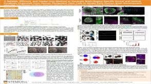 科学海报A Reliable, Efficient, and Feeder-Free Method to Generate Brain-Region-Specific Dorsal and Ventral Forebrain Organoids From Human Pluripotent Stem Cells to Model Early Human Brain Development
 科学海报A Human Pluripotent Stem Cell-Derived Organoid Model for Recapitulation of Central Nervous System (CNS) Barrier and Fluid Secretion Functions of the Choroid Plexus

 沪公网安备31010102008431号