Acoustofluidic bioassembly induced morphogenesis for therapeutic tissue fabrication
To build in vitro tissues for therapeutic applications,it is essential to replicate the spatial distribution of cells that occurs during morphogenesis in vivo. However,it remains technically challenging to simultaneously regulate the geometric alignment and aggregation of cells during tissue fabrication. Here,we introduce the acoustofluidic bioassembly induced morphogenesis,which is the combination of precise arrangement of cells by the mechanical forces produced by acoustofluidic cues,and the morphological and functional changes of cells in the following in vitro and in vivo cultures. The acoustofluidic bioassembly can be used to create tissues with regulated nano-,micro-,and macro-structures. We demonstrate that the neuromuscular tissue fabricated with the acoustofluidic bioassembly exhibits enhanced contraction dynamics,electrophysiology,and therapeutic efficacy. The potential of the acoustofluidic bioassembly as an in situ application is demonstrated by fabricating artificial tissues at the defect sites of living tissues. The acoustofluidic bioassembly induced morphogenesis can provide a pioneering platform to fabricate tissues for biomedical applications. Tissue engineering is essential for drug screening and regenerative medicine. Here,authors developed an acoustofluidic method that can induce morphogenesis of therapeutic tissues at varied dimensions/scales.
View Publication
P. Bank'o et al. (may 2019)
Journal of hematology oncology 12 1 48
Technologies for circulating tumor cell separation from whole blood.
The importance of early cancer diagnosis and improved cancer therapy has been clear for years and has initiated worldwide research towards new possibilities in the care strategy of patients with cancer using technological innovations. One of the key research fields involves the separation and detection of circulating tumor cells (CTC) because of their suggested important role in early cancer diagnosis and prognosis,namely,providing easy access by a liquid biopsy from blood to identify metastatic cells before clinically detectable metastasis occurs and to study the molecular and genetic profile of these metastatic cells. Provided the opportunity to further progress the development of technology for treating cancer,several CTC technologies have been proposed in recent years by various research groups and companies. Despite their potential role in cancer healthcare,CTC methods are currently mainly used for research purposes,and only a few methods have been accepted for clinical application because of the difficulties caused by CTC heterogeneity,CTC separation from the blood,and a lack of thorough clinical validation. Therefore,the standardization and clinical application of various developed CTC technologies remain important subsequent necessary steps. Because of their suggested future clinical benefits,we focus on describing technologies using whole blood samples without any pretreatment and discuss their advantages,use,and significance. Technologies using whole blood samples utilize size-based,immunoaffinity-based,and density-based methods or combinations of these methods as well as positive and negative enrichment during separation. Although current CTC technologies have not been truly implemented yet,they possess high potential as future clinical diagnostic techniques for the individualized therapy of patients with cancer. Thus,a detailed discussion of the clinical suitability of these new advanced technologies could help prepare clinicians for the future and can be a foundation for technologies that would be used to eliminate CTCs in vivo.
View Publication
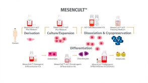
产品类型:
产品号#:
19657
产品名:
EasySep™ Direct 人CTC富集试剂盒
Suvà et al. (DEC 2009)
Cancer research 69 24 9211--8
EZH2 is essential for glioblastoma cancer stem cell maintenance.
Overexpression of the polycomb group protein enhancer of zeste homologue 2 (EZH2) occurs in diverse malignancies,including prostate cancer,breast cancer,and glioblastoma multiforme (GBM). Based on its ability to modulate transcription of key genes implicated in cell cycle control,DNA repair,and cell differentiation,EZH2 is believed to play a crucial role in tissue-specific stem cell maintenance and tumor development. Here,we show that targeted pharmacologic disruption of EZH2 by the S-adenosylhomocysteine hydrolase inhibitor 3-deazaneplanocin A (DZNep),or its specific downregulation by short hairpin RNA (shRNA),strongly impairs GBM cancer stem cell (CSC) self-renewal in vitro and tumor-initiating capacity in vivo. Using genome-wide expression analysis of DZNep-treated GBM CSCs,we found the expression of c-myc,recently reported to be essential for GBM CSCs,to be strongly repressed upon EZH2 depletion. Specific shRNA-mediated downregulation of EZH2 in combination with chromatin immunoprecipitation experiments revealed that c-myc is a direct target of EZH2 in GBM CSCs. Taken together,our observations provide evidence that direct transcriptional regulation of c-myc by EZH2 may constitute a novel mechanism underlying GBM CSC maintenance and suggest that EZH2 may be a valuable new therapeutic target for GBM management.
View Publication
产品类型:
产品号#:
72322
72324
产品名:
3-Deazaneplanocin A
3-Deazaneplanocin A
Oronsky B et al. (OCT 2014)
Translational oncology 7 5 626--31
Rewriting the epigenetic code for tumor resensitization: a review.
In cancer chemotherapy,one axiom,which has practically solidified into dogma,is that acquired resistance to antitumor agents or regimens,nearly inevitable in all patients with metastatic disease,remains unalterable and irreversible,rendering therapeutic rechallenge futile. However,the introduction of epigenetic therapies,including histone deacetylase inhibitors (HDACis) and DNA methyltransferase inhibitors (DNMTIs),provides oncologists,like computer programmers,with new techniques to overwrite" the modifiable software pattern of gene expression in tumors and challenge the "one and done" treatment prescription. Taking the epigenetic code-as-software analogy a step further�
View Publication
M. Wei et al. ( 2022)
Frontiers in oncology 12 835603
Ubiquitin ligase RNF125 targets PD-L1 for ubiquitination and degradation.
As a critical immune checkpoint molecule,PD-L1 is expressed at significantly higher levels in multiple neoplastic tissues compared to normal ones. PD-L1/PD-1 axis is a critical target for tumor immunotherapy,blocking the PD-L1/PD-1 axis is recognized and has achieved unprecedented success in clinical applications. However,the clinical efficacy of therapies targeting the PD-1/PD-L1 pathway remains limited,emphasizing the need for the mechanistic elucidation of PD-1/PD-L1 expression. In this study,we found that RNF125 interacted with PD-L1 and regulated PD-L1 protein expression. Mechanistically,RNF125 promoted K48-linked polyubiquitination of PD-L1 and mediated its degradation. Notably,MC-38 and H22 cell lines with RNF125 knockout,transplanted in C57BL/6 mice,exhibited a higher PD-L1 level and faster tumor growth than their parental cell lines. In contrast,overexpression of RNF125 in MC-38 and H22 cells had the opposite effect,resulting in lower PD-L1 levels and delayed tumor growth compared with parental cell lines. In addition,immunohistochemical analysis of MC-38 tumors with RNF125 overexpression showed significantly increased infiltration of CD4+,CD8+ T cells and macrophages. Consistent with these findings,analyses using The Cancer Genome Atlas (TCGA) public database revealed a positive correlation of RNF125 expression with CD4+,CD8+ T cell and macrophage tumor infiltration. Moreover,RNF125 expression was significantly downregulated in several human cancer tissues,and was negatively correlated with the clinical stage of these tumors,and patients with higher RNF125 expression had better clinical outcomes. Our findings identify a novel mechanism for regulating PD-L1 expression and may provide a new strategy to increase the efficacy of immunotherapy.
View Publication
产品类型:
产品号#:
18000
19853
19853RF
产品名:
EasySep™磁极
EasySep™小鼠CD8+ T细胞分选试剂盒
RoboSep™ 小鼠CD8+ T细胞分选试剂盒
X. Shi et al. (nov 2019)
Molecular therapy : the journal of the American Society of Gene Therapy
Genetically Engineered Cell-Derived Nanoparticles for Targeted Breast Cancer Immunotherapy.
Exosomes are nanosized membranous vesicles secreted by a variety of cells. Due to their unique and pharmacologically important properties,cell-derived exosome nanoparticles have drawn significant interest for drug development. By genetically modifying exosomes with two distinct types of surface-displayed monoclonal antibodies,we have developed an exosome platform termed synthetic multivalent antibodies retargeted exosome (SMART-Exo) for controlling cellular immunity. Here,we apply this approach to human epidermal growth factor receptor 2 (HER2)-expressing breast cancer by engineering exosomes through genetic display of both anti-human CD3 and anti-human HER2 antibodies,resulting in SMART-Exos dually targeting T cell CD3 and breast cancer-associated HER2 receptors. By redirecting and activating cytotoxic T cells toward attacking HER2-expressing breast cancer cells,the designed SMART-Exos exhibited highly potent and specific anti-tumor activity both in vitro and in vivo. This work demonstrates preclinical feasibility of utilizing endogenous exosomes for targeted breast cancer immunotherapy and the SMART-Exos as a broadly applicable platform technology for the development of next-generation immuno-nanomedicines.
View Publication
产品类型:
产品号#:
19844
19844RF
19849
19851
19851RF
19762
19762RF
产品名:
EasySep™小鼠Pan-B细胞分选试剂盒
RoboSep™ 小鼠Pan-B细胞分选试剂盒
EasySep™小鼠/人嵌合体分选试剂盒
EasySep™小鼠T细胞分选试剂盒
RoboSep™ 小鼠T细胞分选试剂盒
EasySep™小鼠中性粒细胞富集试剂盒
RoboSep™ 小鼠中性粒细胞富集试剂盒含滤芯吸头
A. Demchenko et al. (Oct 2025)
PLOS Computational Biology 21 10
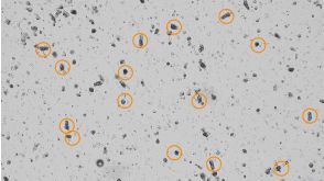
A semi-automated algorithm for image analysis of respiratory organoids
Respiratory organoids have emerged as a powerful in vitro model for studying respiratory diseases and drug discovery. However,the high-throughput analysis of organoid images remains a challenge due to the lack of automated and accurate segmentation tools. This study presents a semi-automatic algorithm for image analysis of respiratory organoids (nasal and lung organoids),employing the U-Net architecture and CellProfiler for organoids segmentation. The algorithm processes bright-field images acquired through z-stack fusion and stitching. The model demonstrated a high level of accuracy,as evidenced by an intersection-over-union metric (IoU) of 0.8856,F1-score = 0.937 and an accuracy of 0.9953. Applied to forskolin-induced swelling assays of lung organoids,the algorithm successfully quantified functional differences in Cystic Fibrosis Transmembrane conductance Regulator (CFTR)-channel activity between healthy donor and cystic fibrosis patient-derived organoids,without fluorescent dyes. Additionally,an open-source dataset of 827 annotated respiratory organoid images was provided to facilitate further research. Our results demonstrate the potential of deep learning to enhance the efficiency and accuracy of high-throughput respiratory organoid analysis for future therapeutic screening applications. Author summaryIn this study,we developed a semi-automated tool to analyze images of respiratory organoids—3D cell structures that mimic the human respiratory system. These organoids are vital for studying diseases like cystic fibrosis and testing potential drugs,but manually analyzing their images is time-consuming and prone to errors. Our tool uses artificial intelligence (AI) to quickly and accurately measure organoid size and shape from bright-field microscope images,eliminating the need for fluorescent dyes that can harm cells. We trained our AI model on a publicly shared dataset of 827 annotated organoid images,achieving high accuracy in detecting and quantifying organoids. When applied to cystic fibrosis research,the tool successfully measured differences in organoid swelling (forskolin-induced swelling - a key test for drug response) between healthy and patient-derived samples. By making our dataset and method openly available,we hope to support further research into respiratory diseases. Our work bridges the gap between complex lab techniques and practical applications,offering a faster,more reliable way to study human health and disease.
View Publication
产品类型:
产品号#:
05040
产品名:
PneumaCult™-Ex Plus 培养基
Mä et al. (AUG 2005)
Blood 106 4 1215--22
Infection of human CD34+ progenitor cells with Bartonella henselae results in intraerythrocytic presence of B. henselae.
Although there is evidence that endothelial cells are important targets for human pathogenic Bartonella species,the primary niche of infection is unknown. Here we elucidated whether human CD34+ hematopoietic progenitor cells (HPCs) internalize B. henselae and may serve as a potential niche of the pathogen. We showed that B. henselae does not adhere to or invade human erythrocytes. In contrast,B. henselae invades and persists in HPCs as shown by gentamicin protection assays,confocal laser scanning microscopy (CLSM),and electron microscopy (EM). Fluorescence-activated cell sorting (FACS) analysis of glycophorin A expression revealed that erythroid differentiation of HPCs was unaffected following infection with B. henselae. The number of intracellular B. henselae continuously increased over a 13-day period. When HPCs were infected with B. henselae immediately after isolation,intracellular bacteria were subsequently detectable in differentiated erythroid cells on day 9 and day 13 after infection,as shown by CLSM,EM,and FACS analysis. Our data provide,for the first time,evidence that a bacterial pathogen is able to infect and persist in differentiating HPCs,and suggest that HPCs might serve as a potential primary niche in Bartonella infections.
View Publication

 EasySep™小鼠TIL(CD45)正选试剂盒


 沪公网安备31010102008431号