
 EasySep™小鼠TIL(CD45)正选试剂盒


搜索结果: 'EasySep Release'
-

产品类型:
产品号#:
03434
03444
产品名:
MethoCult™GF M3434
MethoCult™GF M3434
-
产品类型:
产品号#:
04434
04444
09600
09650
产品名:
MethoCult™H4434经典
MethoCult™H4434经典
StemSpan™ SFEM
StemSpan™ SFEM
-
 技术窍门Isolating Hematopoietic Progenitors Using EasySep™, RoboSep™, RosetteSep™ and SepMate™发布日期: 12/10/2013
 -
产品类型:
产品号#:
06005
产品名:
IntestiCult™ 肠道类器官生长培养基 (小鼠)
-
产品类型:
产品号#:
73352
产品名:
QNZ
-
产品类型:
产品号#:
15024
15064
15023
15063
产品名:
RosetteSep™ 人B细胞富集抗体混合物
RosetteSep™人B细胞富集抗体混合物
RosetteSep™ 人CD8+ T细胞富集抗体混合物
RosetteSep™人CD8+ T细胞富集抗体混合物
-
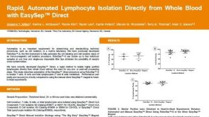 科学海报Rapid, Automated Lymphocyte Isolation Directly from Whole Blood with EasySep™ Direct
产品类型:
Conference:
EFI 2019
产品号#:
21000
20155
20119
19655
19655RF
89671
89671RF
89684
89684RF
19671
19671RF
19684
19684RF
产品名:
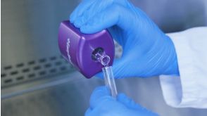
RoboSep™- S
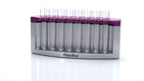
RoboSep™分选管套装(9个塑料管)
RoboSep™ 吸头组件抛光剂
EasySep™ Direct人总淋巴细胞分选试剂盒
RoboSep™ Direct人总淋巴细胞分选试剂盒
EasySep™ Direct HLA交叉配型T细胞分选试剂盒
RoboSep™ Direct HLA交叉配型T细胞分选试剂盒
EasySep™ Direct HLA交叉配型B细胞分选试剂盒
RoboSep™ Direct HLA交叉配型B细胞分选试剂盒
-
产品类型:
产品号#:
18952
18952RF
产品名:
EasySep™ 小鼠CD4正选试剂盒 II
RoboSep™ 小鼠CD4正选试剂盒II


 沪公网安备31010102008431号