
 EasySep™小鼠TIL(CD45)正选试剂盒


搜索结果: 'EasySep Release'
-

产品类型:
产品号#:
73722
73724
产品名:
离子霉素(Ionomycin)
离子霉素(Ionomycin)
-
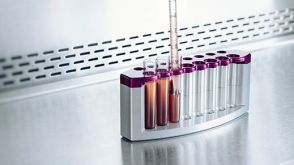 技术公告Simplify Your PBMC Isolations with the EasySep™ Direct Human PBMC Isolation Kit
产品类型:
细胞类型:
B细胞,NK细胞,T细胞,单个核细胞,单核细胞,淋巴细胞
产品号#:
产品名:
发布日期: 02/23/2026 -
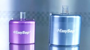
 科学海报Easy 250 EasySep™ Magnet: A Novel Magnetic Platform for Large-Volume Cell Isolation
产品类型:
产品号#:
产品名:
-
产品类型:
产品号#:
18752
18752RF
21000
20119
20155
18758
18758RF
18768
18768RF
产品名:
RoboSep™- S
RoboSep™ 吸头组件抛光剂
RoboSep™分选管套装(9个塑料管)
-
 3:04
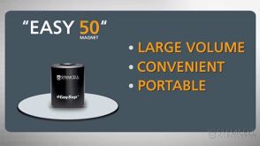
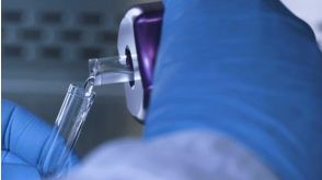
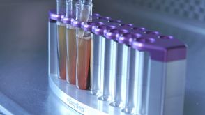
视频The Easy 50 EasySep™ Magnet: Portable Large-Volume Cell Separation In 25 Minutes发布日期: 10/10/2010
3:04
视频The Easy 50 EasySep™ Magnet: Portable Large-Volume Cell Separation In 25 Minutes发布日期: 10/10/2010 -
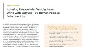 技术公告Isolating Extracellular Vesicles from Urine with EasySep™ EV Human Positive Selection Kits
产品类型:
细胞类型:
其他细胞系
产品号#:
产品名:
发布日期: 01/01/2024 -
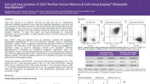 科学海报Fast and Easy Isolation of CD27-Positive Human Memory B Cells Using EasySep™ Releasable RapidSpheres™
产品类型:
Conference:
AAI 2017
产品号#:
17864
产品名:
EasySep™ 人记忆B细胞分选试剂盒

 沪公网安备31010102008431号