Baumann BC et al. (MAY 2004)
Journal of immunology (Baltimore,Md. : 1950) 172 10 6460--7
Lack of galactose-alpha-1,3-galactose expression on porcine endothelial cells prevents complement-induced lysis but not direct xenogeneic NK cytotoxicity.
The galactose-alpha-1,3-galactose (alphaGal) carbohydrate epitope is expressed on porcine,but not human cells,and therefore represents a major target for preformed human anti-pig natural Abs (NAb). Based on results from pig-to-primate animal models,NAb binding to porcine endothelial cells will likely induce complement activation,lysis,and hyperacute rejection in pig-to-human xenotransplantation. Human NK cells may also contribute to innate immune responses against xenografts,either by direct recognition of activating molecules on target cells or by FcgammaRIII-mediated xenogeneic Ab-dependent cellular cytotoxicity (ADCC). The present study addressed the question as to whether the lack of alphaGal protects porcine endothelial cells from NAb/complement-induced lysis,direct xenogeneic NK lysis,NAb-dependent ADCC,and adhesion of human NK cells under shear stress. Homologous recombination,panning,and limiting dilution cloning were used to generate an alphaGal-negative porcine endothelial cell line,PED2*3.51. NAb/complement-induced xenogeneic lysis of PED2*3.51 was reduced by an average of 86% compared with the alphaGal-positive phenotype. PED2*3.51 resisted NK cell-mediated ADCC with a reduction of lysis ranging from 30 to 70%. However,direct xenogeneic lysis of PED2*3.51,mediated either by freshly isolated or IL-2-activated human NK cells or the NK cell line NK92,was not reduced. Furthermore,adhesion of IL-2-activated human NK cells did not rely on alphaGal expression. In conclusion,removal of alphaGal leads to a clear reduction in complement-induced lysis and ADCC,but does not resolve adhesion of NK cells and direct anti-porcine NK cytotoxicity,indicating that alphaGal is not a dominant target for direct human NK cytotoxicity against porcine cells.
View Publication
Kerscher P et al. (MAR 2016)
Biomaterials 83 383--395
Direct hydrogel encapsulation of pluripotent stem cells enables ontomimetic differentiation and growth of engineered human heart tissues
Human engineered heart tissues have potential to revolutionize cardiac development research,drug-testing,and treatment of heart disease; however,implementation is limited by the need to use pre-differentiated cardiomyocytes (CMs). Here we show that by providing a 3D poly(ethylene glycol)-fibrinogen hydrogel microenvironment,we can directly differentiate human pluripotent stem cells (hPSCs) into contracting heart tissues. Our straight-forward,ontomimetic approach,imitating the process of development,requires only a single cell-handling step,provides reproducible results for a range of tested geometries and size scales,and overcomes inherent limitations in cell maintenance and maturation,while achieving high yields of CMs with developmentally appropriate temporal changes in gene expression. We demonstrate that hPSCs encapsulated within this biomimetic 3D hydrogel microenvironment develop into functional cardiac tissues composed of self-aligned CMs with evidence of ultrastructural maturation,mimicking heart development,and enabling investigation of disease mechanisms and screening of compounds on developing human heart tissue.
View Publication
产品类型:
产品号#:
05850
05857
05870
05875
85850
85857
85870
85875
产品名:
mTeSR™1
mTeSR™1
A. Revenko et al. (apr 2022)
Journal for immunotherapy of cancer 10 4
Direct targeting of FOXP3 in Tregs with AZD8701, a novel antisense oligonucleotide to relieve immunosuppression in cancer.
BACKGROUND The Regulatory T cell (Treg) lineage is defined by the transcription factor FOXP3,which controls immune-suppressive gene expression profiles. Tregs are often recruited in high frequencies to the tumor microenvironment where they can suppress antitumor immunity. We hypothesized that pharmacological inhibition of FOXP3 by systemically delivered,unformulated constrained ethyl-modified antisense oligonucleotides could modulate the activity of Tregs and augment antitumor immunity providing therapeutic benefit in cancer models and potentially in man. METHODS We have identified murine Foxp3 antisense oligonucleotides (ASOs) and clinical candidate human FOXP3 ASO AZD8701. Pharmacology and biological effects of FOXP3 inhibitors on Treg function and antitumor immunity were tested in cultured Tregs and mouse syngeneic tumor models. Experiments were controlled by vehicle and non-targeting control ASO groups as well as by use of multiple independent FOXP3 ASOs. Statistical significance of biological effects was evaluated by one or two-way analysis of variance with multiple comparisons. RESULTS AZD8701 demonstrated a dose-dependent knockdown of FOXP3 in primary Tregs,reduction of suppressive function and efficient target downregulation in humanized mice at clinically relevant doses. Surrogate murine FOXP3 ASO,which efficiently downregulated Foxp3 messenger RNA and protein levels in primary Tregs,reduced Treg suppressive function in immune suppression assays in vitro. FOXP3 ASO promoted more than 70% reduction in FOXP3 levels in Tregs in vitro and in vivo,strongly modulated Treg effector molecules (eg,ICOS,CTLA-4,CD25 and 4-1BB),and augmented CD8+ T cell activation and produced antitumor activity in syngeneic tumor models. The combination of FOXP3 ASOs with immune checkpoint blockade further enhanced antitumor efficacy. CONCLUSIONS Antisense inhibitors of FOXP3 offer a promising novel cancer immunotherapy approach. AZD8701 is being developed clinically as a first-in-class FOXP3 inhibitor for the treatment of cancer currently in Ph1a/b clinical trial (NCT04504669).
View Publication
产品类型:
产品号#:
10981
产品名:
ImmunoCult™ XF 人T细胞扩增培养基,500 mL
O'Sullivan S et al. (NOV 2007)
Journal of bone and mineral research 22 11 1679--89
Imatinib promotes osteoblast differentiation by inhibiting PDGFR signaling and inhibits osteoclastogenesis by both direct and stromal cell-dependent mechanisms.
UNLABELLED: Several lines of evidence suggest that imatinib may affect skeletal tissue. We show that inhibition by imatinib of PDGFR signaling in osteoblasts activates osteoblast differentiation and inhibits osteoblast proliferation and that imatinib inhibits osteoclastogenesis by both stromal cell-dependent and direct effects on osteoclast precursors. INTRODUCTION: Imatinib mesylate,an orally active inhibitor of the c-abl,c-kit,and platelet-derived growth factor receptor (PDGFR) tyrosine kinases,is in clinical use for the treatment of chronic myeloid leukemia (CML) and gastrointestinal stromal cell tumors. Interruption of both c-kit and c-abl signaling in mice induces osteopenia,suggesting that imatinib might have adverse effects on the skeleton. However,biochemical markers of bone formation increase in patients with CML starting imatinib therapy,whereas bone resorption is unchanged,despite secondary hyperparathyroidism. We assessed the actions of imatinib on bone cells in vitro to study the cellular and molecular mechanism(s) underlying the skeletal effects we observed in imatinib-treated patients. MATERIALS AND METHODS: Osteoblast differentiation was assessed using a mineralization assay,proliferation by [(3)H]thymidine incorporation,and apoptosis by a TUNEL assay. Osteoclastogenesis was assessed using murine bone marrow cultures and RAW 264.7 cells. RT and multiplex PCR were performed on RNA prepared from human bone marrow samples,osteoblastic cells,and murine bone marrow cultures. Osteoprotegerin was measured by ELISA. RESULTS: The molecular targets of imatinib are expressed in bone cells. In vitro,imatinib increases osteoblast differentiation and prevents PDGF-induced inhibition of this process. Imatinib inhibits proliferation of osteoblast-like cells induced by serum and PDGF. In murine bone marrow cultures,imatinib inhibits osteoclastogenesis stimulated by 1,25-dihydroxyvitamin D(3) and partially inhibits osteoclastogenesis induced by RANKL and macrophage-colony stimulating factor. Imatinib partially inhibited osteoclastogenesis in RANKL-stimulated RAW-264.7 cells. Treatment with imatinib increases the expression of osteoprotegerin in bone marrow from patients with CML and osteoblastic cells. CONCLUSIONS: Taken together with recent in vivo data,these results suggest a role for the molecular targets of imatinib in bone cell function,that inhibition by imatinib of PDGFR signaling in osteoblasts activates bone formation,and that the antiresorptive actions of imatinib are mediated by both stromal cell-dependent and direct effects on osteoclast precursors.
View Publication
Pettinato G et al. (SEP 2016)
Scientific reports 6 32888
Scalable Differentiation of Human iPSCs in a Multicellular Spheroid-based 3D Culture into Hepatocyte-like Cells through Direct Wnt/β-catenin Pathway Inhibition.
Treatment of acute liver failure by cell transplantation is hindered by a shortage of human hepatocytes. Current protocols for hepatic differentiation of human induced pluripotent stem cells (hiPSCs) result in low yields,cellular heterogeneity,and limited scalability. In the present study,we have developed a novel multicellular spheroid-based hepatic differentiation protocol starting from embryoid bodies of hiPSCs (hiPSC-EBs) for robust mass production of human hepatocyte-like cells (HLCs) using two novel inhibitors of the Wnt pathway. The resultant hiPSC-EB-HLCs expressed liver-specific genes,secreted hepatic proteins such as Albumin,Alpha Fetoprotein,and Fibrinogen,metabolized ammonia,and displayed cytochrome P450 activities and functional activities typical of mature primary hepatocytes,such as LDL storage and uptake,ICG uptake and release,and glycogen storage. Cell transplantation of hiPSC-EB-HLC in a rat model of acute liver failure significantly prolonged the mean survival time and resolved the liver injury when compared to the no-transplantation control animals. The transplanted hiPSC-EB-HLCs secreted human albumin into the host plasma throughout the examination period (2 weeks). Transplantation successfully bridged the animals through the critical period for survival after acute liver failure,providing promising clues of integration and full in vivo functionality of these cells after treatment with WIF-1 and DKK-1.
View Publication
产品类型:
产品号#:
05850
05857
05870
05875
85850
85857
85870
85875
产品名:
mTeSR™1
mTeSR™1
Morgan AJ and Jacob R (JUN 1994)
The Biochemical journal 300 ( Pt 3 665--72
Ionomycin enhances Ca2+ influx by stimulating store-regulated cation entry and not by a direct action at the plasma membrane.
In fura-2-loaded ECV304 cells ionomycin elicited a saturable biphasic change in intracellular Ca2+ concentration ([Ca2+]i),where the initial phase represented mobilization of intracellular stores and the sustained component represented Ca2+ influx. To examine whether ionomycin could stimulate influx via a store-dependent mechanism. Mn2+ entry was monitored by the quenching of fura-2 fluorescence: influx was enhanced even after ionomycin wash-out,provided that internal stores were not refilled with Ca2+. Moreover,the maximal rate of histamine-stimulated Mn2+ entry was unaffected by ionomycin,suggesting a common route of entry. The Ca(2+)-entry blocker SK&F 96365 inhibited both the ionomycin-induced Mn2+ entry and the sustained [Ca2+]i response to the ionophore (leaving the initial peak [Ca2+]i response unaffected). In other experiments,although addition of ionomycin further increased the plateau phase induced by 100 microM histamine,the increase was completely abolished by pretreatment with the store Ca(2+)-ATPase inhibitor cyclopiazonic acid (CPA). Furthermore,in store-depleted cells,re-addition of 1 mM extracellular Ca2+ (in the presence of CPA plus histamine) led to a rapid rise in [Ca2+]i,dependent on Ca2+ influx,with kinetics that were not enhanced by ionomycin. These data suggest that ionomycin acts primarily at the level of the internal Ca2+ stores,so that,at the concentrations used here (textless or = 1 microM),it increases Ca2+ (and Mn2+) influx via activation of endogenous entry pathways and not by plasmalemmal translocation.
View Publication

 EasySep™小鼠TIL(CD45)正选试剂盒


 科学海报Easy 250 EasySep™ Magnet: A Novel Magnetic Platform for Large-Volume Cell Isolation
 3:04
视频The Easy 50 EasySep™ Magnet: Portable Large-Volume Cell Separation In 25 Minutes发布日期: 10/10/2010
3:04
视频The Easy 50 EasySep™ Magnet: Portable Large-Volume Cell Separation In 25 Minutes发布日期: 10/10/2010 技术公告Isolating Extracellular Vesicles from Urine with EasySep™ EV Human Positive Selection Kits
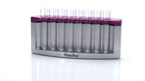


 沪公网安备31010102008431号