RIG-I-like receptor LGP2 is required for tumor control by radiation therapy.
Dendritic cells (DC) play an essential role in innate immunity and radiation-elicited immune responses. LGP2 is a RIG-I like receptor (RLR) involved in cytoplasmic RNA recognition and anti-viral responses. Although LGP2 has also been linked to cell survival of both tumor cells and T cells,the role of LGP2 in mediating DC function and anti-tumor immunity elicited by radiotherapy remains unclear. Here we report that tumor DC are linked to the clinical outcome of breast cancer patients who received radiotherapy (RT) and the presence of DC correlates with gene expression of LGP2 in the tumor microenvironment. In preclinical models,host LGP2 was essential for optimal anti-tumor control by ionizing radiation (IR). The absence of LGP2 in DC dampened type I interferon production and the priming capacity of DC. In the absence of LGP2,MDA5-mediated activation of type I IFN signaling was abrogated. The MDA5/LGP2 agonist high molecular weight poly I: C improved the anti-tumor effect of IR. This study reveals a previously undefined role of LGP2 in host immunity and provides a new strategy to improve the efficacy of radiotherapy.
View Publication
Yu B et al. ( 2002)
Biochemical pharmacology 64 7 1091--1100
SU9516, a cyclin-dependent kinase 2 inhibitor, promotes accumulation of high molecular weight E2F complexes in human colon carcinoma cells.
The E2F family plays a critical role in the expression of genes required for entry into and progression through S phase. E2F-mediated transcription is repressed by the tumor suppressor retinoblastoma protein (pRb),which results in sequestration of E2F in a multiprotein complex that includes pRb. Derepression of E2F results from a series of complex phosphorylation events mediated by cyclin D/cdk4 and cyclin E/cdk2. We have employed a novel 3-substituted indolinone compound,3-[1-(3H-imidazol-4-yl)-meth-(Z)-ylidene]-5-methoxy-1,3-dihydro-indol-2-one (SU9516),which selectively inhibits cdk2 activity (Lane et al.,Cancer Res 2001;61:6170-7) to investigate these events. Electrophoretic mobility gel shift assays were performed on SU9516-treated and -untreated HT-29,SW480,and RKO human colon cancer cell extracts. Treatment with 5 microM SU9516 prevented dissociation of pRb from E2F1 in all cell lines (HT-29textgreaterRKOtextgreaterSW480). Treatment effects were time-dependent,demonstrating greater inhibition at 48 hr versus 24hr in HT-29 cells. Furthermore,E2F species were sequestered in complexes with p107,p130,DP-1,and cyclins A and E. After a 24-hr treatment with 5 microM SU9516,cyclin D1 and cdk2 levels decreased by 10-60%. These findings delineate a previously undescribed mechanism for SU9516-mediated cell growth arrest through down-regulation of cyclin D1,inhibition of cdk2 levels and activity,and pan-sequestration of E2F.
View Publication
产品类型:
产品号#:
73452
产品名:
SU9516
Kohler JJ et al. (MAR 2003)
Journal of leukocyte biology 73 3 407--16
Human immunodeficiency virus type 1 (HIV-1) induces activation of multiple STATs in CD4+ cells of lymphocyte or monocyte/macrophage lineages.
Human immunodeficiency virus type 1 (HIV-1) impacts the activation state of multiple lineages of hematopoietic cells. Chronic HIV-1 infection among individuals with progressive disease can be associated with increased levels of activated signal transducers and activators of transcription (STATs) in peripheral blood mononuclear cells. To investigate interactions between HIV-1 and CD4(+) cells,activated,phosphorylated STAT proteins in nuclear extracts from lymphocytic and promonocytic cell lines as well as primary monocyte-derived macrophages were measured. Levels of activated STATs increased six- to tenfold in HUT78 and U937 cells within 2 h following exposure to virions. The response to virus was dose-dependent,but kinetics of activation was delayed relative to interleukin-2 or interferon-gamma. Activation of STAT1,STAT3,and STAT5 occurred with diverse viral envelope proteins,independent of coreceptor use or viral replication. Envelope-deficient virions had no effect on STAT activation. Monoclonal antibody engagement of CD4 identified a novel role for CD4 as a mediator in the activation of multiple STATs. Results provide a model for HIV-1 pathogenesis in infected and noninfected hematopoietic cells.
View Publication
产品类型:
产品号#:
15028
15068
产品名:
RosetteSep™ 人单核细胞富集抗体混合物
RosetteSep™人单核细胞富集抗体混合物
Xia L et al. (NOV 2004)
Blood 104 10 3091--6
Surface fucosylation of human cord blood cells augments binding to P-selectin and E-selectin and enhances engraftment in bone marrow.
Murine hematopoietic stem and progenitor cells (HSPCs) home to bone marrow in part by rolling on P-selectin and E-selectin expressed on endothelial cells. Human adult CD34(+) cells,which are enriched in HSPCs,roll on endothelial selectins in bone marrow vessels of nonobese diabetic/severe combined immune deficiency (NOD/SCID) mice. Many human umbilical cord blood (CB) CD34(+) cells do not roll in these vessels,in part because of an uncharacterized defect in binding to P-selectin. Selectin ligands must be alpha1-3 fucosylated to form glycan determinants such as sialyl Lewis x (sLe(x)). We found that inadequate alpha1-3 fucosylation of CB CD34(+) cells,particularly CD34(+)CD38(-/low) cells that are highly enriched in HSPCs,caused them to bind poorly to E-selectin as well as to P-selectin. Treatment of CB CD34(+) cells with guanosine diphosphate (GDP) fucose and exogenous alpha1-3 fucosyltransferase VI increased cell-surface sLe(x) determinants,augmented binding to fluid-phase P- and E-selectin,and improved cell rolling on P- and E-selectin under flow. Similar treatment of CB mononuclear cells enhanced engraftment of human hematopoietic cells in bone marrows of irradiated NOD/SCID mice. These observations suggest that alpha1-3 fucosylation of CB cells might be a simple and effective method to improve hematopoietic cell homing to and engraftment in bone marrows of patients receiving CB transplants.
View Publication
产品类型:
产品号#:
产品名:
Li H et al. (SEP 2016)
In vitro cellular & developmental biology. Animal 52 8 885--893
Directed differentiation of human embryonic stem cells into keratinocyte progenitors in vitro: an attempt with promise of clinical use.
Human embryonic stem cells (hESCs) can differentiate into all somatic lineages including stratified squamous epithelia. Thus,efficient methods are required to direct hESC differentiation to obtain a pure subpopulation for tissue engineering. The study aimed to assess the effects of retinoic acid (RA),bone morphogenetic protein-4 (BMP4),and ascorbic acid (AA) on the differentiation of hESCs into keratinocyte progenitors in vitro. The first media contained AA and BMP4; the second contained RA,AA,and BMP4; the third was commercial-defined keratinocyte serum-free medium,which was used to differentiate H9 hESCs (direct approach) or embryoid bodies (EBs) (indirect approach) into keratinocyte progenitors. Real-time RT-PCR,immunofluorescence,and flow-cytometry were used to characterize the differentiated cells. Cells induced by AA + BMP4 + RA showed the typical epithelial morphology,while cells induced by AA + BMP4 showed multiple appearances. CK14 and p63 messenger RNA (mRNA) expressions in the AA + BMP4 + RA-treated cells were higher than those of the AA + BMP4-treated cells (CK14: 22.4-fold; p63: 84.7-fold). Epithelial marker CK18 mRNA expressions at 14 d of differentiation and keratinocyte marker CK14 and transcription factor p63 mRNA expressions at 35 d of differentiation were higher in cells differentiated from hESCs compared with those differentiated from EBs (CK18 10.51 ± 3.26 vs. 6.67 ± 1.28; CK14 9.27 ± 3.61 vs. 5.32 ± 1.86; p63 0.73 ± 0.06 vs. 0.44 ± 0.12,all P textless 0.05) After hESC induction by AA+BMP4+RA,CK14 mRNA expression was upregulated after day 21,peaking by 35 d of differentiation. Combined RA,BMP4,and AA could effectively induce differentiation of hESCs into keratinocyte progenitors in vitro. These keratinocytes could be used for oral mucosa and skin tissue engineering.
View Publication
产品类型:
产品号#:
07923
85850
85857
产品名:
Dispase (1 U/mL)
mTeSR™1
mTeSR™1
Morizane A et al. (FEB 2011)
Journal of neuroscience research 89 2 117--126
Small-molecule inhibitors of bone morphogenic protein and activin/nodal signals promote highly efficient neural induction from human pluripotent stem cells.
The balance of bone morphogenic protein (BMP),transforming growth factor-β (TGFβ)/activin/nodal,and Wnt signals regulates the early lineage segregation of human embryonic stem cells (ESCs). Here we demonstrate that a combination of small-molecule inhibitors of BMP (Dorsomorphin) and TGFβ/activin/nodal (SB431542) signals promotes highly efficient neural induction from both human ESCs and induced pluripotent stem cells (iPSCs). The combination of small molecules had effects on both cell survival and purity of neural differentiation,under conditions of stromal (PA6) cell coculture and feeder-free floating aggregation culture,for all seven pluripotent stem cell lines that we studied,including three ESC and four iPSC lines. Small molecule compounds are stable and cost effective,so our findings provide a promising strategy for controlled production of neurons in regenerative medicine.
View Publication
产品类型:
产品号#:
72102
100-0246
产品名:
Dorsomorphin
白消安(Busulfan)
(Nov 2024)
Antioxidants 13 11
An In Vitro Oxidative Stress Model of the Human Inner Ear Using Human-Induced Pluripotent Stem Cell-Derived Otic Progenitor Cells
The inner ear organs responsible for hearing (cochlea) and balance (vestibular system) are susceptible to oxidative stress due to the high metabolic demands of their sensorineural cells. Oxidative stress-induced damage to these cells can cause hearing loss or vestibular dysfunction,yet the precise mechanisms remain unclear due to the limitations of animal models and challenges of obtaining living human inner ear tissue. Therefore,we developed an in vitro oxidative stress model of the pre-natal human inner ear using otic progenitor cells (OPCs) derived from human-induced pluripotent stem cells (hiPSCs). OPCs,hiPSCs,and HeLa cells were exposed to hydrogen peroxide or ototoxic drugs (gentamicin and cisplatin) that induce oxidative stress to evaluate subsequent cell viability,cell death,reactive oxygen species (ROS) production,mitochondrial activity,and apoptosis (caspase 3/7 activity). Dose-dependent reductions in OPC cell viability were observed post-exposure,demonstrating their vulnerability to oxidative stress. Notably,gentamicin exposure induced ROS production and cell death in OPCs,but not hiPSCs or HeLa cells. This OPC-based human model effectively simulates oxidative stress conditions in the human inner ear and may be useful for modeling the impact of ototoxicity during early pregnancy or evaluating therapies to prevent cytotoxicity.
View Publication
产品类型:
产品号#:
100-0483
100-0484
85850
85857
产品名:
Hausser Scientificᵀᴹ 明线血球计数板
ReLeSR™
mTeSR™1
mTeSR™1
Daga A et al. (MAY 2000)
Experimental hematology 28 5 569--74
The retroviral transduction of HOXC4 into human CD34(+) cells induces an in vitro expansion of clonogenic and early progenitors.
OBJECTIVE: +HOX genes are expressed in the hematopoietic system and increasing data point to their involvement in the control of proliferation and/or differentiation. Genes belonging to the C cluster are preferentially expressed in developing and differentiated lymphoid lineages. However,recent studies demonstrated,by RT-PCR,that the HOXC4 gene is also actively transcribed in the most undifferentiated hematopoietic cells (CD34(+)38(low)) and in more mature myeloid and erythroid progenitors. We evaluated the expression of HOXC4 protein on human CD34(+) cells and the in vitro effect of its overexpression on proliferation and differentiation. MATERIALS AND METHODS: We assessed the expression of HOXC4 on human CD34(+) cells using a polyclonal antibody raised against the C-terminal portion of the protein expressed using the baculovirus system. Overexpression of HOXC4 in human CD34(+) cells was obtained by retroviral gene transfer; its effect on clonogenic (CFU-GM,BFU-E,and CFU-GEMM) and early progenitors (LTC-IC) was evaluated. RESULTS: The HOXC4 protein is indeed expressed in human CD34(+) cells,and its overexpression in human CD34(+) cells increases the proliferation potential of clonogenic and early progenitors. CFU-GM showed a median threefold expansion (range: 1.1-19.4; p textless 0.002) compared with control transduced with the vector alone. The increment of BFU-E was higher (median ninefold,range 2.5-35; p textless 0. 0009) and erythroid colonies presented a larger size with normal morphology. An even more marked effect was observed on LTC-IC (median 13,onefold; range 4.1-102.1,p textless 0.0001). CONCLUSION: We demonstrate that HOXC4 is expressed in CD34(+) cells and that its overexpression induces an in vitro expansion of committed as well as very early hematopoietic progenitors. The most striking effect was obtained on LTC-IC with an expansion of 13.1-fold. The enforced expression of HOXC4 induced a significant increase (p textless 0.009) in the number of erythroid colonies compared with CFU-GM,although without perturbing,at least in vitro,the maturation program of the cells. On the other hand,the effect of the gene overexpression did not induce any skewing in the colony types derived from the myeloid lineage.
View Publication
Wang H et al. (APR 2016)
The Journal of biological chemistry 291 16 8644--8652
Germ Cell Nuclear Factor (GCNF) Represses Oct4 Expression and Globally Modulates Gene Expression in Human Embryonic Stem (hES) Cells.
Oct4 is considered a key transcription factor for pluripotent stem cell self-renewal. It binds to specific regions within target genes to regulate their expression and is downregulated upon induction of differentiation of pluripotent stem cells; however,the mechanisms that regulate the levels of human Oct4 expression remain poorly understood. Here we show that expression of human Oct4 is directly repressed by germ cell nuclear factor (GCNF),an orphan nuclear receptor,in hES cells. Knockdown of GCNF by siRNA resulted in maintenance of Oct4 expression during RA-induced hES cell differentiation. While overexpression of GCNF promoted repression of Oct4 expression in both undifferentiated and differentiated hES cells. The level of Oct4 repression was dependent on the level of GCNF expression in a dose-dependent manner. mRNA microarray analysis demonstrated that overexpression of GCNF globally regulates gene expression in undifferentiated and differentiated hES cells. Within the group of altered genes,GCNF down-regulated 36% of the genes,and up-regulated 64% in undifferentiated hES cells. In addition,GCNF also showed a regulatory gene pattern that is different from RA treatment during hES cell differentiation. These findings increase our understanding of the mechanisms that maintain hES cell pluripotency and regulate gene expression during the differentiation process.
View Publication

 EasySep™小鼠TIL(CD45)正选试剂盒


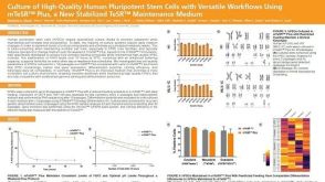 科学海报Culture of High-Quality Human Pluripotent Stem Cells with Versatile Workflows Using mTeSR™ Plus, a New Stabilized TeSR™ Maintenance Medium
 沪公网安备31010102008431号