Wildum S et al. (AUG 2006)
Journal of virology 80 16 8047--59
Contribution of Vpu, Env, and Nef to CD4 down-modulation and resistance of human immunodeficiency virus type 1-infected T cells to superinfection.
Human immunodeficiency virus type 1 (HIV-1) utilizes Vpu,Env,and Nef to down-modulate its primary CD4 receptor from the cell surface,and this function seems to be critical for the pathogenesis of AIDS. The physiological relevance of CD4 down-modulation,however,is currently not well understood. In the present study,we analyzed the kinetics of CD4 down-modulation and the susceptibility of HIV-1-infected T cells to superinfection using proviral HIV-1 constructs containing individual and combined defects in vpu,env,and nef and expressing red or green fluorescent proteins. T cells infected with HIV-1 mutants containing functional nef genes expressed low surface levels of CD4 from the first moment that viral gene expression became detectable. In comparison,Vpu and Env had only minor to moderate effects on CD4 during later stages of infection. Consistent with these quantitative differences,Nef inhibited superinfection more efficiently than Vpu and Env. Notably,nef alleles from AIDS patients were more effective in preventing superinfection than those derived from a nonprogressor of HIV-1 infection. Our data suggest that protection against X4-tropic HIV-1 superinfection involves both CD4-independent and CD4-dependent mechanisms of HIV-1 Nef. X4 was effectively down-regulated by simian immunodeficiency virus and HIV-2 but not by HIV-1 Nef proteins. Thus,maximal protection seems to involve an as-yet-unknown mechanism that is independent of CD4 or coreceptor down-modulation. Finally,we demonstrate that superinfected primary T cells show enhanced levels of apoptosis. Accordingly,one reason that HIV-1 inhibits CD4 surface expression and superinfection is to prevent premature cell death in order to expand the period of effective virus production.
View Publication
产品类型:
产品号#:
15022
15062
产品名:
RosetteSep™人CD4+ T细胞富集抗体混合物
RosetteSep™人CD4+ T细胞富集抗体混合物
Snykers S et al. (JAN 2007)
BMC developmental biology 7 24
Chromatin remodeling agent trichostatin A: a key-factor in the hepatic differentiation of human mesenchymal stem cells derived of adult bone marrow.
BACKGROUND The capability of human mesenchymal stem cells (hMSC) derived of adult bone marrow to undergo in vitro hepatic differentiation was investigated. RESULTS Exposure of hMSC to a cocktail of hepatogenic factors [(fibroblast growth factor-4 (FGF-4),hepatocyte growth factor (HGF),insulin-transferrin-sodium-selenite (ITS) and dexamethasone)] failed to induce hepatic differentiation. Sequential exposure to these factors (FGF-4,followed by HGF,followed by HGF+ITS+dexamethasone),however,resembling the order of secretion during liver embryogenesis,induced both glycogen-storage and cytokeratin (CK)18 expression. Additional exposure of the cells to trichostatin A (TSA) considerably improved endodermal differentiation,as evidenced by acquisition of an epithelial morphology,chronological expression of hepatic proteins,including hepatocyte-nuclear factor (HNF)-3beta,alpha-fetoprotein (AFP),CK18,albumin (ALB),HNF1alpha,multidrug resistance-associated protein (MRP)2 and CCAAT-enhancer binding protein (C/EBP)alpha,and functional maturation,i.e. upregulated ALB secretion,urea production and inducible cytochrome P450 (CYP)-dependent activity. CONCLUSION hMSC are able to undergo mesenchymal-to-epithelial transition. TSA is hereby essential to promote differentiation of hMSC towards functional hepatocyte-like cells.
View Publication
产品类型:
产品号#:
72282
72284
产品名:
曲古抑菌素 A(Trichostatin A)
曲古抑菌素 A(Trichostatin A)
Kovarova M and Koller B (APR 2012)
Current protocols in immunology / edited by John E. Coligan ... [et al.] Chapter 22 Unit 22F.10.1--16
Differentiation of mast cells from embryonic stem cells.
In this unit,we describe a simple coculture-free method for obtaining mast cells from mouse and human embryonic stem (ES) cells. Much of our knowledge regarding the mechanisms by which mast cells are activated comes from studies of mouse bone marrow-derived mast cells. Studies of human mast cells have been hampered by the limited sources from which they can be cultured,the difficulty in introducing specific genetic changes into these cells,and differences between established cultures that reflect the unique genetic makeup of the tissue donor. Derivation of mast cells from embryonic stem cells addresses these limitations. ES-derived mast cells can be generated in numbers sufficient for studies of the pathways involved in mast cell effector functions. These ES cell-derived mast cells respond to antigens and other stimuli by releasing histamine,cytokines,lipids,and other bioactive mediators. The derivation of human mast cells from ES cells carrying mutations introduced by homologous recombination should provide a novel means of testing the function of genes in both the development and the effector functions of mast cells.
View Publication
产品类型:
产品号#:
85850
85857
产品名:
mTeSR™1
mTeSR™1
Gao N et al. ( 2006)
Molecular pharmacology 70 2 645--655
The three-substituted indolinone cyclin-dependent kinase 2 inhibitor 3-[1-(3H-imidazol-4-yl)-meth-(Z)-ylidene]-5-methoxy-1,3-dihydro-indol-2-one (SU9516) kills human leukemia cells via down-regulation of Mcl-1 through a transcriptional mechanism.
Mechanisms of lethality of the three-substituted indolinone and putatively selective cyclin-dependent kinase (CDK)2 inhibitor 3-[1-(3H-imidazol-4-yl)-meth-(Z)-ylidene]-5-methoxy-1,3-dihydro-indol-2-one (SU9516) were examined in human leukemia cells. Exposure of U937 and other leukemia cells to SU9516 concentrations textgreater or =5 microM rapidly (i.e.,within 4 h) induced cytochrome c release,Bax mitochondrial translocation,and apoptosis in association with pronounced down-regulation of the antiapoptotic protein Mcl-1. These effects were associated with inhibition of phosphorylation of the carboxyl-terminal domain (CTD) of RNA polymerase (Pol) II on serine 2 but not serine 5. Reverse transcription-polymerase chain reaction analysis revealed pronounced down-regulation of Mcl-1 mRNA levels in SU9516-treated cells. Similar results were obtained in Jurkat and HL-60 leukemia cells. Furthermore,cotreatment with the proteasome inhibitor N-benzoyloxycarbonyl (Z)-Leu-Leu-leucinal (MG132) blocked SU9516-mediated Mcl-1 down-regulation,implicating proteasomal degradation in diminished expression of this protein. Ectopic expression of Mcl-1 largely blocked SU9516-induced cytochrome c release,Bax translocation,and apoptosis,whereas knockdown of Mcl-1 by small interfering RNA potentiated SU9516 lethality,confirming the functional contribution of Mcl-1 down-regulation to SU9516-induced cell death. It is noteworthy that SU9516 treatment resulted in a marked increase in reactive oxygen species production,which was diminished,along with cell death,by the free radical scavenger N-acetylcysteine (NAC). We were surprised to find that NAC blocked SU9516-mediated inhibition of RNA Pol II CTD phosphorylation on serine 2,reductions in Mcl-1 mRNA levels,and Mcl-1 down-regulation. Together,these findings suggest that SU9516 kills leukemic cells through inhibition of RNA Pol II CTD phosphorylation in association with oxidative damage and down-regulation of Mcl-1 at the transcriptional level,culminating in mitochondrial injury and cell death.
View Publication
产品类型:
产品号#:
73452
产品名:
SU9516
Miller TW et al. (APR 2011)
Clinical cancer research : an official journal of the American Association for Cancer Research 17 7 2024--34
A gene expression signature from human breast cancer cells with acquired hormone independence identifies MYC as a mediator of antiestrogen resistance.
PURPOSE: Although most patients with estrogen receptor α (ER)-positive breast cancer initially respond to endocrine therapy,many ultimately develop resistance to antiestrogens. However,mechanisms of antiestrogen resistance and biomarkers predictive of such resistance are underdeveloped. EXPERIMENTAL DESIGN: We adapted four ER(+) human breast cancer cell lines to grow in an estrogen-depleted medium. A gene signature of estrogen independence was developed by comparing expression profiles of long-term estrogen-deprived (LTED) cells to their parental counterparts. We evaluated the ability of the LTED signature to predict tumor response to neoadjuvant therapy with an aromatase inhibitor and disease outcome following adjuvant tamoxifen. We utilized Gene Set Analysis (GSA) of LTED cell gene expression profiles and a loss-of-function approach to identify pathways causally associated with resistance to endocrine therapy. RESULTS: The LTED gene expression signature was predictive of high tumor cell proliferation following neoadjuvant therapy with anastrozole and letrozole,each in different patient cohorts. This signature was also predictive of poor recurrence-free survival in two studies of patients treated with adjuvant tamoxifen. Bioinformatic interrogation of expression profiles in LTED cells revealed a signature of MYC activation. The MYC activation signature and high MYC protein levels were both predictive of poor outcome following tamoxifen therapy. Finally,knockdown of MYC inhibited LTED cell growth. CONCLUSIONS: A gene expression signature derived from ER(+) breast cancer cells with acquired hormone independence predicted tumor response to aromatase inhibitors and associated with clinical markers of resistance to tamoxifen. Activation of the MYC pathway was associated with this resistance.
View Publication
产品类型:
产品号#:
05620
产品名:
MammoCult™人培养基试剂盒
Sebastiano V et al. (NOV 2011)
Stem Cells 29 11 1717--1726
In situ genetic correction of the sickle cell anemia mutation in human induced pluripotent stem cells using engineered zinc finger nucleases.
The combination of induced pluripotent stem cell (iPSC) technology and targeted gene modification by homologous recombination (HR) represents a promising new approach to generate genetically corrected,patient-derived cells that could be used for autologous transplantation therapies. This strategy has several potential advantages over conventional gene therapy including eliminating the need for immunosuppression,avoiding the risk of insertional mutagenesis by therapeutic vectors,and maintaining expression of the corrected gene by endogenous control elements rather than a constitutive promoter. However,gene targeting in human pluripotent cells has remained challenging and inefficient. Recently,engineered zinc finger nucleases (ZFNs) have been shown to substantially increase HR frequencies in human iPSCs,raising the prospect of using this technology to correct disease causing mutations. Here,we describe the generation of iPSC lines from sickle cell anemia patients and in situ correction of the disease causing mutation using three ZFN pairs made by the publicly available oligomerized pool engineering method (OPEN). Gene-corrected cells retained full pluripotency and a normal karyotype following removal of reprogramming factor and drug-resistance genes. By testing various conditions,we also demonstrated that HR events in human iPSCs can occur as far as 82 bps from a ZFN-induced break. Our approach delineates a roadmap for using ZFNs made by an open-source method to achieve efficient,transgene-free correction of monogenic disease mutations in patient-derived iPSCs. Our results provide an important proof of principle that ZFNs can be used to produce gene-corrected human iPSCs that could be used for therapeutic applications.
View Publication
产品类型:
产品号#:
85850
85857
产品名:
mTeSR™1
mTeSR™1
Pereira RC et al. ( 2016)
Frontiers in immunology 7 415
Human Articular Chondrocytes Regulate Immune Response by Affecting Directly T Cell Proliferation and Indirectly Inhibiting Monocyte Differentiation to Professional Antigen-Presenting Cells.
Autologous chondrocyte implantation is the current gold standard cell therapy for cartilage lesions. However,in some instances,the heavily compromised health of the patient can either impair or limit the recovery of the autologous chondrocytes and a satisfactory outcome of the implant. Allogeneic human articular chondrocytes (hAC) could be a good alternative,but the possible immunological incompatibility between recipient and hAC donor should be considered. Herein,we report that allogeneic hAC inhibited T lymphocyte response to antigen-dependent and -independent proliferative stimuli. This effect was maximal when T cells and hAC were in contact and it was not relieved by the addition of exogenous lymphocyte growth factor interleukin (IL)-2. More important,hAC impaired the differentiation of peripheral blood monocytes induced with granulocyte monocyte colony-stimulating factor and IL-4 (Mo) to professional antigen-presenting cells,such as dendritic cells (DC). Indeed,a marked inhibition of the onset of the CD1a expression and an ineffective downregulation of CD14 antigens was observed in Mo-hAC co-cultures. Furthermore,compared to immature or mature DC,Mo from Mo-hAC co-cultures did not trigger an efficacious allo-response. The prostaglandin (PG) E2 present in the Mo-hAC co-culture conditioned media is a putative candidate of the hAC-mediated inhibition of Mo maturation. Altogether,these findings indicate that allogeneic hAC inhibit,rather than trigger,immune response and strongly suggest that an efficient chondrocyte implantation could be possible also in an allogeneic setting.
View Publication
产品类型:
产品号#:
100-0695
100-0696
17951
17951RF
17952
17952RF
产品名:
EasySep™人T细胞分选试剂盒
EasySep™人CD4+ T细胞分离试剂盒
EasySep™人T细胞分选试剂盒
RoboSep™ 人T细胞分选试剂盒
EasySep™人CD4+ T细胞分选试剂盒
RoboSep™ 人CD4+ T细胞分选试剂盒
Deng X et al. (DEC 2017)
Journal of virology 91 24 1--23
Human Parvovirus Infection of Human Airway Epithelia Induces Pyroptotic Cell Death by Inhibiting Apoptosis.
Human bocavirus 1 (HBoV1) is a human parvovirus that causes acute respiratory tract infections in young children. In this study,we confirmed that,when polarized/well-differentiated human airway epithelia are infected with HBoV1in vitro,they develop damage characterized by barrier function disruption and cell hypotrophy. Cell death mechanism analyses indicated that the infection induced pyroptotic cell death characterized by caspase-1 activation. Unlike infections with other parvoviruses,HBoV1 infection did not activate the apoptotic or necroptotic cell death pathway. When the NLRP3-ASC-caspase-1 inflammasome-induced pathway was inhibited by short hairpin RNA (shRNA),HBoV1-induced cell death dropped significantly; thus,NLRP3 mediated by ASC appears to be the pattern recognition receptor driving HBoV1 infection-induced pyroptosis. HBoV1 infection induced steady increases in the expression of interleukin 1α (IL-1α) and IL-18. HBoV1 infection was also associated with the marked expression of the antiapoptotic genesBIRC5andIFI6When the expression ofBIRC5and/orIFI6was inhibited by shRNA,the infected cells underwent apoptosis rather than pyroptosis,as indicated by increased cleaved caspase-3 levels and the absence of caspase-1.BIRC5and/orIFI6gene inhibition also significantly reduced HBoV1 replication. Thus,HBoV1 infection of human airway epithelial cells activates antiapoptotic proteins that suppress apoptosis and promote pyroptosis. This response may have evolved to confer a replicative advantage,thus allowing HBoV1 to establish a persistent airway epithelial infection. This is the first report of pyroptosis in airway epithelia infected by a respiratory virus.IMPORTANCEMicrobial infection of immune cells often induces pyroptosis,which is mediated by a cytosolic protein complex called the inflammasome that senses microbial pathogens and then activates the proinflammatory cytokines IL-1 and IL-18. While virus-infected airway epithelia often activate NLRP3 inflammasomes,studies to date suggest that these viruses kill the airway epithelial cells via the apoptotic or necrotic pathway; involvement of the pyroptosis pathway has not been reported previously. Here,we show for the first time that virus infection of human airway epithelia can also induce pyroptosis. Human bocavirus 1 (HBoV1),a human parvovirus,causes lower respiratory tract infections in young children. This study indicates that HBoV1 kills airway epithelial cells by activating genes that suppress apoptosis and thereby promote pyroptosis. This strategy appears to promote HBoV1 replication and may have evolved to allow HBoV1 to establish persistent infection of human airway epithelia.
View Publication
Maeda M et al. (JAN 2006)
The Journal of biological chemistry 281 1 59--68
Src activation is not necessary for transforming growth factor (TGF)-beta-mediated epithelial to mesenchymal transitions (EMT) in mammary epithelial cells. PP1 directly inhibits TGF-beta receptors I and II.
Epithelial to mesenchymal transitions (EMTs) are key events during embryonic development and cancer progression. It has been proposed that Src plays a major role in some EMT models,as shown by the overexpression of viral Src (v-Src) in epithelial cells. It is clear that Src family kinases can regulate the integrity of both adherens junctions and focal adhesions; however,their significance in EMT,especially in the physiological context,remains to be elucidated. Here we showed that Src is activated in transforming growth factor-beta1 (TGF-beta1)-mediated EMT in mammary epithelial cells and that the Src family kinase inhibitor,PP1,prevents EMT. However,neither a more specific Src family kinase inhibitor,SU6656,nor a dominant-negative Src inhibited TGF-beta1-mediated EMT,leading us to speculate that Src activation is not an essential component of TGF-beta1-mediated EMT. Unexpectedly,PP1 prevented Smad2/3 activation by TGF-beta1,whereas SU6656 did not. Most interestingly,an in vitro kinase assay showed that PP1 strongly inhibited the TGF-beta receptor type I,and to a lesser extent,the TGF-beta receptor type II. Taken together,our data indicated that PP1 interferes with TGF-beta1-mediated EMT not by inhibiting Src family kinases but by inhibiting the Smad pathway via a direct inhibition of TGF-beta receptor kinase activity.
View Publication
产品类型:
产品号#:
73112
73114
产品名:
PP1
PP1
挂图
Innate Lymphoid Cells
Overview of innate lymphoid cells (ILCs) development, classification, plasticity and functional diversity
挂图
Neural Stem Cells
Overview of the types of NSCs and their potential use as therapeutic agents for disease

 EasySep™小鼠TIL(CD45)正选试剂盒


 挂图Innate Lymphoid Cells Overview of innate lymphoid cells (ILCs) development, classification, plasticity and functional diversity
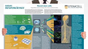 挂图Neural Stem Cells Overview of the types of NSCs and their potential use as therapeutic agents for disease

 沪公网安备31010102008431号