(Mar 2025)
The Journal of Experimental Medicine 222 5
KLF family members control expression of genes required for tissue macrophage identities
This work demonstrates that group 2 KLF family transcription factors are critical for specifying the identity of distinct tissue-resident macrophages. KLF2 directly controls expression of genes previously shown to be necessary in cavity macrophages,while KLF4 may play a similar role in alveolar macrophages. Tissue-resident macrophages adopt distinct gene expression profiles and exhibit functional specialization based on their tissue of residence. Recent studies have begun to define the signals and transcription factors that induce these identities. Here we describe an unexpected and specific role for the broadly expressed transcription factor Krüppel-like factor 2 (KLF2) in the development of embryonically derived large cavity macrophages (LCMs) in the serous cavities. KLF2 not only directly regulates the transcription of genes previously shown to specify LCM identity,such as retinoic acid receptors and GATA6,but also is required for induction of many other transcripts that define the identity of these cells. Our results suggest that KLF4 may similarly regulate the identity of alveolar macrophages in the lung. These data demonstrate that broadly expressed transcription factors,such as group 2 KLFs,can play important roles in the specification of distinct identities of tissue-resident macrophages.
View Publication
产品类型:
产品号#:
19861
19861RF
产品名:
EasySep™小鼠单核细胞分选试剂盒
RoboSep™ 小鼠单核细胞分选试剂盒
M. L. Stone et al. (Nov 2024)
iScience 27 12
Agarose hydrogel-mediated electroporation method for retinal tissue cultured at the air-liquid interface
It is advantageous to culture the ex vivo retina and other tissues at the air-liquid interface to allow for more efficient gas exchange. However,gene delivery to these cultures can be challenging. Electroporation is a fast and robust method of gene delivery,but typically requires submergence in liquid buffer for electrical current flow. We have developed a submergence-free electroporation technique that incorporates an agarose hydrogel disk between the positive electrode and retina. Inner retinal neurons and Müller glia are transfected with increased propensity toward Müller glia transfection after extended time in culture. We also observed an increase in BrdU incorporation in Müller glia following electrical stimulation,and variation in detection of transfected cells from expression vectors with different promoters. This method advances our ability to use ex vivo retinal tissue for genetic studies and should be adaptable for other tissues cultured at an air-liquid interface. Subject areas: Genetic engineering,Methodology in biological sciences,Bioelectrical engineering
View Publication
产品类型:
产品号#:
05790
产品名:
BrainPhys™神经元培养基
H. Sim et al. (may 2020)
International journal of molecular sciences 21 10
Iroquois Homeobox Protein 2 Identified as a Potential Biomarker for Parkinson's Disease.
The diagnosis of Parkinson's disease (PD) is initiated after the occurrence of motor symptoms,such as resting tremors,rigidity,and bradykinesia. According to previous reports,non-motor symptoms,notably gastrointestinal dysfunction,could potentially be early biomarkers in PD patients as such symptoms occur earlier than motor symptoms. However,connecting PD to the intestine is methodologically challenging. Thus,we generated in vitro human intestinal organoids from PD patients and ex vivo mouse small intestinal organoids from aged transgenic mice. Both intestinal organoids (IOs) contained the human LRRK2 G2019S mutation,which is the most frequent genetic cause of familial and sporadic PD. By conducting comprehensive genomic comparisons with these two types of IOs,we determined that a particular gene,namely,Iroquois homeobox protein 2 (IRX2),showed PD-related expression patterns not only in human pluripotent stem cell (PSC)-derived neuroectodermal spheres but also in human PSC-derived neuronal cells containing dopaminergic neurons. We expected that our approach of using various cell types presented a novel technical method for studying the effects of multi-organs in PD pathophysiology as well as for the development of diagnostic markers for PD.
View Publication
Fierro F et al. (JUN 2007)
Cell proliferation 40 3 355--66
Inhibition of platelet-derived growth factor receptorbeta by imatinib mesylate suppresses proliferation and alters differentiation of human mesenchymal stem cells in vitro.
OBJECTIVES: Recent data show that Imatinib mesylate (IM) also affects haematopoietic stem cells (HSC),T lymphocytes and dendritic cells that do not harbour constitutively active tyrosine kinases. MATERIALS AND METHODS: We evaluated possible effects of IM on human bone marrow-derived mesenchymal stem cells (MSC) in vitro. RESULTS: Screening the activity of 42 receptor tyrosine kinases revealed an exclusive inhibition of platelet-derived growth factor receptorbeta (PDGFRbeta). Analysis of downstream targets of PDGFRbeta demonstrated IM-mediated reduction of Akt and Erk1/2 phosphorylation. Culture of MSC with IM led to the reversible development of perinuclear multi-vesicular bodies. The proliferation and clonogenicity of MSC were significantly reduced compared to control cultures. IM favoured adipogenic differentiation of MSC whereas osteogenesis was suppressed. The functional deficits described led to a 50% reduction in the support of clonogenic haematopoietic stem cells,cultured for 1 month on a monolayer of MSC with IM. CONCLUSION: In summary,inhibition of PDGFRbeta and downstream Akt and Erk signalling by IM has a significant impact on proliferation and differentiation of human MSC in vitro.
View Publication
产品类型:
产品号#:
72532
产品名:
Imatinib (Mesylate)
Sokolov MV et al. (MAY 2010)
Gene 455 1-2 8--15
Expression of pluripotency-associated genes in the surviving fraction of cultured human embryonic stem cells is not significantly affected by ionizing radiation.
Human embryonic stem cells (hESC) are capable to give rise to all cell types in the human body during the normal course of development. Therefore,these cells hold a great promise in regenerative cell replacement based therapeutical approaches. However,some controversy exists in literature concerning the ultimate fate of hESC after exposure to genotoxic agents,in particular,regarding the effect of DNA damaging insults on pluripotency of hESC. To comprehensively address this issue,we performed an analysis of the expression of marker genes,associated with pluripotent state of hESC,such as Oct-4,Nanog,Sox-2,SSEA-4,TERT,TRA-1-60 and TRA-1-81 up to 65h after exposure to ionizing radiation (IR) using flow cytometry,immunocytochemistry and quantitative real-time polymerase chain reaction techniques. We show that irradiation with relatively low doses of gamma-radiation (0.2Gy and 1Gy) does not lead to loss of expression of the pluripotency-associated markers in the surviving hESC. While changes in the levels of expression of some of the pluripotency markers were observed at different time points after IR exposure,these alterations were not persistent,and,in most cases,the expression of the pluripotency-associated markers remained significantly higher than that observed in fully differentiated human fibroblasts,and in hESCs differentiated into definitive endodermal lineage. Our data suggest that exposure of hESC to relatively low doses of IR as a model genotoxic agent does not significantly affect pluripotency of the surviving fraction of hESC.
View Publication
Acquisition of neurodegenerative features in isogenic OPTN(E50K) human stem cell-derived retinal ganglion cells associated with autophagy disruption and mTORC1 signaling reduction
The ability to derive retinal ganglion cells (RGCs) from human pluripotent stem cells (hPSCs) has led to numerous advances in the field of retinal research,with great potential for the use of hPSC-derived RGCs for studies of human retinal development,in vitro disease modeling,drug discovery,as well as their potential use for cell replacement therapeutics. Of all these possibilities,the use of hPSC-derived RGCs as a human-relevant platform for in vitro disease modeling has received the greatest attention,due to the translational relevance as well as the immediacy with which results may be obtained compared to more complex applications like cell replacement. While several studies to date have focused upon the use of hPSC-derived RGCs with genetic variants associated with glaucoma or other optic neuropathies,many of these have largely described cellular phenotypes with only limited advancement into exploring dysfunctional cellular pathways as a consequence of the disease-associated gene variants. Thus,to further advance this field of research,in the current study we leveraged an isogenic hPSC model with a glaucoma-associated mutation in the Optineurin (OPTN) protein,which plays a prominent role in autophagy. We identified an impairment of autophagic-lysosomal degradation and decreased mTORC1 signaling via activation of the stress sensor AMPK,along with subsequent neurodegeneration in OPTN(E50K) RGCs differentiated from hPSCs,and have further validated some of these findings in a mouse model of ocular hypertension. Pharmacological inhibition of mTORC1 in hPSC-derived RGCs recapitulated disease-related neurodegenerative phenotypes in otherwise healthy RGCs,while the mTOR-independent induction of autophagy reduced protein accumulation and restored neurite outgrowth in diseased OPTN(E50K) RGCs. Taken together,these results highlighted that autophagy disruption resulted in increased autophagic demand which was associated with downregulated signaling through mTORC1,contributing to the degeneration of RGCs.Supplementary InformationThe online version contains supplementary material available at 10.1186/s40478-024-01872-2.
View Publication
产品类型:
产品号#:
85850
85857
产品名:
mTeSR™1
mTeSR™1
D. Bautista et al. ( 2020)
Frontiers in immunology 11 736
Differential Expression of IgM and IgD Discriminates Two Subpopulations of Human Circulating IgM+IgD+CD27+ B Cells That Differ Phenotypically, Functionally, and Genetically.
The origin and function of blood IgM+IgD+CD27+ B cells is controversial,and they are considered a heterogeneous population. Previous staining of circulating B cells of healthy donors with rotavirus fluorescent virus-like particles allowed us to differentiate two subsets of IgM+IgD+CD27+: IgMhi and IgMlo B cells. Here,we confirmed this finding and compared the phenotype,transcriptome,in vitro function,and Ig gene repertoire of these two subsets. Eleven markers phenotypically discriminated both subsets (CD1c,CD69,IL21R,CD27,MTG,CD45RB,CD5,CD184,CD23,BAFFR,and CD38) with the IgMhi phenotypically resembling previously reported marginal zone B cells and the IgMlo resembling both na{\{i}}ve and memory B cells. Transcriptomic analysis showed that both subpopulations clustered close to germinal center-experienced IgM only B cells with a Principal Component Analysis but differed in expression of 78 genes. Moreover IgMhi B cells expressed genes characteristic of previously reported marginal zone B cells. After stimulation with CpG and cytokines significantly (p {\textless} 0.05) higher frequencies (62.5{\%}) of IgMhi B cells proliferated compared with IgMlo B cells (35.37{\%}) and differentiated to antibody secreting cells (14.22{\%} for IgMhi and 7.19{\%} for IgMlo). IgMhi B cells had significantly (p {\textless} 0.0007) higher frequencies of mutations in IGHV and IGKV regions IgMlo B cells had higher usage of IGHJ6 genes (p {\textless} 0.0001) and both subsets differed in their HCDR3 properties. IgMhi B cells shared most of their shared IGH clonotypes with IgM only memory B cells and IgMlo B cells with IgMhi B cells. These results support the notion that differential expression of IgM and IgD discriminates two subpopulations of human circulating IgM+IgD+CD27+ B cells with the IgMhi B cells having similarities with previously described marginal zone B cells that passed through germinal centers and the IgMlo B cells being the least differentiated amongst the IgM+CD27+ subsets."
View Publication
产品类型:
产品号#:
15024
15064
产品名:
RosetteSep™ 人B细胞富集抗体混合物
RosetteSep™人B细胞富集抗体混合物
Hideshima T et al. (MAY 2006)
Blood 107 10 4053--62
Perifosine, an oral bioactive novel alkylphospholipid, inhibits Akt and induces in vitro and in vivo cytotoxicity in human multiple myeloma cells.
Perifosine is a synthetic novel alkylphospholipid,a new class of antitumor agents which targets cell membranes and inhibits Akt activation. Here we show that baseline phosphorylation of Akt in multiple myeloma (MM) cells is completely inhibited by perifosine [octadecyl-(1,1-dimethyl-piperidinio-4-yl)-phosphate] in a time- and dose-dependent fashion,without inhibiting phosphoinositide-dependent protein kinase 1 phosphorylation. Perifosine induces significant cytotoxicity in both MM cell lines and patient MM cells resistant to conventional therapeutic agents. Perifosine does not induce cytotoxicity in peripheral blood mononuclear cells. Neither exogenous interleukin-6 (IL-6) nor insulinlike growth factor 1 (IGF-1) overcomes Perifosine-induced cytotoxicity. Importantly,Perifosine induces apoptosis even of MM cells adherent to bone marrow stromal cells. Perifosine triggers c-Jun N-terminal kinase (JNK) activation,followed by caspase-8/9 and poly (ADP)-ribose polymerase cleavage. Inhibition of JNK abrogates perifosine-induced cytotoxicity,suggesting that JNK plays an essential role in perifosine-induced apoptosis. Interestingly,phosphorylation of extracellular signal-related kinase (ERK) is increased by perifosine; conversely,MEK inhibitor synergistically enhances Perifosine-induced cytotoxicity in MM cells. Furthermore,perifosine augments dexamethasone,doxorubicin,melphalan,and bortezomib-induced MM cell cytotoxicity. Finally,perifosine demonstrates significant antitumor activity in a human plasmacytoma mouse model,associated with down-regulation of Akt phosphorylation in tumor cells. Taken together,our data provide the rationale for clinical trials of perifosine to improve patient outcome in MM.
View Publication
产品类型:
产品号#:
15129
15169
产品名:
RosetteSep™人多发性骨髓瘤细胞富集抗体混合物
RosetteSep™人多发性骨髓瘤细胞富集抗体混合物
Na YJ et al. (SEP 2007)
Biochemical pharmacology 74 5 780--6
[4-t-butylphenyl]-N-(4-imidazol-1-yl phenyl)sulfonamide (ISCK03) inhibits SCF/c-kit signaling in 501mel human melanoma cells and abolishes melanin production in mice and brownish guinea pigs.
It is well known that c-kit is related to pigmentation as well as to the oncology target protein. The objective of this study was to discover a skin-whitening agent that regulates c-kit activity. We have developed a high-throughput screening system using recombinant human c-kit protein. Approximately 10,000 synthetic compounds were screened for their effect on c-kit activity. Phenyl-imidazole sulfonamide derivatives showed inhibitory activity on c-kit phosphorylation in vitro. The effects of one derivative,[4-t-butylphenyl]-N-(4-imidazol-1-yl phenyl)sulfonamide (ISCK03),on stem-cell factor (SCF)/c-kit cellular signaling in 501mel human melanoma cells were examined further. Pretreatment of 501mel cells with ISCK03 inhibited SCF-induced c-kit phosphorylation dose dependently. ISCK03 also inhibited p44/42 ERK mitogen-activated protein kinase (MAPK) phosphorylation,which is known to be involved in SCF/c-kit downstream signaling. However ISCK03 did not inhibit hepatocyte growth factor (HGF)-induced phosphorylation of p44/42 ERK proteins. To determine the in vivo potency of ISCK03,it was orally administered to depilated C57BL/6 mice. Interestingly,oral administration of ISCK03 induced the dose-dependent depigmentation of newly regrown hair,and this was reversed with cessation of ISCK03 treatment. Finally,to investigate whether the inhibitory effect of ISCK03 on SCF/c-kit signaling abolished UV-induced pigmentation,ISCK03 was applied to UV-induced pigmented spots on brownish guinea pig skin. The topical application of ISCK03 promoted the depigmentation of UV-induced hyperpigmented spots. Fontana-Masson staining analysis showed epidermal melanin was diminished in spots treated with ISCK03. These results indicate that phenyl-imidazole sulfonamide derivatives are potent c-kit inhibitors and might be used as skin-whitening agents.
View Publication
产品类型:
产品号#:
73734
产品名:
ISCK03
Vetter ML and D'Aquila RT (SEP 2009)
Journal of virology 83 17 8646--54
Cytoplasmic APOBEC3G restricts incoming Vif-positive human immunodeficiency virus type 1 and increases two-long terminal repeat circle formation in activated T-helper-subtype cells.
Cytoplasmic APOBEC3G has been reported to block wild-type human immunodeficiency virus type 1 (HIV-1) infection in some primary cells. It is not known whether cytoplasmic APOBEC3G has residual activity in activated T cells,even though virion-packaged APOBEC3G does restrict HIV-1 in activated T cells. Because we found that APOBEC3G expression is greater in activated CD4(+) T-helper type 1 (Th1) lymphocytes than in T-helper type 2 (Th2) lymphocytes,we hypothesized that residual target cell restriction of incoming Vif-positive virions that lack APOBEC3G,if present,would be greater in Th1 than Th2 lymphocytes. Infection of activated Th1 cells with APOBEC3-negative virions did result in decreased amounts of early and late reverse transcription products and integrated virus relative to infection of activated Th2 cells. Two-long terminal repeat (2-LTR) circles,which are formed in the nucleus when reverse transcripts do not integrate,were increased after APOBEC3-negative virus infection of activated Th1 cells relative to infection of activated Th2 cells. In contrast,2-LTR circle forms were decreased after infection of APOBEC3G-negative cells with APOBEC3G-containing virions relative to APOBEC3G-negative virions and with Th1 cell-produced virions relative to Th2 cell-produced virions. Increasing APOBEC3G in Th2 cells and decreasing APOBEC3G in Th1 cells modulated the target cell phenotypes,indicating causation by APOBEC3G. The comparison between activated Th1 and Th2 cells indicates that cytoplasmic APOBEC3G in activated Th1 cells partially restricts reverse transcription and integration of incoming Vif-positive,APOBEC3G-negative HIV-1. The differing effects of cytoplasmic and virion-packaged APOBEC3G on 2-LTR circle formation indicate a difference in their antiviral mechanisms.
View Publication

 EasySep™小鼠TIL(CD45)正选试剂盒


 实验方案How to Process Leukocyte Reduction System (LRS) Cones/Chambers for Downstream Cell Isolation
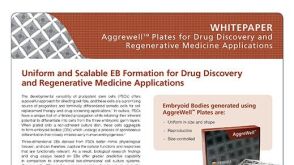 技术公告Uniform and Scalable EB Formation for Drug Discovery and Regenerative Medicine Applications
 沪公网安备31010102008431号