T. Halegua et al. (Jan 2025)
Nature Communications 16
Delivery of Prime editing in human stem cells using pseudoviral NanoScribes particles
Prime Editing can rewrite genes in living cells by allowing point mutations,deletions,or insertion of small DNA sequences with high precision. However,its safe and efficient delivery into human stem cells remains a technical challenge. In this report,we engineer Nanoscribes,virus-like particles that encapsidate ribonucleoprotein complexes of the Prime Editing system and allow their delivery into recipient cells. We identify key features that unlock the potential of Nanoscribes,including the use of multiple fusogens,the improvement of pegRNAs structures,their encoding by a Pol II system and the optimization of Prime-Editors. Nanoscribes edit HEK293T with an efficiency of 68% at the HEK3 locus with increased fidelity over DNA-transfection and support pegRNA-multiplexing. Importantly,Nanoscribes permit editing of myoblasts,hiPSCs and hiPSCs-derived hematopoietic stem cells with an editing efficiency up to 25%. Nanoscribes is an asset for development of next generation genome editing approaches using VLPs. Subject terms: CRISPR-Cas9 genome editing,Genetic vectors,Nanoparticles
View Publication
产品类型:
产品号#:
05310
产品名:
STEMdiff™ 造血试剂盒
K. V. Nerum et al. (Apr 2025)
Nature Cell Biology 27 5
α-Ketoglutarate promotes trophectoderm induction and maturation from naive human embryonic stem cells
Development and lineage choice are driven by interconnected transcriptional,epigenetic and metabolic changes. Specific metabolites,such as α-ketoglutarate (αKG),function as signalling molecules affecting the activity of chromatin-modifying enzymes. However,how metabolism coordinates cell-state changes,especially in human pre-implantation development,remains unclear. Here we uncover that inducing naive human embryonic stem cells towards the trophectoderm lineage results in considerable metabolic rewiring,characterized by αKG accumulation. Elevated αKG levels potentiate the capacity of naive embryonic stem cells to specify towards the trophectoderm lineage. Moreover,increased αKG levels promote blastoid polarization and trophectoderm maturation. αKG supplementation does not affect global histone methylation levels; rather,it decreases acetyl-CoA availability,reduces histone acetyltransferase activity and weakens the pluripotency network. We propose that metabolism functions as a positive feedback loop aiding in trophectoderm fate induction and maturation,highlighting that global metabolic rewiring can promote specificity in cell fate decisions through intricate regulation of signalling and chromatin. Subject terms: Embryonic stem cells,Embryology
View Publication
产品类型:
产品号#:
34411
34415
34421
34425
34450
34460
产品名:
AggreWell™ 400 24孔板,1个
AggreWell™400 24孔板,5个
AggreWell™ 400 6孔板,1个
AggreWell™ 400 6孔板,5个
AggreWell™400 24孔板启动套装
AggreWell™ 400 6孔板启动套装
J. Pesic et al. (Jun 2025)
ERJ Open Research 11 3
Inhibition of IL-4Rα reduces CCL26 in bronchial epithelial cells from COPD patients
Anti-interleukin (IL)-4Rα monoclonal antibodies (mAb) improve lung function and decrease the number of exacerbations in patients with COPD type (T)2 inflammation. However,the involvement of early innate immune responses underlying these treatment effects is not well known. We sought to understand the effect and mechanisms of IL-4Rα mAb treatment on bronchial epithelial cells (BECs) from COPD patients under T2 inflammatory conditions with and without rhinoviral infection. Primary BECs from healthy and COPD patients were grown at an air–liquid interface and stimulated with IL-4 or IL-13 cytokines in the presence of IL-4Rα mAb. Cells were infected with human rhinovirus 1B and collected 24 h after infection. Antiviral mediators ( i.e.,interferons (IFNs) and pattern recognition receptors (PRRs)),as well as chemokine and alarmin expression,were measured by reverse transcriptase quantitative PCR and ELISA. Treatment with IL-4Rα mAb (100 nM) inhibited the eotaxin-3 (CCL26) gene after IL-4/IL-13 induction (p<0.05) in COPD BECs. However,no significant changes in rhinovirus-induced IFN-β,PRRs or thymic stromal lymphopoietin gene responses were observed with IL-4/IL-13 stimulation and IL-4Rα mAb treatment. A significant increase in mucin 5AC gene expression was observed with both IL-4 and IL-13 stimulation,but it was not reduced with IL-4Rα treatment in BECs. Inhibition of IL-4Rα reduced CCL26 levels without affecting antiviral immune responses in BECs from COPD patients. Inhibition of IL-4Rα reduced IL-4/IL-13 signalling without broadly suppressing the immune system,which might suggest that inhibition of the IL-4Rα pathways may prevent COPD exacerbations through reduction of eosinophil chemotaxis.
View Publication
产品类型:
产品号#:
05001
05021
05022
产品名:
PneumaCult™-ALI 培养基
PneumaCult™-ALI 培养基含12 mm Transwell®插件
PneumaCult™-ALI 培养基含6.5 mm Transwell®插件
S. Okabe et al. (Jul 2025)
Discover Oncology 16 Suppl 1
Chronic myeloid leukemia (CML) is a myeloproliferative neoplasm characterized by the uncontrolled proliferation of white blood cells. Tyrosine kinase inhibitors (TKIs) are the standard treatment; however,resistance to BCR::ABL1 mutations remains challenging. WEE1,a checkpoint kinase involved in mitosis and DNA repair,is a potential therapeutic target for CML treatment. Ponatinib-resistant CML cells were screened to identify candidates for overcoming drug resistance. The efficacy of the ABL TKI asciminib and the WEE1 inhibitor MK-1775 was evaluated using proliferation and colony formation assays. Public database analysis ( GSE100026 ) assessed WEE1/PKMYT1 expression in CML. In vitro screening identified MK-1775 as a promising therapeutic candidate. WEE1/PKMYT1 expression was elevated in CML cells compared to healthy cells. Both asciminib and MK-1775 inhibited CML cell proliferation after 72 h,with enhanced cytotoxicity when combined. Co-treatment reduced colony formation and induced G2/M arrest,whereas an increase in the sub-G1 cell population indicated apoptosis. Furthermore,the combination treatment disrupted the mitochondrial membrane potential. The combination of asciminib and WEE1 inhibition demonstrated greater efficacy than either drug alone,suggesting a novel therapeutic strategy for treating CML. These findings provide insights into overcoming TKI resistance and highlight a promising approach for future clinical applications. The online version contains supplementary material available at 10.1007/s12672-025-03036-7.
View Publication
A. E. Herzog et al. (Nov 2025)
Translational Oncology 63 3
Bmi-1 inhibition sensitizes head and neck cancer stem cells to cytotoxic chemotherapy
Cancer stem cells (CSC) drive therapeutic resistance and recurrence in head and neck squamous cell carcinoma (HNSCC). We and others have shown that treatment with cytotoxic chemotherapy agents (e.g. Cisplatin,Carboplatin) induce Bmi-1 expression and increase the fraction of highly tumorigenic CSC in HNSCC. Notably,Bmi-1 is a master regulator of stem cell self-renewal and DNA repair. The purpose of this work was to test whether therapeutic inhibition of Bmi-1 sensitizes HNSCC cancer stem cells to chemotherapy. HNSCC cells (UM-SCC-1,-22A,-22B) were treated with Cisplatin or Carboplatin and subjected to stemness analyses to evaluate the impact of Bmi-1 on chemoresistance. We observed that both,shRNA-mediated Bmi-1 silencing or pharmacologic inhibition of Bmi-1 with the small molecule inhibitor PTC596,blocked chemotherapy-induced cancer stemness (i.e. increase in the fraction of ALDHhighCD44high cells),CSC self-renewal (i.e. orosphere formation) and inhibited protective DNA damage responses in HNSCC. Further,it is known that high IL-6 serum levels correlate with poor HNSCC patient survival,and that platinum-based therapies induce IL-6 signaling. Here,we observed that Bmi-1 silencing (or PTC596 treatment) inhibited the IL-6R/STAT3 signaling pathway even in presence of platinum-based cytotoxic agents (i.e. Cisplatin,Carboplatin). In vivo,Bmi-1 inhibition with PTC596 suppressed Cisplatin-mediated increase in the fraction of ALDHhighCD44high cells (cancer stemness). Collectively,these preclinical results demonstrate that Bmi-1 is a key mediator of head and neck cancer stemness and suggest that HNSCC patients might benefit from treatment with a Bmi-1 inhibitor combined with a conventional chemotherapeutic agent.
View Publication
产品类型:
产品号#:
01700
产品名:
ALDEFLUOR™ 试剂盒
Mou H et al. ( 2016)
Stem Cell 19 4 217--231
Dual SMAD signaling inhibition enables long-term expansion of diverse epithelial basal cells cell stem cell dual SMAD signaling inhibition enables long-term expansion of diverse epithelial basal cells.
Graphical Abstract Highlights d SMAD activity is active in suprabasal cells but is weaker in basal epithelial cells d SMAD signaling activity correlates with mucociliary differentiation in the airway d Dual TGFb/BMP inhibition prevents spontaneous differentiation in culture d Dual TGFb/BMP inhibition allows prolonged culture of diverse epithelial basal cells Correspondence jrajagopal@partners.org In Brief Mou et al. show that small-molecule-mediated SMAD signaling inhibition allows prolonged feeder-free culture of diverse functional epithelial basal stem cells in a 2D format. This methodology provides a facile patient-specific epithelial disease modeling platform,as shown by the expansion of airway epithelium from non-invasively obtained specimens from cystic fibrosis patients.
View Publication
产品类型:
产品号#:
05001
05021
05022
产品名:
PneumaCult™-ALI 培养基
PneumaCult™-ALI 培养基含12 mm Transwell®插件
PneumaCult™-ALI 培养基含6.5 mm Transwell®插件
Tan BL et al. (MAR 2003)
The Journal of biological chemistry 278 13 11686--95
Functional and biochemical consequences of abrogating the activation of multiple diverse early signaling pathways in Kit. Role for Src kinase pathway in Kit-induced cooperation with erythropoietin receptor.
Kit receptor tyrosine kinase and erythropoietin receptor (Epo-R) cooperate in regulating blood cell development. Mice that lack the expression of Kit or Epo-R die in utero of severe anemia. Stimulation of Kit by its ligand,stem cell factor activates several distinct early signaling pathways,including phospholipase C gamma,phosphatidylinositol 3-kinase,Src kinase,Grb2,and Grb7. The role of these pathways in Kit-induced growth,proliferation,or cooperation with Epo-R is not known. We demonstrate that inactivation of any one of these early signaling pathways in Kit significantly impairs growth and proliferation. However,inactivation of the Src pathway demonstrated the most profound defect. Combined stimulation with Epo also resulted in impaired cooperation between Src-defective Kit mutant and Epo-R and,to a lesser extent,with Kit mutants defective in the activation of phosphatidylinositol 3-kinase or Grb2. The impaired cooperation between the Src-defective Kit mutant and Epo-R was associated with reduced transphosphorylation of Epo-R and expression of c-Myc. Remarkably,restoration of only the Src pathway in a Kit receptor defective in the activation of all early signaling pathways demonstrated a 50% correction in proliferation in response to Kit stimulation and completely restored the cooperation with Epo-R. These data demonstrate an essential role for Src pathway in regulating growth,proliferation,and cooperation with Epo-R downstream from Kit.
View Publication
产品类型:
产品号#:
03434
03444
产品名:
MethoCult™GF M3434
MethoCult™GF M3434
Kozikowski AP et al. (AUG 2008)
Journal of medicinal chemistry 51 15 4370--3
Use of the nitrile oxide cycloaddition (NOC) reaction for molecular probe generation: a new class of enzyme selective histone deacetylase inhibitors (HDACIs) showing picomolar activity at HDAC6.
A series of hydroxamate based HDAC inhibitors containing a phenylisoxazole as the CAP group has been synthesized using nitrile oxide cycloaddition chemistry. An HDAC6 selective inhibitor having a potency of approximately 2 picomolar was identified. Some of the compounds were examined for their ability to block pancreatic cancer cell growth and found to be about 10-fold more potent than SAHA. This research provides valuable,new molecular probes for use in exploring HDAC biology.
View Publication
产品类型:
产品号#:
73582
产品名:
CAY10603
E. Lucchinetti et al. (dec 2022)
The American journal of clinical nutrition 116 6 1805--1819
Novel lipid emulsion for total parenteral nutrition based on 18-carbon n-3 fatty acids elicits a superior immunometabolic phenotype in a murine model compared with standard lipid emulsions.
BACKGROUND While lipid emulsions in modern formulations for total parenteral nutrition (TPN) provide essential fatty acids and dense calories,they also promote inflammation and immunometabolic disruptions. OBJECTIVES We aimed to develop a novel lipid emulsion for TPN use with superior immunometabolic actions compared with available standard lipid emulsions. METHODS A novel lipid emulsion [Vegaven (VV)] containing 30% of 18-carbon n-3 fatty acids ($\alpha$-linolenic acid and stearidonic acid) was developed for TPN (VV-TPN) and compared with TPN containing soybean oil-based lipid emulsion (IL-TPN) and fish-oil-based lipid emulsion (OV-TPN). In vivo studies were performed in instrumented male C57BL/6 mice subjected to 7-d TPN prior to analysis of cytokines,indices of whole-body and hepatic glucose metabolism,immune cells,lipid mediators,and mucosal bowel microbiome. RESULTS IL-6 to IL-10 ratios were significantly lower in liver and skeletal muscle of VV-TPN mice when compared with IL-TPN or OV-TPN mice. VV-TPN and OV-TPN each increased hepatic insulin receptor abundance and resulted in similar HOMA-IR values,whereas only VV-TPN increased hepatic insulin receptor substrate 2 and maintained normal hepatic glycogen content,effects that were IL-10-dependent and mediated by glucokinase activation. The percentages of IFN-$\gamma$- and IL-17-expressing CD4+ T cells were increased in livers of VV-TPN mice,and liver macrophages exhibited primed phenotypes when compared with IL-TPN. This immunomodulation was associated with successful elimination of the microinvasive bacterium Akkermansia muciniphila from the bowel mucosa by VV-TPN as opposed to standard lipid emulsions. Assay of hepatic lipid mediators revealed a distinct profile with VV-TPN,including increases in 9(S)-hydroxy-octadecatrienoic acid. When co-administered with IL-TPN,hydroxy-octadecatrienoic acids mimicked the VV-TPN immunometabolic phenotype. CONCLUSIONS We here report the unique anti-inflammatory,insulin-sensitizing,and immunity-enhancing properties of a newly developed lipid emulsion designed for TPN use based on 18-carbon n-3 fatty acids.
View Publication

 EasySep™小鼠TIL(CD45)正选试剂盒


 实验方案Expansion of Hepatic Organoids via Single Cells Using HepatiCult™ Organoid Growth Medium
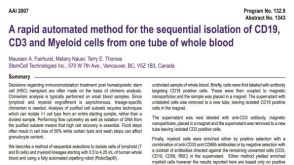 科学海报Isolation of CD19, CD3 and Myeloid Cells from one Tube of Whole Blood

 沪公网安备31010102008431号