Renal cancer cells acquire immune surface protein through trogocytosis and horizontal gene transfer
Trogocytosis is an underappreciated phenomenon that shapes the immune microenvironment surrounding many types of solid tumors. The consequences of membrane-bound proteins being deposited from a donor immune cell to a recipient cancer cell via trogocytosis are still unclear. Here,we report that human clear cell renal carcinoma tumors stably express the lymphoid markers CD45,CD56,CD14,and CD16. Flow cytometry performed on fresh kidney tumors revealed consistent CD45 expression on tumor cells,as well as varying levels of the other markers mentioned previously. These results were consistent with our immunofluorescent analysis,which also revealed colocalization of lymphoid markers with carbonic anhydrase 9,a standard kidney tumor marker. RNA analysis showed a significant upregulation of genes typically associated with immune cells by tumor cells. Finally,we show evidence of chromosomal DNA being transferred from immune cells to tumor cells through physical contact. This horizontal gene transfer has transcriptional consequences in the recipient tumor cell,resulting in a fusion phenotype that expresses both immune and cancer specific proteins. This work demonstrates a novel mechanism by which tumor cell protein expression is altered through the acquisition of surface membrane fragments and genomic DNA from infiltrating lymphocytes. These results alter the way in which we understand tumor-immune cell interactions and may reveal new insights into the mechanisms by which tumors develop. Additionally,further studies into trogocytosis and other mechanisms of contact-mediated cellular transfer will help push the field towards the next generation of immunotherapies and biomarkers for treating renal cell carcinoma and other cancers.
View Publication
产品类型:
产品号#:
100-0785
10970
10990
产品名:
ImmunoCult™ 人CD3/CD28/CD2 T细胞激活剂
ImmunoCult™ 人CD3/CD28/CD2 T细胞激活剂
ImmunoCult™ 人CD3/CD28/CD2 T细胞激活剂
T. Yoshino et al. (Jun 2025)
Engineering in Life Sciences 25 6
Rapid Recovery and Short‐Term Culture of Gastric Circulating Tumor Cells Using Microcavity Array
Circulating tumor cells (CTCs) hold significant promise for cancer diagnosis,prognosis,and treatment monitoring. We previously developed a technique for a single‐cell filtering device known as the microcavity array (MCA),specifically designed for the efficient recovery of CTCs from whole blood samples. Efficient enrichment and release of cells from the MCA remains challenging because of cell adhesion that occurs on the MCA surface during the enrichment phase. This study investigated the effects of surface modification with 2‐methacryloyloxyethyl phosphorylcholine (MPC) on the recovery efficiency of cancer cell lines from MCA. Scanning electron microscope (SEM) demonstrated reduced cell‐substrate interactions,leading to improved recovery efficiency. Comparative analyses showed that the MCA method provided superior recovery efficiency and reduced processing time compared to traditional methods such as density gradient centrifugation (DGC),while maintaining cell viability and proliferative capacity. CTCs were successfully detected in patients with gastric cancer,and short‐term cultures were achieved even when fewer than 20 CTCs per milliliter of blood were isolated. These findings emphasize the importance of surface modification for enhancing CTC isolation and the need for optimized culture conditions. The optimized MCA method offers a promising approach for rapid CTC recovery and potential integration with automated systems. Practical application : The Microcavity array (MCA) is a device specifically designed for efficient recovery of CTCs from whole blood. However cell adhesion on the MCA surface can limit release efficiency. This study demonstrated that surface modification with MPC signigicantly reduces cell‐substrate adhesion,improving recovery efficiency while maintaining cell viability and proliferative capacity. Compared to traditional density gradient centrifugation,the MPC‐modified MCA offers shorter processing time and better performance. CTCs were successfully detected in gastric cancer,and short‐term cultures were achieved even when fewer than 20 CTCs per mL of blood were isolated. The method supports downstearm applications such as cancer cell characterization and treatment monitoring. With potential for integration into automated system,the optimized MCA provides a practical,scalable solution for clinical liquid biopsy and personalized oncology.
View Publication
产品类型:
产品号#:
15122
15162
产品名:
RosetteSep™ 人CD45去除抗体混合物
RosetteSep™人CD45去除抗体混合物
K. E. Gomez et al. (oct 2020)
Cancer research 80 19 4185--4198
Cancer Cell CD44 Mediates Macrophage/Monocyte-Driven Regulation of Head and Neck Cancer Stem Cells.
Tumor-associated macrophages (TAM) in the tumor microenvironment (TME) cooperate with cancer stem cells (CSC) to maintain stemness. We recently identified cluster of differentiation 44 (CD44) as a surface marker defining head and neck squamous cell carcinoma (HNSCC) CSC. PI3K-4EBP1-SOX2 activation and signaling regulate CSC properties,yet the upstream molecular control of this pathway and the mechanisms underlying cross-talk between TAM and CSC in HNSCC remain largely unknown. Because CD44 is a molecular mediator in the TME,we propose here that TAM-influenced CD44 signaling could mediate stemness via the PI3K-4EBP1-SOX2 pathway,possibly by modulating availability of hyaluronic acid (HA),the main CD44 ligand. HNSCC IHC was used to identify TAM/CSC relationships,and in vitro coculture spheroid models and in vivo mouse models were used to identify the influence of TAMs on CSC function via CD44. Patient HNSCC-derived TAMs were positively and negatively associated with CSC marker expression at noninvasive and invasive edge regions,respectively. TAMs increased availability of HA and increased cancer cell invasion. HA binding to CD44 increased PI3K-4EBP1-SOX2 signaling and the CSC fraction,whereas CD44-VCAM-1 binding promoted invasive signaling by ezrin/PI3K. In vivo,targeting CD44 decreased PI3K-4EBP1-SOX2 signaling,tumor growth,and CSC. TAM depletion in syngeneic and humanized mouse models also diminished growth and CSC numbers. Finally,a CD44 isoform switch regulated epithelial-to-mesenchymal plasticity as standard form of CD44 and CD44v8-10 determined invasive and tumorigenic phenotypes,respectively. We have established a mechanistic link between TAMs and CSCs in HNSCC that is mediated by CD44 intracellular signaling in response to extracellular signals. SIGNIFICANCE: These findings establish a mechanistic link between tumor cell CD44,TAM,and CSC properties at the tumor-stroma interface that can serve as a vital area of focus for target and drug discovery.
View Publication
产品类型:
产品号#:
产品名:
C. J. Hanley et al. (nov 2020)
Molecular cancer research : MCR 18 11 1615--1622
Tumor-Resident Stromal Cells Promote Breast Cancer Invasion through Regulation of the Basal Phenotype.
Collective invasion can be led by breast cancer cells expressing basal epithelial markers,typified by keratin-14 (KRT14). We analyzed gene expression data from The Cancer Genome Atlas and demonstrated a significant correlation between a KRT14+ invasion signature and a stromal-mediated extracellular matrix (ECM) organization module. We then developed a novel coculture model of tumor organoids with autologous stromal cells. Coculture significantly increased KRT14 expression and invasion of organoids from both luminal and basal murine breast cancer models. However,stromal cell conditioned medium induced invasion but not KRT14 expression. Cancer cells released TGF$\beta$ and that signaling pathway was required for stromal cell-induced invasion and KRT14 expression. Mechanistically,TGF$\beta$ induced NOX4 expression in stromal cells and NOX4 inhibition reduced invasion and KRT14 expression. In summary,we developed a novel coculture model and revealed dynamic molecular interactions between stromal cells and cancer cells that regulate both basal gene expression and invasive behavior. IMPLICATIONS: Fibroblasts within mammary tumors can regulate the molecular phenotype and invasive behavior of breast cancer cells. VISUAL OVERVIEW: http://mcr.aacrjournals.org/content/molcanres/18/11/1615/F1.large.jpg.
View Publication
产品类型:
产品号#:
19860
19860RF
产品名:
EasySep™ 小鼠Streptavidin RapidSpheres™分选试剂盒
RoboSep™ 小鼠Streptavidin RapidSpheres™分选试剂盒
B. C. Heng et al. (oct 2007)
Bioscience reports 27 5-Apr 257--64
Caspase inhibitor Z-VAD-FMK enhances the freeze-thaw survival rate of human embryonic stem cells.
Previous study demonstrated that the low survival of human embryonic stem cells (hESC) under conventional slow-cooling cryopreservation protocols is predominantly due to apoptosis rather than cellular necrosis. Hence,this study investigated whether a synthetic broad-spectrum irreversible inhibitor of caspase enzymes,Z-VAD-FMK can be used to enhance the post-thaw survival rate of hESC. About 100 mM Z-VAD-FMK was supplemented into either the freezing solution,the post-thaw culture media or both. Intact and adherent hESC colonies were cryopreserved so as to enable subsequent quantitation of the post-thaw cell survival rate through the MTT assay,which can only be performed with adherent cells. Exposure to 100 mM Z-VAD-FMK in the freezing solution alone did not significantly enhance the post-thaw survival rate (10.2{\%} vs. 9.9{\%},p {\textgreater} 0.05). However,when 100 mM Z-VAD-FMK was added to the post-thaw culture media,there was a significant enhancement in the survival rate from 9.9{\%} to 14.4{\%} (p {\textless} 0.05),which was further increased to 18.7{\%} when Z-VAD-FMK was also added to the freezing solution as well (p {\textless} 0.01). Spontaneous differentiation of hESC after cryopreservation was assessed by morphological observations under bright-field microscopy,and by immunocytochemical staining for the pluripotency markers SSEA-3 and TRA-1-81. The results demonstrated that exposure to Z-VAD-FMK did not significantly enhance the spontaneous differentiation of hESC within post-thaw culture.
View Publication
产品类型:
产品号#:
100-0534
100-0535
产品名:
Z-VAD-FMK
Z-VAD-FMK
M. Dastpak et al. (Dec 2025)
PLOS One 20 12
SF3B1K700E mutation in human embryonic stem cells causes aberrant expression of immune-related genes
SF3B1,a component of the U2 snRNP pre-mRNA splicing factor,plays a critical role in splicing and is frequently mutated in cancer,particularly hematologic malignancies. We investigated the effects of the most common SF3B1 mutation,heterozygous substitution of Lysine 700 to Glutamate (K700E),in human embryonic stem cells (hESC),using CRISPR-Cas9 to generate heterozygous SF3B1K700E clones. Interestingly,we observed the upregulation of several key transcription regulators associated with hematopoiesis and a broad range of immune genes in SF3B1K700E hESCs. Despite differences in the transcriptional and splicing profiles between hESC and myelodysplastic syndrome (MDS) cells harboring the SF3B1K700E mutation,several common immune gene programs were identified in both cell types. To elucidate the molecular mechanisms underlying dysregulated gene expression in SF3B1K700E hESCs,we mapped actively engaged RNA polymerase II (RNA Pol II) using Precision Run-On sequencing (PRO-seq). These analyses revealed that the SF3B1K700E mutation alters RNA Pol II elongation properties. Specifically,we observed a general increase in pause release in SF3B1K700E hESCs,consistent with recent work in leukemia cells suggesting that the SF3B1K700E mutation affects early transcription elongation. Taken together,our study identifies several candidate genes that could contribute to the SF3B1 mutated phenotype and clarifies the role for the U2 snRNP and pre-spliceosome assembly on transcription by RNA Pol II. Further,our data suggest that mutations of SF3B1 impact immune gene expression independent of cell type,providing new insights into the role of SF3B1K700E in hematologic malignancies.
View Publication
Mesenchymal stromal cells lower platelet activation and assist in platelet formation in vitro.
The complex process of platelet formation originates with the hematopoietic stem cell,which differentiates through the myeloid lineage,matures,and releases proplatelets into the BM sinusoids. How formed platelets maintain a low basal activation state in the circulation remains unknown. We identify Lepr+ stromal cells lining the BM sinusoids as important contributors to sustaining low platelet activation. Ablation of murine Lepr+ cells led to a decreased number of platelets in the circulation with an increased activation state. We developed a potentially novel culture system for supporting platelet formation in vitro using a unique population of CD51+PDGFRalpha+ perivascular cells,derived from human umbilical cord tissue,which display numerous mesenchymal stem cell (MSC) properties. Megakaryocytes cocultured with MSCs had altered LAT and Rap1b gene expression,yielding platelets that are functional with low basal activation levels,a critical consideration for developing a transfusion product. Identification of a regulatory cell that maintains low baseline platelet activation during thrombopoiesis opens up new avenues for improving blood product production ex vivo.
View Publication
产品类型:
产品号#:
05402
05412
05455
05465
产品名:
MesenCult™ MSC 刺激补充剂(人)
MesenCult™ 脂肪分化试剂盒 (人)
MesenCult™-ACF软骨细胞分化试剂盒
MesenCult™ 成骨细胞分化试剂盒 (人)
Nefedova Y et al. (JAN 2004)
Journal of immunology (Baltimore,Md. : 1950) 172 1 464--74
Hyperactivation of STAT3 is involved in abnormal differentiation of dendritic cells in cancer.
Abnormal differentiation of myeloid cells is one of the hallmarks of cancer. However,the molecular mechanisms of this process remain elusive. In this study,we investigated the effect of tumor-derived factors on Janus kinase (Jak)/STAT signaling in myeloid cells during their differentiation into dendritic cells. Tumor cell conditioned medium induced activation of Jak2 and STAT3,which was associated with an accumulation of immature myeloid cells. Jak2/STAT3 activity was localized primarily in these myeloid cells,which prevented the differentiation of immature myeloid cells into mature dendritic cells. This differentiation was restored after removal of tumor-derived factors. Inhibition of STAT3 abrogated the negative effects of these factors on myeloid cell differentiation,and overexpression of STAT3 reproduced the effects of tumor-derived factors. Thus,this is a first demonstration that tumor-derived factors may affect myeloid cell differentiation in cancer via constitutive activation of Jak2/STAT3.
View Publication
产品类型:
产品号#:
03534
产品名:
MethoCult™GF M3534
Beloti M et al. (JUL 2005)
Cell biology international 29 7 537--41
Purmorphamine enhances osteogenic activity of human osteoblasts derived from bone marrow mesenchymal cells.
Purmorphamine is a novel small molecule with osteogenesis-inducing activity in multipotent mesenchymal progenitor cells,but there has been no evaluation of its effect on human cells to date. The aim of this study was to investigate the induction of osteogenic activity by purmorphamine in human osteoblasts differentiated from bone marrow mesenchymal cells. Cells were cultured in 24-well plates at a density of 2x10(4)/well in medium containing 1,2 or 3 microM purmorphamine,or vehicle. At 7,14 and 21 days,cell proliferation,viability,and alkaline phosphatase (ALP) activity were evaluated. Bone-like nodule formation was evaluated at 21 days. Purmorphamine did not affect cell proliferation or viability,but increased ALP activity and bone-like nodule formation. These results indicate that events related to osteoblast differentiation,including increased ALP activity and bone-like nodule formation,are enhanced by purmorphamine.
View Publication

 EasySep™小鼠TIL(CD45)正选试剂盒


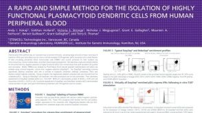 科学海报Rapid Cell Isolation of Highly Functional Plasmacytoid Dendritic Cells from Human Peripheral Blood

 沪公网安备31010102008431号