Ramadan A et al. (SEP 2010)
Genes to cells : devoted to molecular & cellular mechanisms 15 9 983--94
Cells with hematopoietic activity in the mouse placenta reside in side population.
The discovery of a major hematopoietic stem cell pool in midgestation mouse embryo has defined the placenta as an important hematopoietic anatomical site. In this study,we examined the flow cytometric pattern of mouse placenta cells on embryonic days (E) 10.5 to E15.5,in view of CD45 and c-Kit expression. We also determined which population of these cells shows differentiation potential toward multiple hematopoietic lineages by performing coculture with OP9 stromal cells and colony-forming assay in methylcellulose. Only CD45(+)c-Kit(+) population showed the ability to form hematopoietic colonies including multiple lineages. To distinguish which fraction of placenta cells have the hematopoietic activity,we used GFP transgenic mice in which the fetal part of the placenta is GFP positive and the maternal part is GFP negative. E11.5 and E13.5 CD45(+)c-Kit(+) placental cells that have ability to form hematopoietic colonies are the fetal GFP positive placental cells. E11.5 and E13.5 CD45(+)c-Kit(+) placental cells that have an ability to form hematopoietic colonies mainly reside in Hoechst dye-effluxing side population area (SP). Taken together,in the placenta of mouse embryo,we conclude that SP cells in the CD45(+)c-Kit(+) fetal placental cells have the ability to form hematopoietic colonies.
View Publication
Lin S and Talbot P (JAN 2011)
Methods in molecular biology (Clifton,N.J.) 690 31--56
Methods for culturing mouse and human embryonic stem cells
Mouse embryonic stem cells (mESCs) were first derived and cultured almost 30 years ago and ever since have been valuable tools for creating knockout mice and for studying early mammalian development. More recently (1998),human embryonic stem cells (hESCs) have been derived from blastocysts,and numerous methods have evolved to culture hESCs in vitro in both complex and defined media. hESCs are especially important at this time as they could potentially be used to treat degenerative diseases and to access the toxicity of new drugs and environmental chemicals. For both human and mouse ESCs,fibroblast feeder layers are often used at some phase in the culturing protocol. The feeders - often mouse embryonic fibroblasts (mEFs) - provide a substrate that increases plating efficiency,helps maintain pluripotency,and facilitates survival and growth of the stem cells. Various protocols for culturing embryonic stem cells from both species are available with newer trends moving toward feeder-free and serum-free culture. The purpose of this chapter is to provide basic protocol information on the isolation of mouse embryonic fibroblasts and establishment of feeder layers,the culture of mESCs on both mEFs and on gelatin in serum-containing medium,and the culture of hESCs in defined media on both mEFs (hESC culture medium) and Matrigel (mTeSR). These basic protocols are intended for researchers wanting to develop stem cell research in their labs. These protocols have been tested in our laboratory and work well. They can be modified and adapted for any relevant user's particular purpose.
View Publication
Orelio C et al. (APR 2009)
Haematologica 94 4 462--9
Interleukin-1 regulates hematopoietic progenitor and stem cells in the midgestation mouse fetal liver.
BACKGROUND: Hematopoietic progenitors are generated in the yolk sac and aorta-gonad-mesonephros region during early mouse development. At embryonic day 10.5 the first hematopoietic stem cells emerge in the aorta-gonad-mesonephros. Subsequently,hematopoietic stem cells and progenitors are found in the fetal liver. The fetal liver is a potent hematopoietic site,playing an important role in the expansion and differentiation of hematopoietic progenitors and hematopoietic stem cells. However,little is known concerning the regulation of fetal liver hematopoietic stem cells. In particular,the role of cytokines such as interleukin-1 in the regulation of hematopoietic stem cells in the embryo has been largely unexplored. Recently,we observed that the adult pro-inflammatory cytokine interleukin-1 is involved in regulating aorta-gonad-mesonephros hematopoietic progenitor and hematopoietic stem cell activity. Therefore,we set out to investigate whether interleukin-1 also plays a role in regulating fetal liver progenitor cells and hematopoietic stem cells. DESIGN AND METHODS: We examined the interleukin-1 ligand and receptor expression pattern in the fetal liver. The effects of interleukin-1 on hematopoietic progenitor cells and hematopoietic stem cells were studied by FACS and transplantation analyses of fetal liver explants,and in vivo effects on hematopoietic stem cell and progenitors were studied in Il1r1(-/-) embryos. RESULTS: We show that fetal liver hematopoietic progenitor cells express the IL-1RI and that interleukin-1 increases fetal liver hematopoiesis,progenitor cell activity and promotes hematopoietic cell survival. Moreover,we show that in Il1r1(-/-) embryos,hematopoietic stem cell activity is impaired and myeloid progenitor activity is increased. CONCLUSIONS: The IL-1 ligand and receptor are expressed in the midgestation liver and act in the physiological regulation of fetal liver hematopoietic progenitor cells and hematopoietic stem cells.
View Publication
M. Pavel-Dinu et al. ( 2019)
Nature communications 10 1 1634
Gene correction for SCID-X1 in long-term hematopoietic stem cells.
Gene correction in human long-term hematopoietic stem cells (LT-HSCs) could be an effective therapy for monogenic diseases of the blood and immune system. Here we describe an approach for X-linked sSevere cCombined iImmunodeficiency (SCID-X1) using targeted integration of a cDNA into the endogenous start codon to functionally correct disease-causing mutations throughout the gene. Using a CRISPR-Cas9/AAV6 based strategy,we achieve up to 20{\%} targeted integration frequencies in LT-HSCs. As measures of the lack of toxicity we observe no evidence of abnormal hematopoiesis following transplantation and no evidence of off-target mutations using a high-fidelity Cas9 as a ribonucleoprotein complex. We achieve high levels of targeting frequencies (median 45{\%}) in CD34+ HSPCs from six SCID-X1 patients and demonstrate rescue of lymphopoietic defect in a patient derived HSPC population in vitro and in vivo. In sum,our study provides specificity,toxicity and efficacy data supportive of clinical development of genome editing to treat SCID-Xl.
View Publication

 EasySep™小鼠TIL(CD45)正选试剂盒


 技术公告Endothelial Protein C Receptor (EPCR): A New Marker for Identification and Positive Selection of Mouse Hematopoietic Stem Cells
 挂图Identification of Colonies Derived from Mouse Hematopoietic Progenitors Representative colony images and tips for identifying progenitor subtypes in CFU assays
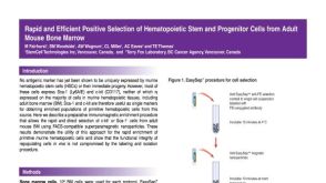 科学海报Positive Selection of Hematopoietic Stem and Progenitor Cells from Adult Mouse Bone Marrow
 产品手册Mammary Epithelial Cells: Standardized Media and Reagents
 技术公告Genome Editing of Human CD34+ Hematopoietic Stem and Progenitor Cells Using the ArciTect™ CRISPR-Cas9 System and StemSpan™ Media


 沪公网安备31010102008431号