Treating activated regulatory T cells with pramipexole protects human dopaminergic neurons from 6?OHDA?induced degeneration
AbstractBackgroundParkinson's disease (PD) is a chronic neurodegenerative disorder characterized by the loss of dopaminergic neurons in the substantia nigra,which promotes a sustained inflammatory environment in the central nervous system. Regulatory T cells (Tregs) play an important role in the control of inflammation and might play a neuroprotective role. Indeed,a decrease in Treg number and function has been reported in PD. In this context,pramipexole,a dopaminergic receptor agonist used to treat PD symptoms,has been shown to increase peripheral levels of Treg cells and improve their suppressive function. The aim of this work was to determine the effect of pramipexole on immunoregulatory Treg cells and its possible neuroprotective effect on human dopaminergic neurons differentiated from human embryonic stem cells.MethodsTreg cells were sorted from white blood cells of healthy human donors. Assays were performed with CD3/CD28?activated and non?activated Treg cells treated with pramipexole at concentrations of 2 or 200 ng/mL. These regulatory cells were co?cultured with in vitro?differentiated human dopaminergic neurons in a cytotoxicity assay with 6?hydroxydopamine (6?OHDA). The role of interleukin?10 (IL?10) was investigated by co?culturing activated IL?10?producing Treg cells with neurons. To further investigate the effect of treatment on Tregs,gene expression in pramipexole?treated,CD3/CD28?activated Treg cells was determined by Fluidigm analysis.ResultsPramipexole?treated CD3/CD28?activated Treg cells showed significant protective effects on dopaminergic neurons when challenged with 6?OHDA. Pramipexole?treated activated Treg cells showed neuroprotective capacity through mechanisms involving IL?10 release and the activation of genes associated with regulation and neuroprotection.ConclusionAnti?CD3/CD28?activated Treg cells protect dopaminergic neurons against 6?OHDA?induced damage. In addition,activated,IL?10?producing,pramipexole?treated Tregs also induced a neuroprotective effect,and the supernatants of these co?cultures promoted axonal growth. Pramipexole?treated,activated Tregs altered their gene expression in a concentration?dependent manner,and enhanced TGF??related dopamine receptor regulation and immune?related pathways. These findings open new perspectives for the development of immunomodulatory therapies for the treatment of PD. Pramipexole?treated,activated regulatory T cells protect dopaminergic neurons against 6 OHDA damage and promote primary neurite length. This could be due to the production of the regulatory cytokine IL?10 and an increased expression of genes related to regulation and neuroprotection.
View Publication
产品类型:
产品号#:
100-0784
10971
10991
15022
15062
85850
85857
产品名:
ImmunoCult™ 人CD3/CD28 T细胞激活剂
ImmunoCult™ 人CD3/CD28 T细胞激活剂
ImmunoCult™ 人CD3/CD28 T细胞激活剂
RosetteSep™人CD4+ T细胞富集抗体混合物
RosetteSep™人CD4+ T细胞富集抗体混合物
mTeSR™1
mTeSR™1
(Dec 2024)
Stem Cell Research & Therapy 15 15
Matrix-free human lung organoids derived from induced pluripotent stem cells to model lung injury
BackgroundOrganoids,as near-physiological 3D culture systems,offer new opportunities to study the pathogenesis of various organs in mimicking the cellular complexity and functionality of human organs.MethodHere we used a quite simple and very practicable method to successfully generate induced pluripotent stem cell (iPSC)-derived human lung organoids (LuOrg) in a matrix-free manner as an alternative to the widely used preclinical mouse models in order to investigate normal lung damage in detail and as close as possible to the patient. We performed detailed morphological and molecular analyses,including bulk and single cell RNA sequencing,of generated lung organoids and evaluated the quality and robustness of our model as a potential in vitro platform for lung diseases,namely radiation-induced lung injury.ResultsA matrix-free method for differentiation of iPSCs can be used to obtain lung organoids that morphologically reflect the target tissue of the human lung very well,especially with regard to the cellular composition. The different cellular fates were investigated following the genotoxic stress induced by radiation and revealed further insights in the radiation-sensitivity of the different lung cells. Finally,we provide cellular gene sets found to be induced in the different lung organoid cellular subsets after irradiation,which could be used as additional RT response and particularly senescence gene sets in future studies.ConclusionBy establishing these free-floating LuOrgs for the investigation of cancer therapeutic approaches as a new and patient-oriented in vitro platform particularly in experimental radiooncology,not only a reduction in the number of experimental animals,but also an adequately and meaningfully replacement of corresponding animal experiments can be achieved.
View Publication
产品类型:
产品号#:
85850
85857
产品名:
mTeSR™1
mTeSR™1
(Apr 2024)
Nature Communications 15
PD-L1- and IL-4-expressing basophils promote pathogenic accumulation of T follicular helper cells in lupus
Systemic lupus erythematosus (SLE) is an autoimmune disease characterized by anti-nuclear autoantibodies whose production is promoted by autoreactive T follicular helper (TFH) cells. During SLE pathogenesis,basophils accumulate in secondary lymphoid organs (SLO),amplify autoantibody production and disease progression through mechanisms that remain to be defined. Here,we provide evidence for a direct functional relationship between TFH cells and basophils during lupus pathogenesis,both in humans and mice. PD-L1 upregulation on basophils and IL-4 production are associated with TFH and TFH2 cell expansions and with disease activity. Pathogenic TFH cell accumulation,maintenance,and function in SLO were dependent on PD-L1 and IL-4 in basophils,which induced a transcriptional program allowing TFH2 cell differentiation and function. Our study establishes a direct mechanistic link between basophils and TFH cells in SLE that promotes autoantibody production and lupus nephritis. Basophils have been implicated in systemic lupus erythematosus (SLE),as evidenced by the fact that basophil-deficient mice do not develop the disease. Here,the authors demonstrate that PD-L1 and IL-4 expression in basophils promotes the pathogenic accumulation of follicular helper T cells in patients with SLE and murine models.
View Publication
产品类型:
产品号#:
19852
19844
19844RF
19852RF
产品名:
EasySep™小鼠CD4+ T细胞分选试剂盒
EasySep™小鼠Pan-B细胞分选试剂盒
RoboSep™ 小鼠Pan-B细胞分选试剂盒
RoboSep™ 小鼠CD4+ T细胞分选试剂盒
(May 2024)
Nature Communications 15
Tlr9 deficiency in B cells leads to obesity by promoting inflammation and gut dysbiosis
Toll-like receptor 9 (TLR9) recognizes bacterial,viral and self DNA and play an important role in immunity and inflammation. However,the role of TLR9 in obesity is less well-studied. Here,we generate B-cell-specific Tlr9-deficient (Tlr9fl/fl/Cd19Cre+/-,KO) B6 mice and model obesity using a high-fat diet. Compared with control mice,B-cell-specific-Tlr9-deficient mice exhibited increased fat tissue inflammation,weight gain,and impaired glucose and insulin tolerance. Furthermore,the frequencies of IL-10-producing-B cells and marginal zone B cells were reduced,and those of follicular and germinal center B cells were increased. This was associated with increased frequencies of IFNγ-producing-T cells and increased follicular helper cells. In addition,gut microbiota from the KO mice induced a pro-inflammatory state leading to immunological and metabolic dysregulation when transferred to germ-free mice. Using 16 S rRNA gene sequencing,we identify altered gut microbial communities including reduced Lachnospiraceae,which may play a role in altered metabolism in KO mice. We identify an important network involving Tlr9,Irf4 and Il-10 interconnecting metabolic homeostasis,with the function of B and T cells,and gut microbiota in obesity. Although the function of Toll-like receptor 9 (TLR9) in immunity and inflammation is well-established,its role in obesity is less well-studied. In this study,the authors demonstrate that TLR9 deficiency in B cells is associated with obesity in mice and results in altered frequencies of T and B lymphocyte subsets and gut microbiome dysbiosis.
View Publication
Autism Spectrum Disorder (ASD) is a neurodevelopmental condition that affects communication,social interaction,and behavior. Calcium (Ca2+) signaling dysregulation has been frequently highlighted in genetic studies as a contributing factor to aberrant developmental processes in ASD. Herein,we used ASD and control induced pluripotent stem cells (iPSCs) to investigate transcriptomic and functional Ca2+ dynamics at various stages of differentiation to cortical neurons. Idiopathic ASD and control iPSC lines underwent the dual SMAD inhibition differentiation protocol to direct their fate toward cortical neurons. Samples from multiple time points along the course of differentiation were processed for bulk RNA sequencing,spanning the following sequential stages: the iPSC stage,neural induction (NI) stage,neurosphere (NSP) stage,and differentiated cortical neuron (Diff) stage. Our transcriptomic analyses suggested that the numbers of Ca2+ signaling-relevant differentially expressed genes between ASD and control samples were higher in the iPSC and Diff stages. Accordingly,samples from the iPSC and Diff stages were processed for Ca2+ imaging studies. Results revealed that iPSC-stage ASD samples displayed elevated maximum Ca2+ levels in response to ATP compared to controls. By contrast,in the Diff stage,ASD neurons showed reduced maximum Ca2+ levels in response to ATP but increased maximum Ca2+ levels in response to KCl and DHPG relative to controls. Considering the distinct functional signaling contexts of these stimuli,this differential profile of receptor- and ionophore-mediated Ca2+ response suggests that aberrant calcium homeostasis underlies the pathophysiology of ASD neurons. Our data provides functional evidence for Ca2+ signaling dysregulation during neurogenesis in idiopathic ASD.
View Publication
产品类型:
产品号#:
05990
产品名:
TeSR™-E8™
D. Zheng et al. (Oct 2025)
Stem Cell Research & Therapy 16
Dynamic molecular and cellular characteristics of VSX2-positive retinal progenitor cells in human retinal organoids
The lack of understanding of the molecular and cellular characteristics of human retinal progenitor cells (RPCs) has hindered their application in cell therapy for retinal degenerative diseases. This study aims to employ retinal organoids (ROs) derived from a VSX2-enhanced green fluorescent protein (eGFP) reporter human induced pluripotent stem cell (hiPSC) line for positive selection of human RPCs,investigate their features,and facilitate their applications. Methods: hiPSCs were differentiated into three-dimensional ROs following established protocols. The fidelity of the VSX2-eGFP reporter was confirmed through immunostaining. Fluorescence-activated cell sorting was employed to select VSX2-eGFP-positive (+) cells at distinct developmental stages,followed by bulk RNA sequencing (RNA-seq) analysis to assess their transcriptome profile. Immunostaining and flow cytometry were utilized to validate the identity of VSX2-eGFP+ cells and potential cluster of differentiation (CD) biomarkers for identifying human RPCs. Results: hiPSCs were successfully differentiated into ROs containing abundant RPCs. The spatiotemporal activity of the VSX2-eGFP reporter recapitulated the dynamic expression of endogenous VSX2 protein. Compared to VSX2-eGFP-negative (-) cells,VSX2-eGFP+ cells mainly exhibited characteristics of RPCs at early stages of retinal development and of bipolar cells at late stages. RNA-seq analysis revealed transcriptional heterogeneity within VSX2-eGFP+ cells across four distinct developmental stages. Moreover,the dynamic expression of 394 known CD biomarkers in VSX2-eGFP+ cells at distinct developmental stages was analyzed herein for the first time. One CD biomarker,TNFRSF1B,which has never been reported to be expressed in RPCs,was found to be highly expressed in RPCs at the early stages and might serve as a candidate CD biomarker for sorting RPCs. Conclusions: This study provides valuable insights into the molecular and cellular characteristics of human RPCs,especially their expression profiles of CD biomarkers,laying a foundation for research on retinal development and the clinical translation of hiPSC-derived RPCs.
View Publication
Schlecht G et al. (OCT 2001)
Journal of immunology (Baltimore,Md. : 1950) 167 8 4215--21
Induction of CTL and nonpolarized Th cell responses by CD8alpha(+) and CD8alpha(-) dendritic cells.
Two distinct dendritic cell (DC) subpopulations have been evidenced in mice on the basis of their differential CD8alpha expression and their localization in lymphoid organs. Several reports suggest that CD8alpha(+) and CD8alpha(-) DC subsets could be functionally different. In this study,using a panel of MHC class I- and/or class II-restricted peptides,we analyzed CD4(+) and CD8(+) T cell responses obtained after i.v. injection of freshly purified peptide-pulsed DC subsets. First,we showed that both DC subsets efficiently induce specific CTL responses and Th1 cytokine production in the absence of CD4(+) T cell priming. Second,we showed that in vivo activation of CD4(+) T cells by CD8alpha(+) or CD8alpha(-) DC,injected i.v.,leads to a nonpolarized Th response with production of both Th1 and Th2 cytokines. The CD8alpha(-) subset induced a higher production of Th2 cytokines such as IL-4 and IL-10 than the CD8alpha(+) subset. However,IL-5 was produced by CD4(+) T cells activated by both DC subsets. When both CD4(+) and CD8(+) T cells were primed by DC injected i.v.,a similar pattern of cytokines was observed,but,under these conditions,Th1 cytokines were mainly produced by CD8(+) T cells,while Th2 cytokines were produced by CD4(+) T cells. Thus,this study clearly shows that CD4(+) T cell responses do not influence the development of specific CD8(+) T cell cytotoxic responses induced either by CD8alpha(+) or CD8alpha(-) DC subsets.
View Publication
产品类型:
产品号#:
09600
09650
产品名:
StemSpan™ SFEM
StemSpan™ SFEM
Tay SS et al. (MAR 2003)
Journal of immunology (Baltimore,Md. : 1950) 170 6 3315--22
IFN-gamma reverses the stop signal allowing migration of antigen-specific T cells into inflammatory sites.
In humans the majority of endothelial cells (EC) constitutively express MHC class II Ags. We know that in vitro ECs can activate CD45RO(+) B7-independent CD4(+) T cells to proliferate and produce IL-2. The in vivo correlate of this T cell response is not known,and here we have explored whether endothelial expression of MHC class II Ags affects the transendothelial migration of alloreactive CD4(+) CD45RO(+) B7-independent T cells. Alloreactive CD4(+) T cell clones and lines were generated against HLA-DR11,DR13,DR4,and DR1 MHC Ags,and their rates of migration across untreated EC line Eahy.926 (MHC class II negative) or Eahy.926 transfected with CIITA (EahyCIITA) to express DR11 and DR13 were investigated. The migrations of EahyCIITA-specific T cell clones and lines were retarded in a DR-specific manner,and retardation was reversed in the presence of mAb to DR Ag. When investigating the ability of T cells to proliferate in response to EahyCIITA before and after transmigration,migrated cells were still able to proliferate,but the frequency of EahyCIITA-specific cells was much reduced compared with that of nonmigrated cells. The use of fluorescently labeled T cells revealed that specific cells become trapped within the endothelial monolayer. Pretreatment of EahyCIITA with IFN-gamma restored the ability of DR11- or DR13-specific T cells to transmigrate and proliferate,thus abrogating DR-specific retardation. We conclude that cognate interaction between T cells and endothelial MHC class II initiates a stop signal possibly similar to an immunological synapse,but this is overcome in an inflammatory milieu.
View Publication
产品类型:
产品号#:
15022
15062
产品名:
RosetteSep™人CD4+ T细胞富集抗体混合物
RosetteSep™人CD4+ T细胞富集抗体混合物
Mak SK et al. (JAN 2012)
Stem cells international 2012 140427
Small molecules greatly improve conversion of human-induced pluripotent stem cells to the neuronal lineage.
Efficient in vitro differentiation into specific cell types is more important than ever after the breakthrough in nuclear reprogramming of somatic cells and its potential for disease modeling and drug screening. Key success factors for neuronal differentiation are the yield of desired neuronal marker expression,reproducibility,length,and cost. Three main neuronal differentiation approaches are stromal-induced neuronal differentiation,embryoid body (EB) differentiation,and direct neuronal differentiation. Here,we describe our neurodifferentiation protocol using small molecules that very efficiently promote neural induction in a 5-stage EB protocol from six induced pluripotent stem cells (iPSC) lines from patients with Parkinson's disease and controls. This protocol generates neural precursors using Dorsomorphin and SB431542 and further maturation into dopaminergic neurons by replacing sonic hedgehog with purmorphamine or smoothened agonist. The advantage of this approach is that all patient-specific iPSC lines tested in this study were successfully and consistently coaxed into the neural lineage.
View Publication
产品类型:
产品号#:
73412
73414
产品名:
SAG
SAG
Nishida S et al. (JUL 2012)
The Journal of urology 188 1 294--9
Gene expression profiles of prostate cancer stem cells isolated by aldehyde dehydrogenase activity assay.
PURPOSE: Prostate cancer cells include a small population of cancer stem-like/cancer initiating cells,which have roles in cancer initiation and progression. Recently aldehyde dehydrogenase activity was used to isolate stem cells of various cancer and normal cells. We evaluated the aldehyde dehydrogenase activity of the human prostate cancer cell line 22Rv1 (ATCC®) with the ALDEFLUOR® assay and determined its potency as prostate cancer stem-like/cancer initiating cells. MATERIALS AND METHODS: The human prostate cancer cell line 22Rv1 was labeled with ALDEFLUOR reagent and analyzed by flow cytometry. ALDH1(high) and ALDH1(low) cells were isolated and tumorigenicity was evaluated by xenograft transplantation into NOD/SCID mice. Tumor sphere forming ability was evaluated by culturing in a floating condition. Invasion capability was evaluated by the Matrigel™ invasion assay. Gene expression profiling was assessed by microarrays and reverse transcriptase-polymerase chain reaction. RESULTS: ALDH1(high) cells were detected in 6.8% of 22Rv1 cells,which showed significantly higher tumorigenicity than ALDH1(low) cells in NOD/SCID mice (p textless 0.05). Gene expression profiling revealed higher expression of the stem cell related genes PROM1 and NKX3-1 in ALDH1(high) cells than in ALDH1(low) cells. ALDH1(high) cells also showed higher invasive capability and sphere forming capability than ALDH1(low) cells. CONCLUSIONS: Results indicate that cancer stem-like/cancer initiating cells are enriched in the ALDH1(high) population of the prostate cancer cell line 22Rv1. This approach may provide a breakthrough to further clarify prostate cancer stem-like/cancer initiating cells. To our knowledge this is the first report of cancer stem-like/cancer initiating cells of 22Rv1 using the aldehyde dehydrogenase activity assay.
View Publication

 EasySep™小鼠TIL(CD45)正选试剂盒


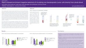 科学海报Rapid Unbiased Enrichment (Negative Selection) of Circulating Non-Hematopoietic Tumor Cells Directly from Whole Blood
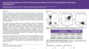 科学海报Fast and Easy Isolation of CD27-Positive Human Memory B Cells Using EasySep™ Releasable RapidSpheres™
 沪公网安备31010102008431号