Takashima Y et al. (SEP 2014)
Cell 158 6 1254--1269
Resetting transcription factor control circuitry toward ground-state pluripotency in human.
Current human pluripotent stem cells lack the transcription factor circuitry that governs the ground state of mouse embryonic stem cells (ESC). Here,we report that short-term expression of two components,NANOG and KLF2,is sufficient to ignite other elements of the network and reset the human pluripotent state. Inhibition of ERK and protein kinase C sustains a transgene-independent rewired state. Reset cells self-renew continuously without ERK signaling,are phenotypically stable,and are karyotypically intact. They differentiate in vitro and form teratomas in vivo. Metabolism is reprogrammed with activation of mitochondrial respiration as in ESC. DNA methylation is dramatically reduced and transcriptome state is globally realigned across multiple cell lines. Depletion of ground-state transcription factors,TFCP2L1 or KLF4,has marginal impact on conventional human pluripotent stem cells but collapses the reset state. These findings demonstrate feasibility of installing and propagating functional control circuitry for ground-state pluripotency in human cells.
View Publication
产品类型:
产品号#:
85850
85857
产品名:
mTeSR™1
mTeSR™1
Guan BX et al. (MAY 2014)
IEEE/ACM transactions on computational biology and bioinformatics / IEEE,ACM 11 3 604--611
Bio-Driven Cell Region Detection in Human Embryonic Stem Cell Assay.
This paper proposes a bio-driven algorithm that detects cell regions automatically in the human embryonic stem cell (hESC) images obtained using a phase contrast microscope. The algorithm uses both statistical intensity distributions of foreground/hESCs and background/substrate as well as cell property for cell region detection. The intensity distributions of foreground/hESCs and background/substrate are modeled as a mixture of two Gaussians. The cell property is translated into local spatial information. The algorithm is optimized by parameters of the modeled distributions and cell regions evolve with the local cell property. The paper validates the method with various videos acquired using different microscope objectives. In comparison with the state-of-the-art methods,the proposed method is able to detect the entire cell region instead of fragmented cell regions. It also yields high marks on measures such as Jacard similarity,Dice coefficient,sensitivity and specificity. Automated detection by the proposed method has the potential to enable fast quantifiable analysis of hESCs using large data sets which are needed to understand dynamic cell behaviors.
View Publication
产品类型:
产品号#:
85850
85857
产品名:
mTeSR™1
mTeSR™1
Xia N et al. (FEB 2016)
Scientific Reports 6 20270
Transcriptional comparison of human induced and primary midbrain dopaminergic neurons
Generation of induced dopaminergic (iDA) neurons may provide a significant step forward towards cell replacement therapy for Parkinson's disease (PD). To study and compare transcriptional programs of induced cells versus primary DA neurons is a preliminary step towards characterizing human iDA neurons. We have optimized a protocol to efficiently generate iDA neurons from human pluripotent stem cells (hPSCs). We then sequenced the transcriptomes of iDA neurons derived from 6 different hPSC lines and compared them to that of primary midbrain (mDA) neurons. We identified a small subset of genes with altered expression in derived iDA neurons from patients with Parkinson's Disease (PD). We also observed that iDA neurons differ significantly from primary mDA neurons in global gene expression,especially in genes related to neuron maturation level. Results suggest iDA neurons from patient iPSCs could be useful for basic and translational studies,including in vitro modeling of PD. However,further refinement of methods of induction and maturation of neurons may better recapitulate full development of mDA neurons from hPSCs.
View Publication
Lin S et al. (JAN 2010)
Journal of visualized experiments : JoVE 39 11330
Video bioinformatics analysis of human embryonic stem cell colony growth.
Because video data are complex and are comprised of many images,mining information from video material is difficult to do without the aid of computer software. Video bioinformatics is a powerful quantitative approach for extracting spatio-temporal data from video images using computer software to perform dating mining and analysis. In this article,we introduce a video bioinformatics method for quantifying the growth of human embryonic stem cells (hESC) by analyzing time-lapse videos collected in a Nikon BioStation CT incubator equipped with a camera for video imaging. In our experiments,hESC colonies that were attached to Matrigel were filmed for 48 hours in the BioStation CT. To determine the rate of growth of these colonies,recipes were developed using CL-Quant software which enables users to extract various types of data from video images. To accurately evaluate colony growth,three recipes were created. The first segmented the image into the colony and background,the second enhanced the image to define colonies throughout the video sequence accurately,and the third measured the number of pixels in the colony over time. The three recipes were run in sequence on video data collected in a BioStation CT to analyze the rate of growth of individual hESC colonies over 48 hours. To verify the truthfulness of the CL-Quant recipes,the same data were analyzed manually using Adobe Photoshop software. When the data obtained using the CL-Quant recipes and Photoshop were compared,results were virtually identical,indicating the CL-Quant recipes were truthful. The method described here could be applied to any video data to measure growth rates of hESC or other cells that grow in colonies. In addition,other video bioinformatics recipes can be developed in the future for other cell processes such as migration,apoptosis,and cell adhesion.
View Publication
产品类型:
产品号#:
85850
85857
产品名:
mTeSR™1
mTeSR™1
Bentley C et al. (NOV 2011)
Nutrition,metabolism,and cardiovascular diseases : NMCD 21 11 871--8
Influence of chylomicron remnants on human monocyte activation in vitro.
BACKGROUND AND AIMS: Atherosclerosis is known to be an inflammatory disease and there is increasing evidence that chylomicron remnants (CMR),the lipoproteins which carry dietary fats in the blood,cause macrophage foam cell formation and inflammation. In early atherosclerosis the frequency of activated monocytes in the peripheral circulation is increased,and clearance of CMR from blood may be delayed,however,whether CMR contribute directly to monocyte activation and subsequent egress into the arterial wall has not been established. Here,the contribution of CMR to activation of monocyte pro-inflammatory pathways was assessed using an in vitro model. METHODS AND RESULTS: Primary human monocytes and CMR-like particles (CRLP) were used to measure several endpoints of monocyte activation. Treatment with CRLP caused rapid and prolonged generation of reactive oxygen species by monocytes. The pro-inflammatory chemokines MCP-1 and IL-8 were secreted in nanogram quantities by the cells in the absence of CRLP. IL-8 secretion was transiently increased after CRLP treatment,and CRLP maintained secretion in the presence of pharmacological inhibitors of IL-8 production. In contrast,exposure to CRLP significantly reduced MCP-1 secretion. Chemotaxis towards MCP-1 was increased in monocytes pre-exposed to CRLP and was reversed by addition of exogenous MCP-1. CONCLUSION: Our findings indicate that CRLP activate human monocytes and augment their migration in vitro by reducing cellular MCP-1 expression. Our data support the current hypothesis that CMR contribute to the inflammatory milieu of the arterial wall in early atherosclerosis,and suggest that this may reflect direct interaction with circulating blood monocytes.
View Publication
Human neural stem cell-derived artificial organelles to improve oxidative phosphorylation
Oxidative phosphorylation (OXPHOS) in the mitochondrial inner membrane is a therapeutic target in many diseases. Neural stem cells (NSCs) show progress in improving mitochondrial dysfunction in the central nervous system (CNS). However,translating neural stem cell-based therapies to the clinic is challenged by uncontrollable biological variability or heterogeneity,hindering uniform clinical safety and efficacy evaluations. We propose a systematic top-down design based on membrane self-assembly to develop neural stem cell-derived oxidative phosphorylating artificial organelles (SAOs) for targeting the central nervous system as an alternative to NSCs. We construct human conditionally immortal clone neural stem cells (iNSCs) as parent cells and use a streamlined closed operation system to prepare neural stem cell-derived highly homogenous oxidative phosphorylating artificial organelles. These artificial organelles act as biomimetic organelles to mimic respiration chain function and perform oxidative phosphorylation,thus improving ATP synthesis deficiency and rectifying excessive mitochondrial reactive oxygen species production. Conclusively,we provide a framework for a generalizable manufacturing procedure that opens promising prospects for disease treatment. Regulating oxidative phosphorylation and restoring redox homeostasis are crucial in neurological disorders. Here,the authors develop a top-down membrane self-assembly strategy to develop stem cell-derived artificial organelles (SAOs) that mimic mitochondrial oxidative phosphorylation without the risks associated with stem cell therapy.
View Publication

 EasySep™小鼠TIL(CD45)正选试剂盒


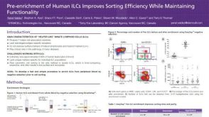 科学海报Pre-enrichment of Human ILCs Improves Sorting Efficiency While Maintaining Functionality
 15:48
线上讲座Generating Pancreatic Organoids from Healthy Murine and Human Pancreatic Ducts发布日期: 07/07/2023
15:48
线上讲座Generating Pancreatic Organoids from Healthy Murine and Human Pancreatic Ducts发布日期: 07/07/2023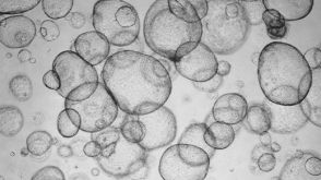 实验方案Detection of CYP3A4 Activity in Human Hepatic Organoids by LC-MS


 沪公网安备31010102008431号