Modulating notochordal differentiation of human induced pluripotent stem cells using natural nucleus pulposus tissue matrix
Human induced pluripotent stem cells (hiPSCs) can differentiate into notochordal cell (NC)-like cells when cultured in the presence of natural porcine nucleus pulposus (NP) tissue matrix. The method promises massive production of high-quality,functional cells to treat degenerative intervertebral discs (IVDs). Based on our previous work,we further examined the effect of cell-NP matrix contact and culture medium on the differentiation,and further assessed the functional differentiation ability of the generated NC-like. The study showed that direct contact between hiPSCs and NP matrix can promote the differentiation yield,whilst both the contact and non-contact cultures can generate functional NC-like cells. The generated NC-like cells are highly homogenous regarding the expression of notochordal marker genes. A culture medium containing a cocktail of growth factors (FGF,EGF,VEGF and IGF-1) also supported the notochordal differentiation in the presence of NP matrix. The NC-like cells showed excellent functional differentiation ability to generate NP-like tissue which was rich in aggrecan and collagen type II; and particularly,the proteoglycan to collagen content ratio was as high as 12.5-17.5 which represents a phenotype close to NP rather than hyaline cartilage. Collectively,the present study confirmed the effectiveness and flexibility of using natural NP tissue matrix to direct notochordal differentiation of hiPSCs,and the potential of using the generated NC-like cells for treating IVD degeneration.
View Publication
产品类型:
产品号#:
85850
85857
产品名:
mTeSR™1
mTeSR™1
Gomez AM et al. (MAR 2015)
The Journal of Immunology 194 5 2300--8
HIV-1-triggered release of type I IFN by plasmacytoid dendritic cells induces BAFF production in monocytes.
HIV-1 infection leads to numerous B cell abnormalities,including hypergammaglobulinemia,nonspecific B cell activation,nonspecific class switching,increased cell turnover,breakage of tolerance,increased immature/transitional B cells,B cell malignancies,as well as a loss of capacity to generate and maintain memory,all of which contribute to a global impairment of the immune humoral compartment. Several cytokines and soluble factors,which are increased in sera of HIV-1-infected individuals,have been suggested to directly or indirectly contribute to these B cell dysfunctions,and one of these is the B cell-activating factor (BAFF). We report in this study that HIV-1 (X4- and R5-tropic) upregulates BAFF expression and secretion by human monocytes. Moreover,we show that the virus-mediated production of BAFF by monocytes relies on a type I IFN response by a small percentage of plasmacytoid dendritic cells (pDCs) present in the monocyte cultures. HIV-1-induced type I IFN by pDCs triggers BAFF production in both classical and intermediate monocytes,but not in nonclassical monocytes,which nonetheless display a very strong basal BAFF production. We report also that basal BAFF secretion was higher in monocytes obtained from females compared with those from male donors. This study provides a novel mechanistic explanation for the increased BAFF levels observed during HIV-1 infection and highlights the importance of pDC/monocyte crosstalk to drive BAFF secretion.
View Publication
RNA-binding protein SAMD4A targets FGF2 to regulate cardiomyocyte lineage specification from human embryonic stem cells
BackgroundRNA-binding proteins (RBPs) are essential in cardiac development. However,a large of them have not been characterized during the process.MethodsWe applied the human embryonic stem cells (hESCs) differentiated into cardiomyocytes model and constructed SAMD4A-knockdown/overexpression hESCs to investigate the role of SAMD4A in cardiomyocyte lineage specification.ResultsSAMD4A,an RBP,exhibits increased expression during early heart development. Suppression of SAMD4A inhibits the proliferation of hESCs,impedes cardiac mesoderm differentiation,and impairs the function of hESC-derived cardiomyocytes. Correspondingly,forced expression of SAMD4A enhances proliferation and promotes cardiomyogenesis. Mechanistically,SAMD4A specifically binds to FGF2 via a specific CNGG/CNGGN motif,stabilizing its mRNA and enhancing translation,thereby upregulating FGF2 expression,which subsequently modulates the AKT signaling pathway and regulates cardiomyocyte lineage differentiation. Additionally,supplementation of FGF2 can rescue the proliferation defect of hESCs in the absence of SAMD4A.ConclusionsOur study demonstrates that SAMD4A orchestrates cardiomyocyte lineage commitment through the post-transcriptional regulation of FGF2 and modulation of AKT signaling. These findings not only underscore the essential role of SAMD4A in cardiac organogenesis,but also provide critical insights into the molecular mechanisms underlying heart development,thereby informing potential therapeutic strategies for congenital heart disease.Supplementary InformationThe online version contains supplementary material available at 10.1186/s13287-025-04269-7.
View Publication
产品类型:
产品号#:
100-0276
100-1130
产品名:
mTeSR™ Plus
mTeSR™ Plus
(May 2024)
Nature Communications 15
Priming with LSD1 inhibitors promotes the persistence and antitumor effect of adoptively transferred T cells
The antitumor efficacy of adoptively transferred T cells is limited by their poor persistence,in part due to exhaustion,but the underlying mechanisms and potential interventions remain underexplored. Here,we show that targeting histone demethylase LSD1 by chemical inhibitors reshapes the epigenome of in vitro activated and expanded CD8+ T cells,and potentiates their antitumor efficacy. Upon T cell receptor activation and IL-2 signaling,a timely and transient inhibition of LSD1 suffices to improve the memory phenotype of mouse CD8+ T cells,associated with a better ability to produce multiple cytokines,resist exhaustion,and persist in both antigen-dependent and -independent manners after adoptive transfer. Consequently,OT1 cells primed with LSD1 inhibitors demonstrate an enhanced antitumor effect in OVA-expressing solid tumor models implanted in female mice,both as a standalone treatment and in combination with PD-1 blockade. Moreover,priming with LSD1 inhibitors promotes polyfunctionality of human CD8+ T cells,and increases the persistence and antitumor efficacy of human CD19-CAR T cells in both leukemia and solid tumor models. Thus,pharmacological inhibition of LSD1 could be exploited to improve adoptive T cell therapy. Phenotypic changes in exhausted T cells are linked to chromatin remodeling. Here the authors show that pharmacological inhibition of the H3K4me1/2 demethylase LSD1 promotes the persistence and enhances the therapeutic activity of adoptively transferred T cells for cancer therapy.
View Publication
产品类型:
产品号#:
19853
19853RF
产品名:
EasySep™小鼠CD8+ T细胞分选试剂盒
RoboSep™ 小鼠CD8+ T细胞分选试剂盒
(Jul 2025)
European Journal of Immunology 55 8
Spatial Organisation of Tumour cDC1 States Correlates with Effector and Stem‐Like CD8+ T Cells Location
CD8+ T cells are central to targeting and eliminating cancer cells. Their function is critically supported by type 1 conventional dendritic cells (cDC1s),which both prime antigen‐specific CD8+ T cells in tumour‐draining lymph nodes (tdLNs) and sustain primed CD8+ T cells within tumours. Despite their importance,the spatiotemporal organisation of cDC1s within tumours and their diverse functional roles remain poorly understood. Here,we use scRNAseq and unbiased spatial analysis to construct a detailed map of cDC1 states and distribution within immunogenic mouse tumours during CD8+ T‐cell‐mediated rejection. We reveal two distinct cDC1 activation states characterised by differential expression of genes linked to anti‐tumour immunity,including Cxcl9 and Il12b. Strikingly,Il12b‐expressing cDC1s are CCR7+ and enriched at tumour borders,where they closely associate with stem‐like TCF1+ CD8+ T cells. In contrast,CCR7–Cxcl9‐expressing cDC1s are preferentially found within the tumour parenchyma alongside effector CD8+ T cells. Analysis of a published dataset of human tumours similarly reveals a spatial association between CCR7+ cDC1 and stem‐like TCF1+ CD8+ T cells. These findings uncover a highly spatially coordinated interaction between cDC1s and CD8+ T cells within tumours,shedding light on the intricate cellular dynamics that underpin effective anti‐tumour immunity. Using scRNAseq and spatial analysis,we analyse cDC1 states and spatial distribution in tumours during immune‐mediated rejection. We identify two cDC1 activation states,each occupying different regions and associated with distinct CD8+ T cell populations. This reveals the spatial organisation of cDC1 states that may be key to anti‐tumour immunity.
View Publication
产品类型:
产品号#:
18000
产品名:
EasySep™磁极
J. C. Buitrago et al. (Oct 2024)
Scientific Reports 14 5
Unveiling the Immunomodulatory and regenerative potential of iPSC-derived mesenchymal stromal cells and their extracellular vesicles
Induced pluripotent stem cell (iPSC)-derived mesenchymal stromal cells (iMSCs) offer a promising alternative to primary mesenchymal stromal cells (MSCs) and their derivatives,particularly extracellular vesicles (EVs),for use in advanced therapy medicinal products. In this study we evaluated the immunomodulatory and regenerative potential of iMSCs as well as iMSC-EVs,alongside primary human umbilical cord-derived mesenchymal stromal cells (hUCMSCs). Our findings demonstrate that iMSCs exhibit comparable abilities to hUCMSCs in regulating lymphocyte proliferation and inducing an anti-inflammatory phenotype in monocytes. We also observed decreased TNFα levels and increased IL-10 induction,indicating a potential mechanism for their immunomodulatory effects. Furthermore,iMSC-EVs also showed effective immunomodulation by inhibiting T cell proliferation and inducing macrophage polarization similar to their parental cells. Additionally,iMSC-EVs exhibited pro-regenerative potential akin to hUCMSC-EVs in in vitro scratch assays. Notably,priming iMSCs with pro-inflammatory cytokines significantly enhanced the immunomodulatory potential of iMSC-EVs. These results underscore the considerable promise of iMSCs and iMSCs-EVs as an alternate source for MSC-derived therapeutics,given their potent immunomodulatory and regenerative properties. The online version contains supplementary material available at 10.1038/s41598-024-75956-3.
View Publication
产品类型:
产品号#:
10961
产品名:
ImmunoCult™ -SF人巨噬细胞培养基
A. Leonteva et al. (Jul 2025)
Cells 14 14
The Activity of Human NK Cells Towards 3D Heterotypic Cellular Tumor Model of Breast Cancer
Due to the complexity of modeling tumor-host interactions within the tumor microenvironment in vitro,we developed a 3D heterotypic cellular breast cancer (BC) model. We generated spheroid models using MCF7,MDA-MB-231,and SK-BR-3 cell lines alongside cancer-associated (BrC4f) and normal (BN120f) fibroblasts in ultra-low attachment plates. Stromal spheroids (3Df) were formed using a liquid overlay technique (graphical abstract). The YT cell line and peripheral blood NK (PB-NK) cells were used as immune components in our 3D model. In this study,we showed that stromal cells promoted tumor cell aggregation into spheroids,regardless of the initial proliferation rates,with NK cells accumulating in fibroblast-rich regions. The presence of CAFs within the model induced alterations in the expression levels of MICA/B and PD-L1 by tumor cells within the 3D-2 model. The feasibility of utilizing a 3D cell BC model in combination with cytokines and PB-NKs was evaluated. We observed that IL-15 and IL-2 enhanced NK cell activity within spheroids,whereas TGFβ had varying effects on proliferation depending on the cell type. Stimulation with IL-2 and IL-15 or TGFβ1 altered PB-NK markers and stimulated their differentiation into ILC1-like cells in 3D models. These findings underscore the regulatory function of CAFs in shaping the response of the tumor microenvironment to immunotherapeutic interventions.
View Publication
产品类型:
产品号#:
15025
15065
产品名:
RosetteSep™人NK细胞富集抗体混合物
RosetteSep™人NK细胞富集抗体混合物
S. Dolma et al. (mar 2003)
Cancer cell 3 3 285--96
Identification of genotype-selective antitumor agents using synthetic lethal chemical screening in engineered human tumor cells.
We used synthetic lethal high-throughput screening to interrogate 23,550 compounds for their ability to kill engineered tumorigenic cells but not their isogenic normal cell counterparts. We identified known and novel compounds with genotype-selective activity,including doxorubicin,daunorubicin,mitoxantrone,camptothecin,sangivamycin,echinomycin,bouvardin,NSC146109,and a novel compound that we named erastin. These compounds have increased activity in the presence of hTERT,the SV40 large and small T oncoproteins,the human papillomavirus type 16 (HPV) E6 and E7 oncoproteins,and oncogenic HRAS. We found that overexpressing hTERT and either E7 or LT increased expression of topoisomerase 2alpha and that overexpressing RAS(V12) and ST both increased expression of topoisomerase 1 and sensitized cells to a nonapoptotic cell death process initiated by erastin.
View Publication
产品类型:
产品号#:
100-0544
100-0545
产品名:
Erastin
Erastin
V. T. Gaddy et al. (aug 2004)
Clinical cancer research : an official journal of the American Association for Cancer Research 10 15 5215--25
Mifepristone induces growth arrest, caspase activation, and apoptosis of estrogen receptor-expressing, antiestrogen-resistant breast cancer cells.
PURPOSE A major clinical problem in the treatment of breast cancer is the inherent and acquired resistance to antiestrogen therapy. In this study,we sought to determine whether antiprogestin treatment,used as a monotherapy or in combination with antiestrogen therapy,induced growth arrest and active cell death in antiestrogen-resistant breast cancer cells. EXPERIMENTAL DESIGN MCF-7 sublines were established from independent clonal isolations performed in the absence of drug selection and tested for their response to the antiestrogens 4-hydroxytamoxifen (4-OHT) and ICI 182,780 (fulvestrant),and the antiprogestin mifepristone (MIF). The cytostatic (growth arrest) effects of the hormones were assessed with proliferation assays,cell counting,flow cytometry,and a determination of the phosphorylation status of the retinoblastoma protein. The cytotoxic (apoptotic) effects were analyzed by assessing increases in caspase activity and cleavage of poly(ADP-ribose) polymerase. RESULTS All of the clonally derived MCF-7 sublines expressed estrogen receptor and progesterone receptor but showed a wide range of antiestrogen sensitivity,including resistance to physiological levels of 4-OHT. Importantly,all of the clones were sensitive to the antiprogestin MIF,whether used as a monotherapy or in combination with 4-OHT. MIF induced retinoblastoma activation,G(1) arrest,and apoptosis preceded by caspase activation. CONCLUSIONS We demonstrate that: (a) estrogen receptor(+)progesterone receptor(+),4-OHT-resistant clonal variants can be isolated from an MCF-7 cell line in the absence of antiestrogen selection; and (b) MIF and MIF plus 4-OHT combination therapy induces growth arrest and active cell death of the antiestrogen-resistant breast cancer cells. These preclinical findings show potential for a combined hormonal regimen of an antiestrogen and an antiprogestin to combat the emergence of antiestrogen-resistant breast cancer cells and,ultimately,improve the therapeutic index of antiestrogen therapy.
View Publication
产品类型:
产品号#:
产品名:
A. Haddad et al. (oct 2019)
Respiratory research 20 1 234
Neutrophils from severe asthmatic patients induce epithelial to mesenchymal transition in healthy bronchial epithelial cells.
BACKGROUND Asthma is a heterogenous disease characterized by chronic inflammation and airway remodeling. An increase in the severity of airway remodeling is associated with a more severe form of asthma. There is increasing interest in the epithelial to mesenchymal transition process and mechanisms involved in the differentiation and repair of the airway epithelium,especially as they apply to severe asthma. Growing evidence suggests that Epithelial-Mesenchymal transition (EMT) could contribute to airway remodeling and fibrosis in asthma. Severe asthmatic patients with remodeled airways have a neutrophil driven inflammation. Neutrophils are an important source of TGF-$\beta$1,which plays a role in recruitment and activation of inflammatory cells,extracellular matrix (ECM) production and fibrosis development,and is a potent inducer of EMT. OBJECTIVE As there is little data examining the contribution of neutrophils and/or their mediators to the induction of EMT in airway epithelial cells,the objective of this study was to better understand the potential role of neutrophils in severe asthma in regards to EMT. METHODS We used an in vitro system to investigate the neutrophil-epithelial cell interaction. We obtained peripheral blood neutrophils from severe asthmatic patients and control subjects and examined for their ability to induce EMT in primary airway epithelial cells. RESULTS Our data indicate that neutrophils from severe asthmatic patients induce changes in morphology and EMT marker expression in bronchial epithelial cells consistent with the EMT process when co-cultured. TGF-$\beta$1 levels in the culture medium of severe asthmatic patients were increased compared to that from co-cultures of non-asthmatic neutrophils and epithelial cells. CONCLUSIONS AND CLINICAL RELEVANCE As an inducer of EMT and an important source of TGF-$\beta$1,neutrophils may play a significant role in the development of airway remodeling and fibrosis in severe asthmatic airways.
View Publication
产品类型:
产品号#:
05040
19656
产品名:
PneumaCult™-Ex Plus 培养基
EasySep™ Direct人嗜酸性粒细胞分选试剂盒
H-S. Kim et al. (Nov 2025)
Journal of Hematology & Oncology 18 1
Directly reprogrammed NK cells driven by BCL11B depletion enhance targeted immunotherapy against pancreatic ductal adenocarcinoma
Pancreatic ductal adenocarcinoma (PDAC) is a lethal malignancy characterized by desmoplastic stroma,immunosuppressive tumor microenvironment (TME),and resistance to standard therapies. Natural killer (NK) cell-based immunotherapies have shown limited efficacy due to impaired persistence,infiltration,and function in PDAC. Methods: We established a direct reprogramming strategy to generate cytotoxic NK cells (1 F-NKs) by targeting BCL11B,a transcription factor essential for T cell lineage commitment,using shRNA or CRISPR/Cas9 in peripheral blood mononuclear cells (PBMCs). A genome-wide CRISPR/Cas9 screen identified tumor-intrinsic modulators of NK resistance. Functional and in vivo studies assesses the efficacy of 1 F-NKs alone and in combination with mesothelin (MSLN)-CAR engineering and PKMYT1 inhibition. Results: BCL11B depletion enabled the generation of CD56brightCD16bright 1 F-NKs with potent cytotoxicity and elevated NKG2D and CX3CR1 expression. Site-specific integration of a mesothelin (MSLN)-CAR into BCL11B locus generated MSLN-1 F-NKs with stable antigen specific activity. A genome-wide screen identified PKMYT1 as a modulator of tumor resistance to NK cell-mediated killing; its inhibition by RP6306 upregulated NKG2D ligands (MICA/B) and CX3CL1,sensitizing PDACs to 1 F-NK cytotoxicity. In PDAC xenograft models,1 F-NKs alone or combined with CAR engineering and RP6306 significantly reduced tumor growth and prolonged survival. Notably,this triple combination elicited a synergistic antitumor effect,outperforming each monotherapy or dual combination. Conclusions: This study presents a synergistic immunotherapy platform that integrates NK reprogramming,CAR engineering,and tumor sensitization. The combinatorial approach significantly enhances antitumor efficacy in PDAC and offers a promising strategy for overcoming immune resistance in solid tumors.
View Publication

 EasySep™小鼠TIL(CD45)正选试剂盒


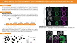 科学海报A Reliable, Efficient, and Matrix-Free Method to Generate Midbrain Organoids from Human Pluripotent Stem Cells
 沪公网安备31010102008431号