S. Hasgur et al. (jul 2022)
American journal of transplantation : official journal of the American Society of Transplantation and the American Society of Transplant Surgeons 22 7 1779--1790
Macrophage-inducible C-type lectin activates B cells to promote T cell reconstitution in heart allograft recipients.
Diminishing homeostatic proliferation of memory T cells is essential for improving the efficacy of lymphoablation in transplant recipients. Our previous studies in a mouse heart transplantation model established that B lymphocytes secreting proinflammatory cytokines are critical for T cell recovery after lymphoablation. The goal of the current study was to identify mediators of B cell activation following lymphoablation in allograft recipients. Transcriptome analysis revealed that macrophage-inducible C-type lectin (Mincle,Clec4e) expression is up-regulated in B cells from heart allograft recipients treated with murine anti-thymocyte globulin (mATG). Recipient Mincle deficiency diminishes B cell production of pro-inflammatory cytokines and impairs T lymphocyte reconstitution. Mixed bone marrow chimeras lacking Mincle only in B lymphocytes have similar defects in T cell recovery. Conversely,treatment with a synthetic Mincle ligand enhances T cell reconstitution after lymphoablation in non-transplanted mice. Treatment with agonistic CD40 mAb facilitates T cell reconstitution in CD4 T cell-depleted,but not in Mincle-deficient,recipients indicating that CD40 signaling induces T cell proliferation via a Mincle-dependent pathway. These findings are the first to identify an important function of B cell Mincle as a sensor of damage-associated molecular patterns released by the graft and demonstrate its role in clinically relevant settings of organ transplantation.
View Publication
产品类型:
产品号#:
18000
产品名:
EasySep™磁极
(Mar 2025)
Cancers 17 6
Effects of Induced Pluripotent Stem Cell-Derived Astrocytes on Cisplatin Sensitivity in Pediatric Brain Cancer Cells
Simple SummaryAtypical teratoid rhabdoid tumors (ATRTs) and diffuse intrinsic pontine gliomas (DIPGs) are lethal pediatric brain tumors that can resist chemotherapy and be influenced by their microenvironment. Astrocytes are the key components of the brain tumor microenvironment and can support tumor growth. We investigated the effects of astrocytes on cisplatin sensitivity in pediatric brain cancer cells. The crosstalk between astrocytes and cancer cells activated astrocytes and promoted cancer cell proliferation. Moreover,the tumor cells expressed elevated levels of drug resistance genes in the presence of astrocytes. In conclusion,astrocytes can significantly improve the growth of these tumor cells and modulate their chemosensitivity,highlighting their role in therapeutic resistance. AbstractBackground: ATRTs and DIPGs are deadly pediatric brain tumors with poor prognosis. These tumors can develop resistance to chemotherapies,which may be significantly influenced by their microenvironment. Since astrocytes are the most abundant glial cell type in the brain microenvironment and may support tumor growth and chemoresistance,this study investigated the effects of induced pluripotent stem cell-derived astrocytes (iPSC-astrocytes) on cisplatin sensitivity in CHLA-05-ATRT and SF8628 (DIPG) cells. iPSCs provide an unlimited and standardized source of nascent astrocytes,which enables modeling the interaction between childhood brain tumor cells and iPSC-astrocytes within a controlled coculture system. Methods: To study the effects on tumor growth,the iPSC-astrocytes were cocultured with tumor cells. Additionally,the tumor cells were exposed to various concentrations of cisplatin to evaluate their chemosensitivity in the presence of astrocytes. Results: The paracrine interaction of iPSC-astrocytes with tumor cells upregulated astrocyte activation markers GFAP and STAT3 and promoted tumor cell proliferation. Moreover,the cisplatin treatment significantly decreased the viability of CHLA-05-ATRT and SF8628 cells. However,tumor cells exhibited reduced sensitivity to cisplatin in the coculture with iPSC-astrocytes. During cisplatin treatment,DIPG cells in particular showed upregulation of resistance markers,ERK1,STAT3,and MTDH,which are associated with enhanced proliferation and invasion. They also had increased expression of APEX1,which is involved in the base excision repair pathway following cisplatin-induced DNA damage. Conclusion: These findings underscore the significance of the tumor microenvironment in modulating tumor cell survival and chemosensitivity.
View Publication
产品类型:
产品号#:
100-0276
100-1130
产品名:
mTeSR™ Plus
mTeSR™ Plus
(Jun 2025)
PLOS Biology 23 6
Multinucleated giant cells are hallmarks of ovarian aging with unique immune and degradation-associated molecular signatures
The ovary is one of the first organs to exhibit signs of aging,characterized by reduced tissue function,chronic inflammation,and fibrosis. Multinucleated giant cells (MNGCs),formed by macrophage fusion,typically occur in chronic immune pathologies,including infectious and non-infectious granulomas and the foreign body response,but are also observed in the aging ovary. The function and consequence of ovarian MNGCs remain unknown as their biological activity is highly context-dependent,and their large size has limited their isolation and analysis through technologies such as single-cell RNA sequencing. In this study,we define ovarian MNGCs through a deep analysis of their presence across age and species using advanced imaging technologies as well as their unique transcriptome using laser capture microdissection. MNGCs form complex interconnected networks that increase with age in both mouse and nonhuman primate ovaries. MNGCs are characterized by high Gpnmb expression,a putative marker of ovarian and non-ovarian MNGCs. Pathway analysis highlighted functions in apoptotic cell clearance,lipid metabolism,proteolysis,immune processes,and increased oxidative phosphorylation and antioxidant activity. Thus,MNGCs have signatures related to degradative processes,immune function,and high metabolic activity. These processes were enriched in MNGCs compared to primary ovarian macrophages,suggesting discrete functionality. MNGCs express CD4 and colocalize with T-cells,which were enriched in regions of MNGCs,indicative of a close interaction between these immune cell types. These findings implicate MNGCs in modulation of the ovarian immune landscape during aging given their high penetrance and unique molecular signature that supports degradative and immune functions. Ovarian multinucleated giant cells are a unique macrophage population that arise within the aging mammalian ovary. This study characterizes their transcriptome in mice,uncovering a potential role in degradation of cellular debris and immune signaling,suggesting a potential contribution to ovarian inflammation during aging.
View Publication
产品类型:
产品号#:
20144
100-0659
产品名:
EasySep™缓冲液
EasySep™ 小鼠F4/80正选试剂盒
S. L. Calzi et al. (Aug 2025)
Cells 14 17
Targeting Diabetic Retinopathy with Human iPSC-Derived Vascular Reparative Cells in a Type 2 Diabetes Model
Purpose: To investigate the therapeutic potential of inducible pluripotent stem cell (hiPSC)-based vascular repair,we evaluated two vascular reparative cell populations,CD34+ cells derived from hiPSC (hiPSC-CD34+) and endothelial colony forming cells (ECFCs) derived from hiPSC (iPS-ECFCs),alone and in combination,in a type 2 diabetic (db/db) mouse model of DR. Methods: hiPSC-CD34+ cells (1 × 10⁴) or iPSC- ECFCs (1 × 10⁵) alone or in combination (1.1 × 10⁵) were injected into the vitreous of immunosuppressed db/db mice with six months of established diabetes. One month post-injection,mice underwent electroretinography (ERG) and optical coherence tomography (OCT) to evaluate functional and structural retinal recovery with iPSC administration. Immunohistochemistry (IHC) was used to assess recruitment and incorporation of cells into the retinal vasculature. Retinas from the experimental groups were analyzed using Functional Proteomics via Reverse Phase Protein Array (RPPA). Results: Functional assessment via ERG demonstrated significant improvements in retinal response in the diabetic cohorts treated with either hiPSC-derived CD34+ cells or hiPSC-ECFCs. Retinal thickness,assessed by OCT,was restored to near-nondiabetic levels in mice treated with hiPSC-CD34+ cells alone and the combination group,whereas hiPSC-ECFCs alone did not significantly affect retinal thickness. One month following intravitreal injection,hiPSC-CD34+ cells were localized to perivascular regions,whereas hiPSC-ECFCs were observed to integrate directly into the retinal vasculature. RPPA analysis revealed interaction-significant changes,and this was interpreted as a combination-specific,non-additive host responses (m6A,PI3K–AKT–mTOR,glycolysis,endothelial junction pathways). Conclusions: The studies support that injection of hiPSC-CD34+ cells and hiPSC-ECFCs,both individually and in combination,showed benefit; however,iPSC combination-specific effects were identified by measurement of retinal thickness and by RPPA.
View Publication
产品类型:
产品号#:
100-1569
17856
17856RF
产品名:
EasySep™人CD34正选试剂盒 II
EasySep™人CD34正选试剂盒 II
EasySep™人CD34正选试剂盒 II
Y. Numata et al. (May 2025)
Cell Death & Disease 16 1
Digoxin promotes anoikis of circulating cancer cells by targeting Na + /K + -ATPase α3-isoform
Circulating cancer cells (CCCs) are closely related to the process of distant metastasis. In early step of the metastasis cascade,CCCs must evade the detachment-induced cell death (anoikis) for their survival. Here,we examined whether Na + /K + -ATPase α3-isoform (α3NaK) in CCCs contributes to avoidance of anoikis. In CCCs isolated from gastric cancer patients,α3NaK was predominantly localized in the plasma membrane (PM),but it moved to the cytoplasm when the CCCs were attached to culture dishes. The CCCs showed significant expression of integrin α5 but not fibronectin,one of components of the extracellular matrix (ECM). In human gastric cancer MKN45 cells,digoxin (20 and 50 nM),a cardiac glycoside,significantly inhibited the enzyme activity and translocation (from cytoplasm to PM) of α3NaK,while they had no significant effect on ubiquitous Na + /K + -ATPase α1-isoform (α1NaK) in the PM. The translocation of α3NaK required the loss of ECM components from the cells. Additionally,digoxin significantly enhanced caspase 3/7 activity,as well as the expression of cleaved caspase 3,while reducing the viability of detached (floating) cells. In the MKN45 xenograft mouse model,intraperitoneal administration of digoxin (2 mg/kg/day) significantly decreased the number of CCCs and suppressed their liver metastasis. Our results suggest that α3NaK plays an essential role in the survival of CCCs in gastric cancer,and that digoxin enhances anoikis in detached (metastatic) gastric cancer cells by inhibiting the α3NaK translocation from cytoplasm to PM,thereby reducing CCCs. Targeting α3NaK may be a promising therapeutic strategy against CCC survival. Subject terms: Metastasis,Gastric cancer,Apoptosis
View Publication
Prenatal and postnatal myeloid cells demonstrate stepwise progression in the pathogenesis of MLL fusion gene leukemia.
The steps to leukemia following an in utero fusion of MLL (HRX,ALL-1) to a partner gene in humans are not known. Introduction of the Mll-AF9 fusion gene into embryonic stem cells results in leukemia in mice with cell-type specificity similar to humans. In this study we used myeloid colony assays,immunophenotyping,and transplantation to evaluate myelopoiesis in Mll-AF9 mice. Colony assays demonstrated that both prenatal and postnatal Mll-AF9 tissues have significantly increased numbers of CD11b(+)/CD117(+)/Gr-1(+/-) myeloid cells,often in compact clusters. The self-renewal capacity of prenatal myeloid progenitors was found to decrease following serial replating of colony-forming cells. In contrast,early postnatal myeloid progenitors increased following replating; however,the enhanced self-renewal of early postnatal myeloid progenitor cells was limited and did not result in long-term cell lines or leukemia in vivo. Unlimited replating,long-term CD11b/Gr-1(+) myeloid cell lines,and the ability to produce early leukemia in vivo in transplantation experiments,were found only in mice with overt leukemia. Prenatal Mll-AF9 tissues had reduced total (mature and progenitor) CD11b/Gr-1(+) cells compared with wild-type tissues. Colony replating,immunophenotyping,and cytochemistry suggest that any perturbation of cellular differentiation from the prenatal stage onward is partial and largely reversible. We describe a novel informative in vitro and in vivo model system that permits study of the stages in the pathogenesis of Mll fusion gene leukemia,beginning in prenatal myeloid cells,progressing to a second stage in the postnatal period and,finally,resulting in overt leukemia in adult animals.
View Publication
产品类型:
产品号#:
03534
产品名:
MethoCult™GF M3534
Abadier M et al. (DEC 2017)
Cell reports 21 13 3885--3899
Effector and Regulatory T Cells Roll at High Shear Stress by Inducible Tether and Sling Formation.
The adaptive immune response involves T cell differentiation and migration to sites of inflammation. T cell trafficking is initiated by rolling on inflamed endothelium. Tethers and slings,discovered in neutrophils,facilitate cell rolling at high shear stress. Here,we demonstrate that the ability to form tethers and slings during rolling is highly inducible in T helper 1 (Th1),Th17,and regulatory T (Treg) cells but less in Th2 cells. In vivo,endogenous Treg cells rolled stably in cremaster venules at physiological shear stress. Quantitative dynamic footprinting nanoscopy of Th1,Th17,and Treg cells uncovered the formation of multiple tethers per cell. Human Th1 cells also showed tethers and slings. RNA sequencing (RNA-seq) revealed the induction of cell migration and cytoskeletal genes in sling-forming cells. We conclude that differentiated CD4 T cells stabilize rolling by inducible tether and sling formation. These phenotypic changes approximate the adhesion phenotype of neutrophils and support CD4 T cell access to sites of inflammation.
View Publication
产品类型:
产品号#:
19762
19762RF
产品名:
EasySep™小鼠中性粒细胞富集试剂盒
RoboSep™ 小鼠中性粒细胞富集试剂盒含滤芯吸头
Zhou S et al. ( 2017)
PloS one 12 1 e0169899
Reprogramming Malignant Cancer Cells toward a Benign Phenotype following Exposure to Human Embryonic Stem Cell Microenvironment.
The embryonic microenvironment is well known to be non-permissive for tumor development because early developmental signals naturally suppress the expression of proto-oncogenes. In an analogous manner,mimicking an early embryonic environment during embryonic stem cell culture has been shown to suppress oncogenic phenotypes of cancer cells. Exosomes derived from human embryonic stem cells harbor substances that mirror the content of the cells of origin and have been reported to reprogram hematopoietic stem/progenitor cells via horizontal transfer of mRNA and proteins. However,the possibility that these embryonic stem cells-derived exosomes might be the main effectors of the anti-tumor effect mediated by the embryonic stem cells has not been explored yet. The present study aims to investigate whether exosomes derived from human embryonic stem cells can reprogram malignant cancer cells to a benign stage and reduce their tumorigenicity. We show that the embryonic stem cell-conditioned medium contains factors that inhibit cancer cell growth and tumorigenicity in vitro and in vivo. Moreover,we demonstrate that exosomes derived from human embryonic stem cells display anti-proliferation and pro-apoptotic effects,and decrease tumor size in a xenograft model. These exosomes are also able to transfer their cargo into target cancer cells,inducing a dose-dependent increase in SOX2,OCT4 and Nanog proteins,leading to a dose-dependent decrease of cancer cell growth and tumorigenicity. This study shows for the first time that human embryonic stem cell-derived exosomes play an important role in the tumor suppressive activity displayed by human embryonic stem cells.
View Publication
Y.-H. Chang et al. ( 2017)
Immunity 47 5 943--958.e9
Dichotomous Expression of TNF Superfamily Ligands on Antigen-Presenting Cells Controls Post-priming Anti-viral CD4+ T Cell Immunity.
T cell antigen-presenting cell (APC) interactions early during chronic viral infection are crucial for determining viral set point and disease outcome,but how and when different APC subtypes contribute to these outcomes is unclear. The TNF receptor superfamily (TNFRSF) member GITR is important for CD4+ T cell accumulation and control of chronic lymphocytic choriomeningitis virus (LCMV). We found that type I interferon (IFN-I) induced TNFSF ligands GITRL,4-1BBL,OX40L,and CD70 predominantly on monocyte-derived APCs and CD80 and CD86 predominantly on classical dendritic cells (cDCs). Mice with hypofunctional GITRL in Lyz2+ cells had decreased LCMV-specific CD4+ T cell accumulation and increased viral load. GITR signals in CD4+ T cells occurred after priming to upregulate OX40,CD25,and chemokine receptor CX3CR1. Thus IFN-I (signal 3) induced a post-priming checkpoint (signal 4) for CD4+ T cell accumulation,revealing a division of labor between cDCs and monocyte-derived APCs in regulating T cell expansion.
View Publication
产品类型:
产品号#:
19765
19765RF
19852
19852RF
产品名:
EasySep™小鼠Naïve CD4+ T细胞分选试剂盒
RoboSep™ 小鼠Naïve CD4+ T细胞分选试剂盒
EasySep™小鼠CD4+ T细胞分选试剂盒
RoboSep™ 小鼠CD4+ T细胞分选试剂盒
Matsumura-Takeda K et al. (APR 2007)
Stem cells (Dayton,Ohio) 25 4 862--70
CD41+/CD45+ cells without acetylcholinesterase activity are immature and a major megakaryocytic population in murine bone marrow.
Murine megakaryocytes (MKs) are defined by CD41/CD61 expression and acetylcholinesterase (AChE) activity; however,their stages of differentiation in bone marrow (BM) have not been fully elucidated. In murine lineage-negative (Lin(-))/CD45(+) BM cells,we found CD41(+) MKs without AChE activity (AChE(-)) except for CD41(++) MKs with AChE activity (AChE(+)),in which CD61 expression was similar to their CD41 level. Lin(-)/CD41(+)/CD45(+)/AChE(-) MKs could differentiate into AChE(+),with an accompanying increase in CD41/CD61 during in vitro culture. Both proplatelet formation (PPF) and platelet (PLT) production for Lin(-)/CD41(+)/CD45(+)/AChE(-) MKs were observed later than for Lin(-)/CD41(++)/CD45(+)/AChE(+) MKs,whereas MK progenitors were scarcely detected in both subpopulations. GeneChip and semiquantitative polymerase chain reaction analyses revealed that the Lin(-)/CD41(+)/CD45(+)/AChE(-) MKs are assigned at the stage between the progenitor and PPF preparation phases in respect to the many MK/PLT-specific gene expressions,including beta1-tubulin. In normal mice,the number of Lin(-)/CD41(+)/CD45(+)/AChE(-) MKs was 100 times higher than that of AChE(+) MKs in BM. When MK destruction and consequent thrombocytopenia were caused by an antitumor agent,mitomycin-C,Lin(-)/CD41(+)/CD45(+)/AChE(-) MKs led to an increase in AChE(+) MKs and subsequent PLT recovery with interleukin-11 administration. It was concluded that MKs in murine BM at least in part consist of immature Lin(-)/CD41(+)/CD45(+)/AChE(-) MKs and more differentiated Lin(-)/CD41(++)/CD45(+)/AChE(+) MKs. Immature Lin(-)/CD41(+)/CD45(+)/AChE(-) MKs are a major MK population compared with AChE(+) MKs in BM and play an important role in rapid PLT recovery in vivo.
View Publication

 EasySep™小鼠TIL(CD45)正选试剂盒


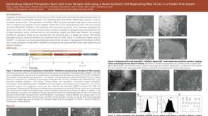 科学海报Generating iPSCs from Somatic Cells using a Novel Synthetic Self-Replicating RNA Vector in a Feeder-Free System
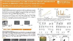 科学海报A Feeder-Independent Culture System to Convert and Maintain Human Pluripotent Stem Cells in a Naïve-Like State
 沪公网安备31010102008431号