Chagraoui J et al. (APR 2003)
Blood 101 8 2973--82
Fetal liver stroma consists of cells in epithelial-to-mesenchymal transition.
Liver becomes the predominant site of hematopoiesis by 11.5 dpc (days after coitus) in the mouse and 15 gestational weeks in humans and stays so until the end of gestation. The reason the liver is the major hematopoietic site during fetal life is not clear. In this work,we tried to define which of the fetal liver microenvironmental cell populations would be associated with the development of hematopoiesis and found that a population of cells with mixed endodermal and mesodermal features corresponded to hematopoietic-supportive fetal liver stroma. Stromal cells generated from primary cultures or stromal lines from mouse or human fetal liver in the hematopoietic florid phase expressed both mesenchymal markers (vimentin,osteopontin,collagen I,alpha smooth muscle actin,thrombospondin-1,EDa fibronectin,calponin,Stro-1 antigens,myocyte-enhancer factor 2C) and epithelial (alpha-fetoprotein,cytokeratins 8 and 18,albumin,E-cadherin,hepatocyte nuclear factor 3 alpha) markers. Such a cell population fits with the description of cells in epithelial-to-mesenchymal transition (EMT),often observed during development,including that of the liver. The hematopoietic supportive capacity of EMT cells was lost after hepatocytic maturation,induced by oncostatin M in the cell line AFT024. EMT cells were observed in the fetal liver microenvironment during the hematopoietic phase but not in nonhematopoietic liver by the end of gestation and in the adult. EMT cells represent a novel stromal cell type that may be generated from hepatic endodermal or mesenchymal stem cells or even from circulating hematopoietic stem cells (HSCs) seeding the liver rudiment.
View Publication
产品类型:
产品号#:
05150
产品名:
MyeloCult™H5100
Kim G-H et al. ( 2014)
Angewandte Chemie (International ed. in English) 53 35 9271--9274
Imidazole-based small molecules that promote neurogenesis in pluripotent cells.
Reported herein are two imidazole-based small molecules,termed neurodazine (Nz) and neurodazole (Nzl),which induce neuronal differentiation of pluripotent P19 cells. Their ability to induce neurogenesis of P19 cells is comparable to that of retinoic acid. However,Nz and Nzl were found to be more selective neurogenesis inducers than retinoic acid owing to their unique ability to suppress astrocyte differentiation of P19 cells. Our results also show that Nz and Nzl promote production of physiologically active neurons because P19-cell-derived neurons induced by these substances have functional glutamate responsiveness. The present study suggests that Nz and Nzl could serve as important chemical tools to induce formation of specific populations of neuronal cell types from pluripotent cells.
View Publication
产品类型:
产品号#:
73292
产品名:
Neurodazine
Viale A et al. (OCT 2014)
Nature 514 7524 628--632
Oncogene ablation-resistant pancreatic cancer cells depend on mitochondrial function.
Pancreatic ductal adenocarcinoma (PDAC) is one of the deadliest cancers in western countries,with a median survival of 6 months and an extremely low percentage of long-term surviving patients. KRAS mutations are known to be a driver event of PDAC,but targeting mutant KRAS has proved challenging. Targeting oncogene-driven signalling pathways is a clinically validated approach for several devastating diseases. Still,despite marked tumour shrinkage,the frequency of relapse indicates that a fraction of tumour cells survives shut down of oncogenic signalling. Here we explore the role of mutant KRAS in PDAC maintenance using a recently developed inducible mouse model of mutated Kras (Kras(G12D),herein KRas) in a p53(LoxP/WT) background. We demonstrate that a subpopulation of dormant tumour cells surviving oncogene ablation (surviving cells) and responsible for tumour relapse has features of cancer stem cells and relies on oxidative phosphorylation for survival. Transcriptomic and metabolic analyses of surviving cells reveal prominent expression of genes governing mitochondrial function,autophagy and lysosome activity,as well as a strong reliance on mitochondrial respiration and a decreased dependence on glycolysis for cellular energetics. Accordingly,surviving cells show high sensitivity to oxidative phosphorylation inhibitors,which can inhibit tumour recurrence. Our integrated analyses illuminate a therapeutic strategy of combined targeting of the KRAS pathway and mitochondrial respiration to manage pancreatic cancer.
View Publication
产品类型:
产品号#:
01700
01705
01702
产品名:
ALDEFLUOR™ 试剂盒
ALDEFLUOR™ DEAB试剂
ALDEFLUOR™测定缓冲液
Naujok O et al. ( 2015)
1341 67--85
Gene transfer into pluripotent stem cells via lentiviral transduction
Recombinant lentiviral vectors are powerful tools to stably manipulate human pluripotent stem cells. They can be used to deliver ectopic genes,shRNAs,miRNAs,or any possible genetic DNA sequence into diving and nondividing cells. Here we describe a general protocol for the production of self-inactivating lentiviral vector particles and their purification to high titers by either ultracentrifugation or ultrafiltration. Next we provide a basic procedure to transduce human pluripotent stem cells and propagate clonal cell lines.
View Publication
产品类型:
产品号#:
07923
85850
85857
产品名:
Dispase (1 U/mL)
mTeSR™1
mTeSR™1
Kerosuo L et al. (DEC 2008)
Journal of cell science 121 Pt 23 3941--50
Myc increases self-renewal in neural progenitor cells through Miz-1.
The mechanisms underlying the decision of a stem or progenitor cell to either self-renew or differentiate are incompletely understood. To address the role of Myc in this process,we expressed different forms of the proto-oncogene Myc in multipotent neural progenitor cells (NPCs) using retroviral transduction. Expression of Myc in neurospheres increased the proportion of self-renewing cells fivefold,and 1% of the Myc-overexpressing cells,but none of the control cells,retained self-renewal capacity even under differentiation-inducing conditions. A Myc mutant (MycV394D) deficient in binding to Miz-1,did not increase the percentage of self-renewing cells but was able to stimulate proliferation of NPCs as efficiently as wild-type Myc,indicating that these two cellular phenomena are regulated by at least partially different pathways. Our results suggest that Myc,through Miz-1,enhances self-renewal of NPCs and influences the way progenitor cells react to the environmental cues that normally dictate the cellular identity of tissues containing self-renewing cells.
View Publication
产品类型:
产品号#:
05707
产品名:
NeuroCult™化学解离试剂盒(小鼠)
Zhong B et al. (MAY 2011)
Stem cells and development 20 5 795--807
Efficient generation of nonhuman primate induced pluripotent stem cells.
Induced pluripotent stem (iPS) cells have great potential for regenerative medicine and gene therapy. Thus far,iPS cells have typically been generated using integrating viral vectors expressing various reprogramming transcription factors; nonintegrating methods have been less effective and efficient. Because there is a significant risk of malignant transformation and cancer involved with the use of iPS cells,careful evaluation of transplanted iPS cells will be necessary in small and large animal studies before clinical application. Here,we have generated and characterized nonhuman primate iPS cells with the goal of evaluating iPS cell transplantation in a clinically relevant large animal model. We developed stable Phoenix-RD114-based packaging cell lines that produce OCT4,SOX2,c-MYC,and KLF4 (OSCK) expressing gammaretroviral vectors. Using these vectors in combination with small molecules,we were able to efficiently and reproducibly generate nonhuman primate iPS cells from pigtailed macaques (Macaca nemestrina). The established nonhuman primate iPS cells exhibited pluripotency and extensive self-renewal capacity. The facile and reproducible generation of nonhuman primate iPS cells using defined producer cells as a source of individual reprogramming factors should provide an important resource to optimize and evaluate iPS cell technology for studies involving stem cell biology and regenerative medicine.
View Publication
产品类型:
产品号#:
27100
27150
85850
85857
产品名:
35 mm培养皿
35 mm培养皿
mTeSR™1
mTeSR™1
Saito T et al. (JUL 2013)
PLoS ONE 8 7 e70010
Metformin, a Diabetes Drug, Eliminates Tumor-Initiating Hepatocellular Carcinoma Cells
Metformin has been widely used as an oral drug for diabetes mellitus for approximately 60 years. Interestingly,recent reports showed that metformin exhibited an anti-tumor action in a wide range of malignancies including hepatocellular carcinoma (HCC). In the present study,we investigated its impact on tumor-initiating HCC cells. Metformin suppressed cell growth and induced apoptosis in a dose-dependent manner. Flow cytometric analysis showed that metformin treatment markedly reduced the number of tumor-initiating epithelial cell adhesion molecule (EpCAM)(+) HCC cells. Non-adherent sphere formation assays of EpCAM(+) cells showed that metformin impaired not only their sphere-forming ability,but also their self-renewal capability. Consistent with this,immunostaining of spheres revealed that metformin significantly decreased the number of component cells positive for hepatic stem cell markers such as EpCAM and α-fetoprotein. In a xenograft transplantation model using non-obese diabetic/severe combined immunodeficient mice,metformin and/or sorafenib treatment suppressed the growth of tumors derived from transplanted HCC cells. Notably,the administration of metformin but not sorafenib decreased the number of EpCAM(+) cells and impaired their self-renewal capability. As reported,metformin activated AMP-activated protein kinase (AMPK) through phosphorylation; however its inhibitory effect on the mammalian target of rapamycin (mTOR) pathway did not necessarily correlate with its anti-tumor activity toward EpCAM(+) tumor-initiating HCC cells. These results indicate that metformin is a promising therapeutic agent for the elimination of tumor-initiating HCC cells and suggest as-yet-unknown functions other than its inhibitory effect on the AMPK/mTOR pathway.
View Publication
Keller KC et al. (MAR 2016)
Stem Cells and Development 25 13 scd.2015.0367
Wnt5a Supports Osteogenic Lineage Decisions in Embryonic Stem Cells
The specification of pluripotent stem cells into the bone-forming osteoblasts has been explored in a number of studies. However,the current body of literature has yet to adequately address the role of Wnt glycoproteins in the differentiation of pluripotent stem cells along the osteogenic lineage. During mouse embryonic stem cell (ESC) in vitro osteogenesis,the non-canonical WNT5a is expressed early on. Cells either sorted by their positive WNT5a expression or when supplemented with recombinant WNT5a (rWNT5a) during a two-day window showed significantly enhanced osteogenic yield. Mechanistically,rWNT5a supplementation up-regulated PKC,CamKII and JNK activity while antagonizing the key effector of canonical Wnt signaling: beta-catenin. Conversely,when recombinant WNT3a (rWNT3a) or other positive regulators of �?�-catenin were employed during this same time-window there was a decrease in osteogenic marker expression. However,if rWNT3a was supplemented during a time-window following rWNT5a treatment,osteogenic differentiation was enhanced both in murine and human ESCs. Elucidating the role of these WNT ligands in directing the early stages of osteogenesis has the potential to considerably improve tissue engineering protocols and applications for regenerative medicine.
View Publication
X. Xia et al. (Nov 2025)
Cell Communication and Signaling : CCS 23 10
Netrin-1-UNC5B/neogenin axis enhances the stemness of colorectal cancer cells
Cancer stem cells were prominent responsible for cancer initiation,metastasis,and invasion as well as therapeutic resistance in colorectal cancer (CRC). The extracellular axon guidance factor netrin-1 has been found to be overexpressed in several malignant cancers such as glioma,lung cancers,and colorectal cancer. However,the role of netrin-1 on cancer stemness in CRC remains unveiled. Our study revealed high expression of netrin-1 in colorectal cancer tissues and its ability to promote cancer stemness by interacting with receptors UNC5B and neogenin on murine colorectal cancer cell. Mechanistically,the netrin-1-UNC5B/neogenin axis activates the downstream NF-κB and ERK1/2 signaling pathways,reinforcing the stemness properties of tumor cells,and further exacerbating tumor progression. Clinically,netrin-1 expression associated with poor survival and high CD133 expression in patients with CRC. Taken together,these results suggest that netrin-1 blockade could be a compelling therapeutic strategy to improve the poor outcomes and trigger cancer stemness inhibition in CRC treatment.
View Publication
产品类型:
产品号#:
01700
产品名:
ALDEFLUOR™ 试剂盒
(Jun 2025)
Nature Communications 16 Suppl 2
Iron deficiency causes aspartate-sensitive dysfunction in CD8+ T cells
Iron is an irreplaceable co-factor for metabolism. Iron deficiency affects >1 billion people and decreased iron availability impairs immunity. Nevertheless,how iron deprivation impacts immune cell function remains poorly characterised. We interrogate how physiologically low iron availability affects CD8+ T cell metabolism and function,using multi-omic and metabolic labelling approaches. Iron limitation does not substantially alter initial post-activation increases in cell size and CD25 upregulation. However,low iron profoundly stalls proliferation (without influencing cell viability),alters histone methylation status,gene expression,and disrupts mitochondrial membrane potential. Glucose and glutamine metabolism in the TCA cycle is limited and partially reverses to a reductive trajectory. Previous studies identified mitochondria-derived aspartate as crucial for proliferation of transformed cells. Despite aberrant TCA cycling,aspartate is increased in stalled iron deficient CD8+ T cells but is not utilised for nucleotide synthesis,likely due to trapping within depolarised mitochondria. Exogenous aspartate markedly rescues expansion and some functions of severely iron-deficient CD8+ T cells. Overall,iron scarcity creates a mitochondrial-located metabolic bottleneck,which is bypassed by supplying inhibited biochemical processes with aspartate. These findings reveal molecular consequences of iron deficiency for CD8+ T cell function,providing mechanistic insight into the basis for immune impairment during iron deficiency. Iron has been shown to be necessary for the activation and differentiation of CD8+ T cells. Here the authors investigate changes in CD8+ T cell metabolism in iron limiting conditions and find that aspartate is increased yet downstream nucleotide synthesis is suppressed and addition of exogenous aspartate partially rescues T cell function.
View Publication

 EasySep™小鼠TIL(CD45)正选试剂盒


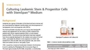 技术公告Culturing Leukemic Stem & Progenitor Cells with StemSpan™ Medium

 沪公网安备31010102008431号