Akatsuka A et al. (SEP 2010)
International immunology 22 9 783--90
Tumor cells of non-hematopoietic and hematopoietic origins express activation-induced C-type lectin, the ligand for killer cell lectin-like receptor F1.
Killer cell lectin-like receptor F1 (KLRF1) is an activating C-type lectin-like receptor expressed on human NK cells and subsets of T cells. In this study,we show that activation-induced C-type lectin (AICL) is a unique KLRF1 ligand expressed on tumor cell lines of hematopoietic and non-hematopoietic origins. We screened a panel of human tumor cell lines using the KLRF1 reporter cells and found that several tumor lines expressed KLRF1 ligands. We characterized a putative KLRF1 ligand expressed on the U937 cell line. The molecular mass for the deglycosylated ligand was 28 kDa under non-reducing condition and 17 kDa under reducing condition,suggesting that the KLRF1 ligand is a homodimer. By expression cloning from a U937 cDNA library,we identified AICL as a KLRF1 ligand. We generated mAbs against AICL to identify the KLRF1 ligands on non-hematopoietic tumor lines. The anti-AICL mAbs stained the tumor lines that express the KLRF1 ligands and importantly the interaction of KLRF1 with the KLRF1 ligand on non-hematopoietic tumors was completely blocked by the two anti-AICL mAbs. Moreover,NK cell degranulation triggered by AICL-expressing targets was partially inhibited by the anti-AICL mAb. Finally,we demonstrate that AICL is expressed in human primary liver cancers. These results suggest that AICL is expressed on tumor cells of non-hematopoietic origins and raise the possibility that AICL may contribute to NK cell surveillance of tumor cells.
View Publication
Forward RNAi screens in primary human hematopoietic stem/progenitor cells.
The mechanisms regulating key fate decisions such as self-renewal and differentiation in hematopoietic stem and progenitor cells (HSPC) remain poorly understood. We report here a screening strategy developed to assess modulators of human hematopoiesis using a lentiviral short hairpin RNA (shRNA) library transduced into cord blood-derived stem/progenitor cells. To screen for modifiers of self-renewal/differentiation,we used the limited persistence of HSPCs under ex vivo culture conditions as a baseline for functional selection of shRNAs conferring enhanced maintenance or expansion of the stem/progenitor potential. This approach enables complex,pooled screens in large numbers of cells. Functional selection identified novel specific gene targets (exostoses 1) or shRNA constructs capable of altering human hematopoietic progenitor differentiation or stem cell expansion,respectively,thereby demonstrating the potential of this forward screening approach in primary human stem cell populations.
View Publication
Enhanced fetal hemoglobin production via dual-beneficial mutation editing of the HBG promoter in hematopoietic stem and progenitor cells for β-hemoglobinopathies
BackgroundSickle cell disease (SCD) and β-thalassemia patients with elevated gamma globin (HBG1/G2) levels exhibit mild or no symptoms. To recapitulate this natural phenomenon,the most coveted gene therapy approach is to edit the regulatory sequences of HBG1/G2 to reactivate them. By editing more than one regulatory sequence in the HBG promoter,the production of fetal hemoglobin (HbF) can be significantly increased. However,achieving this goal requires precise nucleotide conversions in hematopoietic stem and progenitor cells (HSPCs) at therapeutic efficiency,which remains a challenge.MethodsWe employed Cas9 RNP-ssODN-mediated homology-directed repair (HDR) gene editing to mimic two naturally occurring HBG promoter point mutations; -175T > C,associated with high HbF levels,and −158 C > T,a common polymorphism in the Indian population that induces HbF under erythropoietic stress,in HSPCs.ResultsAsymmetric,nontarget ssODN induced high rates of complete HDR conversions,with at least 15% of HSPCs exhibiting both the −175T > C and −158 C > T mutations. Optimized conditions and treatment with the small molecule AZD-7648 increased this rate,with up to 57% of long-term engrafting human HSPCs in NBSGW mice containing at least one beneficial mutation. Functionally,in vivo erythroblasts exhibited high levels of HbF,which was sufficient to reverse the cellular phenotype of β-thalassemia. Further support through bone marrow MSC co-culture boosted complete HDR conversion rates to exceed 80%,with minimal InDels,improved cell viability,and induced fetal hemoglobin levels similar to those of Cas9 RNP-mediated indels at BCL11A enhancer and HBG promoter.ConclusionsCas9 RNP-ssODN-based nucleotide conversion at the HBG promoter offers a promising gene therapy approach to ameliorate the phenotypes of β-thalassemia and SCD. The developed approach can simplify and broaden applications that require the cointroduction of multiple nucleotide modifications in HSPCs.Supplementary InformationThe online version contains supplementary material available at 10.1186/s13287-024-04117-0.
View Publication
产品类型:
产品号#:
09600
09605
09650
09655
17856
17856RF
100-1569
产品名:
StemSpan™ SFEM
StemSpan™ SFEM II
StemSpan™ SFEM
StemSpan™ SFEM II
EasySep™人CD34正选试剂盒 II
EasySep™人CD34正选试剂盒 II
EasySep™人CD34正选试剂盒 II
Rebel VI et al. (JAN 1994)
Blood 83 1 128--36
Amplification of Sca-1+ Lin- WGA+ cells in serum-free cultures containing steel factor, interleukin-6, and erythropoietin with maintenance of cells with long-term in vivo reconstituting potential.
Normal murine bone marrow (BM) cells were sorted on the basis of low forward and orthogonal light scatter properties,Sca-1 expression (Sca-1+),lack of staining with a cocktail of mature hematopoietic lineage markers (Lin-),and binding of wheat germ agglutinin (WGA+). This approach allowed the reproducible isolation of a very small subpopulation (0.037% +/- 0.023% of all nucleated BM cells) that was approximately 400-fold enriched in cells capable of reconstituting both lymphoid and myeloid lineages in lethally irradiated recipients. Transplantation of 30 or 10 of these Sca-1+Lin-WGA+ cells resulted in textgreater or = to 20% donor-derived nucleated peripheral blood cells 3 months posttransplantation in 100% and 22% of the recipients,respectively. When Sca-1+Lin-WGA+ cells were cultured in serum-free medium supplemented with Steel factor,interleukin-6 (IL-6),and erythropoietin (with or without IL-3),a large increase in total cell number,including cells with an Sca-1+Lin-WGA+ phenotype was observed. Single cell cultures showed that 90% to 95% of the input cells underwent at least one division during the first 2 weeks and the remainder died. Interestingly,this proliferative response was not accompanied by a parallel increase in the number of cells with both lymphoid and myeloid repopulating potential in vivo,as quantitation of these by limiting dilution analysis showed they had decreased slightly (1.3-fold) but not significantly below the number initially present. These results demonstrate that Sca-1+Lin-WGA+ cells with long-term repopulating potential can be maintained for 2 weeks in a serum- and stroma cell-free culture,providing a simple in vitro system to study their behavior under well-defined conditions. The observed expansion of Sca-1+Lin-WGA+ cells in vitro without a concomitant increase in reconstituting cells also shows that extensive functional heterogeneity exists within populations of cells with this surface phenotype.
View Publication
Sequences within and upstream of the mouse Ets1 gene drive high level expression in B cells, but are not sufficient for consistent expression in T cells
The levels of transcription factor Ets1 are high in resting B and T cells,but are downregulated by signaling through antigen receptors and Toll-like receptors (TLRs). Loss of Ets1 in mice leads to excessive immune cell activation and development of an autoimmune syndrome and reduced Ets1 expression has been observed in human PBMCs in the context of autoimmune diseases. In B cells,Ets1 serves to prevent premature activation and differentiation to antibody-secreting cells. Given these important roles for Ets1 in the immune response,stringent control of Ets1 gene expression levels is required for homeostasis. However,the genetic regulatory elements that control expression of the Ets1 gene remain relatively unknown. Here we identify a topologically-associating domain (TAD) in the chromatin of B cells that includes the mouse Ets1 gene locus and describe an interaction hub that extends over 100 kb upstream and into the gene body. Additionally,we compile epigenetic datasets to find several putative regulatory elements within the interaction hub by identifying regions of high DNA accessibility and enrichment of active enhancer histone marks. Using reporter constructs,we determine that DNA sequences within this interaction hub are sufficient to direct reporter gene expression in lymphoid tissues of transgenic mice. Further analysis indicates that the reporter construct drives faithful expression of the reporter gene in mouse B cells,but variegated expression in T cells,suggesting the existence of T cell regulatory elements outside this region. To investigate how the downregulation of Ets1 transcription is associated with alterations in the epigenetic landscape of stimulated B cells,we performed ATAC-seq in resting and BCR-stimulated primary B cells and identified four regions within and upstream of the Ets1 locus that undergo changes in chromatin accessibility that correlate to Ets1 gene expression. Interestingly,functional analysis of several putative Ets1 regulatory elements using luciferase constructs suggested a high level of functional redundancy. Taken together our studies reveal a complex network of regulatory elements and transcription factors that coordinate the B cell-specific expression of Ets1.
View Publication
产品类型:
产品号#:
19854
19854RF
产品名:
EasySep™小鼠B细胞分选试剂盒
RoboSep™ 小鼠B细胞分选试剂盒
S. L. Rogers et al. (JUL 2006)
Journal of immunology (Baltimore,Md. : 1950) 177 1 414--21
A role for DNA hypomethylation and histone acetylation in maintaining allele-specific expression of mouse NKG2A in developing and mature NK cells.
The repertoire of receptors that is expressed by NK cells is critical for their ability to kill virally infected or transformed cells. However,the molecular mechanisms that determine whether and when NK receptor genes are transcribed during hemopoiesis remain unclear. In this study,we show that hypomethylation of a CpG-rich region in the mouse NKG2A gene is associated with transcription of NKG2A in ex vivo NK cells and NK cell lines. This observation was extended to various developmental stages of NK cells sorted from bone marrow,in which we demonstrate that the CpGs are methylated in the NKG2A-negative stages (hemopoietic stem cells,NK progenitors,and NKG2A-negative NK cells),and hypomethylated specifically in the NKG2A-positive NK cells. Furthermore,we provide evidence that DNA methylation is important in maintaining the allele-specific expression of NKG2A. Finally,we show that acetylated histones are associated with the CpG-rich region in NKG2A positive,but not negative,cell lines,and that treatment with the histone deacetylase inhibitor trichostatin A alone is sufficient to induce NKG2A expression. Treatment with the methyltransferase inhibitor 5-azacytidine only is insufficient to induce transcription,but cotreatment with both drugs resulted in a significantly greater induction,suggesting a cooperative role for DNA methylation and histone acetylation status in regulating gene expression. These results enhance our understanding of the formation and maintenance of NK receptor repertoires in developing and mature NK cells.
View Publication
A 3D sphere culture system containing functional polymers for large-scale human pluripotent stem cell production
Utilizing human pluripotent stem cells (hPSCs) in cell-based therapy and drug discovery requires large-scale cell production. However,scaling up conventional adherent cultures presents challenges of maintaining a uniform high quality at low cost. In this regard,suspension cultures are a viable alternative,because they are scalable and do not require adhesion surfaces. 3D culture systems such as bioreactors can be exploited for large-scale production. However,the limitations of current suspension culture methods include spontaneous fusion between cell aggregates and suboptimal passaging methods by dissociation and reaggregation. 3D culture systems that dynamically stir carrier beads or cell aggregates should be refined to reduce shearing forces that damage hPSCs. Here,we report a simple 3D sphere culture system that incorporates mechanical passaging and functional polymers. This setup resolves major problems associated with suspension culture methods and dynamic stirring systems and may be optimal for applications involving large-scale hPSC production. ?? 2014 The Authors.
View Publication

 EasySep™小鼠TIL(CD45)正选试剂盒


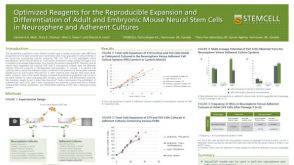 科学海报Optimized Reagents for the Reproducible Expansion and Differentiation of Adult and Embryonic Mouse Neural Stem Cells in Neurosphere and Adherent Cultures


 沪公网安备31010102008431号