J. Jahan et al. (Mar 2024)
Biochemical pharmacology 222
The role of telomerase reverse transcriptase in the mitochondrial protective functions of Angiotensin-(1–7) in diabetic CD34 + cells
Angiotensin (Ang)-(1–7) stimulates vasoprotective functions of diabetic (DB) CD34 + hematopoietic stem/progenitor cells partly by decreasing reactive oxygen species (ROS),increasing nitric oxide (NO) levels and decreasing TGFβ1 secretion. Telomerase reverse transcriptase (TERT) translocates to mitochondria and regulates ROS generation. Alternative splicing of TERT results in variants α-,β- and α-β-TERT,which may oppose functions of full-length (FL) TERT. This study tested if the protective functions of Ang-(1–7) or TGFβ1-silencing are mediated by mitoTERT and that diabetes decreases FL-TERT expression by inducing splicing. CD34 + cells were isolated from the peripheral blood mononuclear cells of nondiabetic (ND,n = 68) or DB (n = 74) subjects. NO and mitoROS levels were evaluated by flow cytometry. TERT splice variants and mitoDNA-lesions were characterized by qPCR. TRAP assay was used for telomerase activity. Decoy peptide was used to block mitochondrial translocation (mitoXTERT). TERT inhibitor or mitoXTERT prevented the effects of Ang-(1–7) on NO or mitoROS levels in DB-CD34 + cells. FL-TERT expression and telomerase activity were lower and mitoDNA-lesions were higher in DB cells compared to ND and were reversed by Ang-(1–7) or TGFβ1-silencing. The prevalence of TERT splice variants,with predominant β-TERT expression,was higher and the expression of FL-TERT was lower in DB cells (n = 25) compared to ND (n = 30). Ang-(1–7) or TGFβ1-silencing decreased TERT-splicing and increased FL-TERT. Blocking of β-splicing increased FL-TERT and protected mitoDNA in DB-cells. The findings suggest that diabetes induces TERT-splicing in CD34 + cells and that β-TERT splice variant largely contributes to the mitoDNA oxidative damage.
View Publication
产品类型:
产品号#:
09600
09650
产品名:
StemSpan™ SFEM
StemSpan™ SFEM
Bu et al. (Jul 2025)
World Journal of Gastroenterology 31 26
Paneth cells inhibit intestinal stem cell proliferation through the bone morphogenic protein 7 pathway under rotavirus-mediated intestinal injury
Rotavirus (RV),a primary cause of diarrhea-related mortality in 2021,has been shown to damage intestinal epithelial cells while upregulating intestinal stem cells (ISCs) activities. ISCs within the crypt niche drive the continuous self-renewal of intestinal epithelium,preserving its barrier functions. Paneth cells secrete antimicrobial peptide and signaling molecules within the intestine crypt,thereby playing a crucial role in intestinal immune defense and providing ISCs functional support. However,the regulatory function of Paneth cells under pathological conditions,such as RV infection,remains unclear. To determine the impact of RV infection on Paneth cells and how Paneth cells regulate ISCs during intestinal injury repair. We constructed a reference genome for the RV enteric cytopathogenic human orphan virus strain and reanalyzed published single-cell RNA sequencing data to investigate Paneth cell responses to RV-induced intestinal injury. We derived Paneth-ISC communication networks using CellChat,tracked ISC differentiation with pseudotime analysis,and validated our findings in leucine-rich repeat-containing G protein-coupled receptor 5-enhanced green fluorescent protein-internal ribosomal entry site-Cre recombinase estrogen receptor variant 2 mice and organoids via immunofluorescence,flow cytometry,and reverse transcription quantitative polymerase chain reaction. We found that RV directly infects Paneth cells,leading to a reduction in mature Paneth cells and an increase in kallikrein 1-high immature Paneth cells. Paneth-ISC communication was significantly enhanced. In particular,the bone morphogenic protein 7 (BMP7)-activin A receptor type 2B/BMP receptor type 1A-Smad pathway was upregulated post-infection,suggesting that Paneth cells suppress excessive ISC proliferation. Functional validation confirmed activation of this pathway. Paneth cells regulate ISC proliferation during RV infection by activating BMP7 signaling,limiting excessive stem cell expansion and preserving crypt homeostasis for effective epithelial repair.
View Publication
产品类型:
产品号#:
06005
产品名:
IntestiCult™ 肠道类器官生长培养基 (小鼠)
M. S. Tavangar et al. (may 2020)
Clinical and experimental dental research
Differential expression of drug resistance genes in CD146 positive dental pulp derived stem cells and CD146 negative fibroblasts.
INTRODUCTION The stem cell portion of the dental pulp derived cultures (DPSCs) showed a higher resistance to cytotoxic effect of restorative dental materials compared to pulpal fibroblasts (DPFs). Here,we aimed to compare the expression of some drug resistant genes between these cells. METHODS AND MATERIALS To separate DPSCs from DPFs,we used magnetic cell sorting technique based on CD146 expression. To assess the stem cell properties,the positive and negative portions underwent colony forming assays and were induced to be differentiated into the adipocytes,osteoblasts,hepatocytes,and neural cells. Cell surface antigen panels were checked using immune fluorescence and flow-cytometry techniques. The mRNA expression of 14 ABC transporters including ABCA2,ABCB1,ABCB11,ABCC1,ABCC2,ABCC3,ABCC4,ABCC5-2,ABCC5-4,ABCC5-13,ABCC6,ABCC10,ABCC11,and ABCG2 genes was assessed,using quantitative RT-PCR technique. RESULTS Only the CD146 positive portion could be differentiated into the desired fates,and they formed higher colonies (16.7 ± 3.32 vs. 1.7 ± 1.67,p {\textless} .001). The cell surface antigen panels were the same,except for CD146 and STRO-1 markers which were expressed only in the positive portion. Among the ABC transporter genes studied,the positive portion showed a higher expression (approximately two-fold) of ABCA2,ABCC5-13,and ABCC5-2 genes. CONCLUSION Dental pulp stem cells which can be separated from dental pulp fibroblasts based on CD146 expression,express higher levels of some drug resistance genes which probably accounts for their features of more resistance to cytotoxic effects of some dental materials. This needs to be more validated in future.
View Publication
产品类型:
产品号#:
05412
产品名:
MesenCult™ 脂肪分化试剂盒 (人)
D. Shishkova et al. (Dec 2025)
International Journal of Molecular Sciences 26 24
Palmitic but Not Oleic Acid Induces Pro-Inflammatory Dysfunction of Human Endothelial Cells from Different Vascular Beds In Vitro
Palmitic acid (PA) is the most common dietary saturated fatty acid,and is abundant in palm and cottonseed oil,butter,and cheese,whereas oleic acid (OA) is a monounsaturated omega-9 fatty acid found in olive oil. The differences in the cytotoxic and pro-inflammatory effects of PA and OA across endothelial cells (ECs) isolated from different vascular beds have not been investigated in detail. Here,we incubated primary human aortic valve (HAVEC),saphenous vein (HSaVEC),internal thoracic artery (HITAEC),and microvascular (HMVEC) ECs with albumin-bound PA or OA for 24 h and found that PA induced a considerable cytotoxic response,accompanied by an elevated expression of the genes encoding cell adhesion molecules (VCAM1,ICAM1,SELE,and SELP) and pro-inflammatory cytokines (MIF,PTX3,CSF2,CSF3,IL1A,IL6,CCL2,CCL5,CCL20,CSF2,CSF3,CXCL1,CXCL2,CXCL3,CXCL5,CXCL6,CXCL8,and CXCL10),followed by an increased release of interleukin-6 and interleukin-8. HAVEC and HSaVEC were more susceptible to PA,whereas OA had mild-to-moderate cytotoxic effects on HAVEC and HMVEC but did not induce generalized EC activation. Compared with other EC types,HITAEC was the most resistant to PA and OA treatment. Collectively,these results indicate considerable heterogeneity across the ECs of distinct origin in response to PA.
View Publication
Human immunodeficiency virus-driven expansion of CD4+CD25+ regulatory T cells, which suppress HIV-specific CD4 T-cell responses in HIV-infected patients.
The present study demonstrates that CD4(+)CD25(+) T cells,expanded in peripheral blood of HIV-infected patients receiving highly active antiretroviral therapy (HAART),exhibit phenotypic,molecular,and functional characteristics of regulatory T cells. The majority of peripheral CD4(+)CD25(+) T cells from HIV-infected patients expressed a memory phenotype. They were found to constitutively express transcription factor forkhead box P3 (Foxp3) messengers. CD4(+)CD25(+) T cells weakly proliferated to immobilized anti-CD3 monoclonal antibody (mAb) and addition of soluble anti-CD28 mAb significantly increased proliferation. In contrast to CD4(+)CD25(-) T cells,CD4(+)CD25(+) T cells from HIV-infected patients did not proliferate in response to recall antigens and to p24 protein. The proliferative capacity of CD4 T cells to tuberculin,cytomegalovirus (CMV),and p24 significantly increased following depletion of CD4(+)CD25(+) T cells. Furthermore,addition of increasing numbers of CD4(+)CD25(+) T cells resulted in a dose-dependent inhibition of CD4(+)CD25(-) T-cell proliferation to tuberculin and p24. CD4(+)CD25(+) T cells responded specifically to p24 antigen stimulation by expressing transforming growth factor beta (TGF-beta) and interleukin 10 (IL-10),thus indicating the presence of p24-specific CD4(+) T cells among the CD4(+)CD25(+) T-cell subset. Suppressive activity was not dependent on the secretion of TGF-beta or IL-10. Taken together,our results suggest that persistence of HIV antigens might trigger the expansion of CD4(+)CD25(+) regulatory T cells,which might induce a tolerance to HIV in vivo.
View Publication
产品类型:
产品号#:
15022
15062
产品名:
RosetteSep™人CD4+ T细胞富集抗体混合物
RosetteSep™人CD4+ T细胞富集抗体混合物
Zang Y et al. (MAR 2008)
The Journal of biological chemistry 283 10 6201--8
AICAR induces astroglial differentiation of neural stem cells via activating the JAK/STAT3 pathway independently of AMP-activated protein kinase.
Neural stem cell differentiation and the determination of lineage decision between neuronal and glial fates have important implications in the study of developmental,pathological,and regenerative processes. Although small molecule chemicals with the ability to control neural stem cell fate are considered extremely useful tools in this field,few were reported. AICAR is an adenosine analog and extensively used to activate AMP-activated protein kinase (AMPK),a metabolic fuel gauge" of the biological system. In the present study�
View Publication
产品类型:
产品号#:
72704
产品名:
AICAR
Kunishima S et al. (MAR 2008)
Blood 111 6 3015--23
Differential expression of wild-type and mutant NMMHC-IIA polypeptides in blood cells suggests cell-specific regulation mechanisms in MYH9 disorders.
MYH9 disorders such as May-Hegglin anomaly are characterized by macrothrombocytopenia and cytoplasmic granulocyte inclusion bodies that result from mutations in MYH9,the gene for nonmuscle myosin heavy chain-IIA (NMMHC-IIA). We examined the expression of mutant NMMHC-IIA polypeptide in peripheral blood cells from patients with MYH9 5770delG and 5818delG mutations. A specific antibody to mutant NMMHC-IIA (NT629) was raised against the abnormal carboxyl-terminal residues generated by 5818delG. NT629 reacted to recombinant 5818delG NMMHC-IIA but not to wild-type NMMHC-IIA,and did not recognize any cellular components of normal peripheral blood cells. Immunofluorescence and immunoblotting revealed that mutant NMMHC-IIA was present and sequestrated only in inclusion bodies within neutrophils,diffusely distributed throughout lymphocyte cytoplasm,sparsely localized on a diffuse cytoplasmic background in monocytes,and uniformly distributed at diminished levels only in large platelets. Mutant NMMHC-IIA did not translocate to lamellipodia in surface activated platelets. Wild-type NMMHC-IIA was homogeneously distributed among megakaryocytes derived from the peripheral blood CD34(+) cells of patients,but coarse mutant NMMHC-IIA was heterogeneously scattered without abnormal aggregates in the cytoplasm. We show the differential expression of mutant NMMHC-IIA and postulate that cell-specific regulation mechanisms function in MYH9 disorders.
View Publication
产品类型:
产品号#:
09600
09650
产品名:
StemSpan™ SFEM
StemSpan™ SFEM
Adamo L et al. (JAN 2009)
BMC pharmacology 9 2
AICAR activates the pluripotency transcriptional network in embryonic stem cells and induces KLF4 and KLF2 expression in fibroblasts.
BACKGROUND Pluripotency,the property of a cell to differentiate into all cellular types of a given organism,is central to the development of stem cell-based therapies and regenerative medicine. Stem cell pluripotency is the result of the orchestrated activation of a complex transcriptional network characterized by the expression of a set of transcription factors including the master regulators of pluripotency Nanog and Oct4. Recently,it has been shown that pluripotency can be induced in somatic cells by viral-mediated expression of the transcription factors Oct3/4,Sox2,Klf4,and c-Myc. RESULTS Here we show that 5-Aminoimidazole-4-carboxamide-1-b-riboside (AICAR) is able to activate the molecular circuitry of pluripotency in mouse embryonic stem cells (mESC) and maintain Nanog and Oct4 expression in mESC exposed to the differentiating agent retinoic acid. We also show that AICAR is able to induce Klf4,Klf2 and Myc expression in both mESC and murine fibroblasts. CONCLUSION AICAR is able to activate the molecular circuitry of pluripotency in mESC and to induce the expression of several key regulators of pluripotency in somatic cells. AICAR is therefore a useful pharmacological entity for studying small molecule mediated induction of pluripotency.
View Publication
产品类型:
产品号#:
72704
产品名:
AICAR
Porayette P et al. (AUG 2009)
The Journal of Biological Chemistry 284 35 23806--17
Differential Processing of Amyloid-β Precursor Protein Directs Human Embryonic Stem Cell Proliferation and Differentiation into Neuronal Precursor Cells
The amyloid-beta precursor protein (AbetaPP) is a ubiquitously expressed transmembrane protein whose cleavage product,the amyloid-beta (Abeta) protein,is deposited in amyloid plaques in neurodegenerative conditions such as Alzheimer disease,Down syndrome,and head injury. We recently reported that this protein,normally associated with neurodegenerative conditions,is expressed by human embryonic stem cells (hESCs). We now report that the differential processing of AbetaPP via secretase enzymes regulates the proliferation and differentiation of hESCs. hESCs endogenously produce amyloid-beta,which when added exogenously in soluble and fibrillar forms but not oligomeric forms markedly increased hESC proliferation. The inhibition of AbetaPP cleavage by beta-secretase inhibitors significantly suppressed hESC proliferation and promoted nestin expression,an early marker of neural precursor cell (NPC) formation. The induction of NPC differentiation via the non-amyloidogenic pathway was confirmed by the addition of secreted AbetaPPalpha,which suppressed hESC proliferation and promoted the formation of NPCs. Together these data suggest that differential processing of AbetaPP is normally required for embryonic neurogenesis.
View Publication
产品类型:
产品号#:
85850
85857
产品名:
mTeSR™1
mTeSR™1
Jä et al. (SEP 2010)
Proceedings of the National Academy of Sciences of the United States of America 107 37 16280--5
Isolation and killing of candidate chronic myeloid leukemia stem cells by antibody targeting of IL-1 receptor accessory protein.
Chronic myeloid leukemia (CML) is genetically characterized by the Philadelphia (Ph) chromosome,formed through a reciprocal translocation between chromosomes 9 and 22 and giving rise to the constitutively active tyrosine kinase P210 BCR/ABL1. Therapeutic strategies aiming for a cure of CML will require full eradication of Ph chromosome-positive (Ph(+)) CML stem cells. Here we used gene-expression profiling to identify IL-1 receptor accessory protein (IL1RAP) as up-regulated in CML CD34(+) cells and also in cord blood CD34(+) cells as a consequence of retroviral BCR/ABL1 expression. To test whether IL1RAP expression distinguishes normal (Ph(-)) and leukemic (Ph(+)) cells within the CML CD34(+)CD38(-) cell compartment,we established a unique protocol for conducting FISH on small numbers of sorted cells. By using this method,we sorted cells directly into drops on slides to investigate their Ph-chromosome status. Interestingly,we found that the CML CD34(+)CD38(-)IL1RAP(+) cells were Ph(+),whereas CML CD34(+)CD38(-)IL1RAP(-) cells were almost exclusively Ph(-). By performing long-term culture-initiating cell assays on the two cell populations,we found that Ph(+) and Ph(-) candidate CML stem cells could be prospectively separated. In addition,by generating an anti-IL1RAP antibody,we provide proof of concept that IL1RAP can be used as a target on CML CD34(+)CD38(-) cells to induce antibody-dependent cell-mediated cytotoxicity. This study thus identifies IL1RAP as a unique cell surface biomarker distinguishing Ph(+) from Ph(-) candidate CML stem cells and opens up a previously unexplored avenue for therapy of CML.
View Publication
产品类型:
产品号#:
09600
09650
04435
04445
产品名:
StemSpan™ SFEM
StemSpan™ SFEM
MethoCult™H4435富集
MethoCult™H4435富集
Zhang L-Z et al. (JUN 2010)
Zhonghua xue ye xue za zhi = Zhonghua xueyexue zazhi 31 6 398--402
[In vitro effects of anti-CD44 monoclonal antibody on the adhesion and migration of chronic myeloid leukemia stem cells.]
OBJECTIVE: To explore the effects of anti-CD44 monoclonal antibody-IM7 on the in vitro adhesion and migration of chronic myeloid leukemia stem cell (CML-LSC) and its mechanism. METHODS: CD34(+)CD38(-)CD123(+) leukemic stem cells (LSC) from 20 newly-diagnosed chronic myeloid leukemia (CML) patients BM cells and CD34(+)CD38(-) hematopoietic stem cells (HSC) from 20 full-term newborn cord blood cells were isolated with EasySep(TM) magnet beads. The CD44 expression of the LSC and HSC was detected by flow cytometry (FCM),and the adhesion and migration ability of the LSC and HSC pre- and post-incubated with IM7 in vitro by MTT assay and transendothelial migration assay,respectively. RESULTS: (1) After incubated with IM7,the LSC and HSC CD44 expression rates were (86.60 ± 2.10)% vs. (25.40 ± 1.70)% (P textless 0.05),respectively. (2) The adhesive ability of the LSC to endothelial cells was decreased markedly after incubated with IM7,the OD value (A(570)) changing from pre-incubation of (0.62 ± 0.11) to post-incubation of (0.34 ± 0.07),while there was little change of A(570) in the HSC group. (3) The migration ability of the LSC group was inhibited evidently after incubated with IM7,the inhibition rate being 46% ∼ 63%,while little change of that in HSC group was detected. (4) The adhesive ability of the LSC group to marrow stromal cells was decreased markedly after incubated with IM7,while little change was found in that of HSC group. CONCLUSION: The anti-CD44 monoclonal antibody-IM7 can effectively inhibit the adhesion and migration abilities of the LSC in vitro,which might provide a theoretical evidence for targeting therapy.
View Publication

 EasySep™小鼠TIL(CD45)正选试剂盒


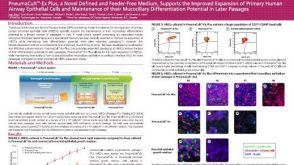 科学海报PneumaCult™-Ex Plus, a Novel Defined and Feeder-Free Medium, Supports the Improved Expansion of Primary Human Airway Epithelial Cells
 沪公网安备31010102008431号