E. Gabriel et al. (JAN 2016)
Stem cell reports 7 4 678--692
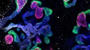
Development and Dynamic Regulation of Mitochondrial Network in Human Midbrain Dopaminergic Neurons Differentiated from iPSCs.
Mitochondria are critical to neurogenesis,but the mechanisms of mitochondria in neurogenesis have not been well explored. We fully characterized mitochondrial alterations and function in relation to the development of human induced pluripotent stem cell (hiPSC)-derived dopaminergic (DA) neurons. Following directed differentiation of hiPSCs to DA neurons,mitochondria in these neurons exhibit pronounced changes during differentiation,including mature neurophysiology characterization and functional synaptic network formation. Inhibition of mitochondrial respiratory chains via application of complex IV inhibitor KCN (potassium cyanide) or complex I inhibitor rotenone restricted neurogenesis of DA neurons. These results demonstrated the direct importance of mitochondrial development and bioenergetics in DA neuronal differentiation. Our study also provides a neurophysiologic model of mitochondrial involvement in neurogenesis,which will enhance our understanding of the role of mitochondrial dysfunctions in neurodegenerative diseases.
View Publication
产品类型:
产品号#:
05832
05835
05839
08581
08582
05833
05790
05792
05794
05795
05793
产品名:
STEMdiff™ 神经花环选择试剂
STEMdiff™ 神经诱导培养基
STEMdiff™ 神经诱导培养基
STEMdiff™SMADi神经诱导试剂盒
STEMdiff™SMADi神经诱导试剂盒,2套
STEMdiff™神经前体细胞培养基
BrainPhys™神经元培养基
BrainPhys™神经元培养基和SM1试剂盒
BrainPhys™原代神经元试剂盒
BrainPhys™ hPSC 神经元试剂盒
BrainPhys™ 神经元培养基N2-A和SM1试剂盒
Newby BN et al. ( 2017)
Diabetes 66 12 3061--3071
Type 1 Interferons Potentiate Human CD8+ T-Cell Cytotoxicity Through a STAT4- and Granzyme B-Dependent Pathway.
Events defining the progression to human type 1 diabetes (T1D) have remained elusive owing to the complex interaction between genetics,the immune system,and the environment. Type 1 interferons (T1-IFN) are known to be a constituent of the autoinflammatory milieu within the pancreas of patients with T1D. However,the capacity of IFNα/β to modulate human activated autoreactive CD8+ T-cell (cytotoxic T lymphocyte) responses within the islets of patients with T1D has not been investigated. Here,we engineer human β-cell-specific cytotoxic T lymphocytes and demonstrate that T1-IFN augments cytotoxicity by inducing rapid phosphorylation of STAT4,resulting in direct binding at the granzyme B promoter within 2 h of exposure. The current findings provide novel insights concerning the regulation of effector function by T1-IFN in human antigen-experienced CD8+ T cells and provide a mechanism by which the presence of T1-IFN potentiates diabetogenicity within the autoimmune islet.
View Publication
Lippmann ES et al. (APR 2014)
Stem Cells 32 4 1032--1042
Defined human pluripotent stem cell culture enables highly efficient neuroepithelium derivation without small molecule inhibitors.
The embryonic neuroepithelium gives rise to the entire central nervous system in vivo,making it an important tissue for developmental studies and a prospective cell source for regenerative applications. Current protocols for deriving homogenous neuroepithelial cultures from human pluripotent stem cells (hPSCs) consist of either embryoid body-mediated neuralization followed by a manual isolation step or adherent differentiation using small molecule inhibitors. Here,we report that hPSCs maintained under chemically defined,feeder-independent,and xeno-free conditions can be directly differentiated into pure neuroepithelial cultures ([mt]90% Pax6(+)/N-cadherin(+) with widespread rosette formation) within 6 days under adherent conditions,without small molecule inhibitors,and using only minimalistic medium consisting of Dulbecco's modified Eagle's medium/F-12,sodium bicarbonate,selenium,ascorbic acid,transferrin,and insulin (i.e.,E6 medium). Furthermore,we provide evidence that the defined culture conditions enable this high level of neural conversion in contrast to hPSCs maintained on mouse embryonic fibroblasts (MEFs). In addition,hPSCs previously maintained on MEFs could be rapidly converted to a neural compliant state upon transfer to these defined conditions while still maintaining their ability to generate all three germ layers. Overall,this fully defined and scalable protocol should be broadly useful for generating therapeutic neural cells for regenerative applications.
View Publication
产品类型:
产品号#:
85850
85857
产品名:
mTeSR™1
mTeSR™1
Krivega M et al. (NOV 2014)
Reproduction 148 5 531--544
Car expression in human embryos and hesc illustrates its role in pluripotency and tight junctions
Coxsackie virus and adenovirus receptor,CXADR (CAR),is present during embryogenesis and is involved in tissue regeneration,cancer and intercellular adhesion. We investigated the expression of CAR in human preimplantation embryos and embryonic stem cells (hESC) to identify its role in early embryogenesis and differentiation. CAR protein was ubiquitously present during preimplantation development. It was localised in the nucleus of uncommitted cells,from the cleavage stage up to the precursor epiblast,and corresponded with the presence of soluble CXADR3/7 splice variant. CAR was displayed on the membrane,involving in the formation of tight junction at compaction and blastocyst stages in both outer and inner cells,and CAR corresponded with the full-length CAR-containing transmembrane domain. In trophectodermal cells of hatched blastocysts,CAR was reduced in the membrane and concentrated in the nucleus,which correlated with the switch in RNA expression to the CXADR4/7 and CXADR2/7 splice variants. The cells in the outer layer of hESC colonies contained CAR on the membrane and all the cells of the colony had CAR in the nucleus,corresponding with the transmembrane CXADR and CXADR4/7. Upon differentiation of hESC into cells representing the three germ layers and trophoblast lineage,the expression of CXADR was downregulated. We concluded that CXADR is differentially expressed during human preimplantation development. We described various CAR expressions: i) soluble CXADR marking undifferentiated blastomeres; ii) transmembrane CAR related with epithelial-like cell types,such as the trophectoderm (TE) and the outer layer of hESC colonies; and iii) soluble CAR present in TE nuclei after hatching. The functions of these distinct forms remain to be elucidated.
View Publication
产品类型:
产品号#:
85850
85857
产品名:
mTeSR™1
mTeSR™1
Gallegos-Cá et al. (AUG 2015)
Stem cells and development 24 16 1901--1911
For diseases of the brain,the pig (Sus scrofa) is increasingly being used as a model organism that shares many anatomical and biological similarities with humans. We report that pig induced pluripotent stem cells (iPSC) can recapitulate events in early mammalian neural development. Pig iPSC line (POU5F1(high)/SSEA4(low)) had a higher potential to form neural rosettes (NR) containing neuroepithelial cells than either POU5F1(low)/SSEA4(low) or POU5F1(low)/SSEA4(high) lines. Thus,POU5F1 and SSEA4 pluripotency marker profiles in starting porcine iPSC populations can predict their propensity to form more robust NR populations in culture. The NR were isolated and expanded in vitro,retaining their NR morphology and neuroepithelial molecular properties. These cells expressed anterior central nervous system fate markers OTX2 and GBX2 through at least seven passages,and responded to retinoic acid,promoting a more posterior fate (HOXB4+,OTX2-,and GBX2-). These findings offer insight into pig iPSC development,which parallels the human iPSC in both anterior and posterior neural cell fates. These in vitro similarities in early neural differentiation processes support the use of pig iPSC and differentiated neural cells as a cell therapy in allogeneic porcine neural injury and degeneration models,providing relevant translational data for eventual human neural cell therapies.
View Publication
产品类型:
产品号#:
07923
85850
85857
产品名:
Dispase (1 U/mL)
mTeSR™1
mTeSR™1
D. Nag et al. (aug 2019)
Clinical cancer research : an official journal of the American Association for Cancer Research 25 15 4791--4807
Auranofin Protects Intestine against Radiation Injury by Modulating p53/p21 Pathway and Radiosensitizes Human Colon Tumor.
PURPOSE The radiosensitivity of the normal intestinal epithelium is the major limiting factor for definitive radiotherapy against abdominal malignancies. Radiosensitizers,which can be used without augmenting radiation toxicity to normal tissue,are still an unmet need. Inhibition of proteosomal degradation is being developed as a major therapeutic strategy for anticancer therapy as cancer cells are more susceptible to proteasomal inhibition-induced cytotoxicity compared with normal cells. Auranofin,a gold-containing antirheumatoid drug,blocks proteosomal degradation by inhibiting deubiquitinase inhibitors. In this study,we have examined whether auranofin selectively radiosensitizes colon tumors without promoting radiation toxicity in normal intestine. EXPERIMENTAL DESIGN The effect of auranofin (10 mg/kg i.p.) on the radiation response of subcutaneous CT26 colon tumors and the normal gastrointestinal epithelium was determined using a mouse model of abdominal radiation. The effect of auranofin was also examined in a paired human colonic organoid system using malignant and nonmalignant tissues from the same patient. RESULTS Both in the mouse model of intestinal injury and in the human nonmalignant colon organoid culture,auranofin pretreatment prevented radiation toxicity and improved survival with the activation of p53/p21-mediated reversible cell-cycle arrest. However,in a mouse model of abdominal tumor and in human malignant colonic organoids,auranofin inhibited malignant tissue growth with inhibition of proteosomal degradation,induction of endoplasmic reticulum stress/unfolded protein response,and apoptosis. CONCLUSIONS Our data suggest that auranofin is a potential candidate to be considered as a combination therapy with radiation to improve therapeutic efficacy against abdominal malignancies.
View Publication
产品类型:
产品号#:
15128
15168
产品名:
RosetteSep™人间充质干细胞富集抗体混合物
RosetteSep™人间充质干细胞富集抗体混合物
(Aug 2024)
ACS Omega 9 34
LSD Modulates Proteins Involved in Cell Proteostasis, Energy Metabolism and Neuroplasticity in Human Cerebral Organoids
Proteomic analysis of human cerebral organoids may reveal how psychedelics regulate biological processes,shedding light on drug-induced changes in the brain. This study elucidates the proteomic alterations induced by lysergic acid diethylamide (LSD) in human cerebral organoids. By employing high-resolution mass spectrometry-based proteomics,we quantitatively analyzed the differential abundance of proteins in cerebral organoids exposed to LSD. Our findings indicate changes in proteostasis,energy metabolism,and neuroplasticity-related pathways. Specifically,LSD exposure led to alterations in protein synthesis,folding,autophagy,and proteasomal degradation,suggesting a complex interplay in the regulation of neural cell function. Additionally,we observed modulation in glycolysis and oxidative phosphorylation,crucial for cellular energy management and synaptic function. In support of the proteomic data,complementary experiments demonstrated LSD’s potential to enhance neurite outgrowth in vitro,confirming its impact on neuroplasticity. Collectively,our results provide a comprehensive insight into the molecular mechanisms through which LSD may affect neuroplasticity and potentially contribute to therapeutic effects for neuropsychiatric disorders.
View Publication
产品类型:
产品号#:
85850
85857
产品名:
mTeSR™1
mTeSR™1
(Apr 2025)
Journal of Neuroinflammation 22 1788–1805
A 3D human iPSC-derived multi-cell type neurosphere system to model cellular responses to chronic amyloidosis
Background: Alzheimer's disease (AD) is characterized by progressive amyloid beta (Aβ) deposition in the brain,with eventual widespread neurodegeneration. While the cell-specific molecular signature of end-stage AD is reasonably well characterized through autopsy material,less is known about the molecular pathways in the human brain involved in the earliest exposure to Aβ. Human model systems that not only replicate the pathological features of AD but also the transcriptional landscape in neurons,astrocytes and microglia are crucial for understanding disease mechanisms and for identifying novel therapeutic targets. Methods: In this study,we used a human 3D iPSC-derived neurosphere model to explore how resident neurons,microglia and astrocytes and their interplay are modified by chronic amyloidosis induced over 3-5 weeks by supplementing media with synthetic Aβ1 - 42 oligomers. Neurospheres under chronic Aβ exposure were grown with or without microglia to investigate the functional roles of microglia. Neuronal activity and oxidative stress were monitored using genetically encoded indicators,including GCaMP6f and roGFP1,respectively. Single nuclei RNA sequencing (snRNA-seq) was performed to profile Aβ and microglia driven transcriptional changes in neurons and astrocytes,providing a comprehensive analysis of cellular responses. Results: Microglia efficiently phagocytosed Aβ inside neurospheres and significantly reduced neurotoxicity,mitigating amyloidosis-induced oxidative stress and neurodegeneration following different exposure times to Aβ. The neuroprotective effects conferred by the presence of microglia was associated with unique gene expression profiles in astrocytes and neurons,including several known AD-associated genes such as APOE. These findings reveal how microglia can directly alter the molecular landscape of AD. Conclusions: Our human 3D neurosphere culture system with chronic Aβ exposure reveals how microglia may be essential for the cellular and transcriptional responses in AD pathogenesis. Microglia are not only neuroprotective in neurospheres but also act as key drivers of Aβ-dependent APOE expression suggesting critical roles for microglia in regulating APOE in the AD brain. This novel,well characterized,functional in vitro platform offers unique opportunities to study the roles and responses of microglia to Aβ modelling key aspects of human AD. This tool will help identify new therapeutic targets,accelerating the transition from discovery to clinical applications.
View Publication
产品类型:
产品号#:
100-0276
100-1130
产品名:
mTeSR™ Plus
mTeSR™ Plus
M. Gijsbertsen et al. (Sep 2025)
Disease Models & Mechanisms 18 10
Generation of human induced pluripotent stem cell lines from patients with FGFR2 -linked syndromic craniosynostosis
Craniosynostosis is a multigenic congenital condition in which one or more calvarial sutures have prematurely fused during the development of the fetus. Pathogenic variants in FGFR2 are associated with the development of syndromic craniosynostosis,such as Crouzon,Apert and Pfeifer syndromes. Investigation of FGFR2 -linked craniosynostosis is hindered by the lack of appropriate in vitro models. Patient-derived human induced pluripotent stem cell (hiPSC) in vitro disease models provide the opportunity to investigate the disease,identify molecular targets for pharmaceutical treatments,and enable the generation of autologous pluripotent stem cell catalogues. Here,we report three patient-derived hiPSC lines carrying the C342Y,S252W or E565G FGFR2 pathogenic variant. The patient hiPSC lines express characteristic pluripotency markers and display distinct phosphorylation profiles under unstimulated conditions. FGFR2 C342Y showed autophosphorylation in the absence of bFGF ligand,although downstream docking proteins PLCγ and FRS2α were not phosphorylated. FGFR2 S252W and FGFR2 E565G hiPSCs showed increased phosphorylation of docking proteins PLCγ and FRS2α,whereas FGFR2 was not phosphorylated. These patient hiPSC lines provide molecular and cellular options to investigate FGFR2 -linked craniosynostosis in the patient-specific genomic context and develop therapeutic modalities.
View Publication
产品类型:
产品号#:
05230
100-0483
100-0484
100-0276
100-1130
05946
85850
85857
产品名:
STEMdiff™ 三谱系分化试剂盒
Hausser Scientificᵀᴹ 明线血球计数板
ReLeSR™
mTeSR™ Plus
mTeSR™ Plus
TeSR™-E6
mTeSR™1
mTeSR™1
F. Huang et al. (Jul 2025)
Journal of Nanobiotechnology 23
Early-life exposure to polypropylene nanoplastics induces neurodevelopmental toxicity in mice and human iPSC-derived cerebral organoids
Nanoplastics (NPs) are emerging environmental pollutants that pose growing concerns due to their potential health risks. However,the effects of inhaled NP exposure during pregnancy on fetal brain development remain poorly understood. In this study,we investigated the impact of maternal exposure to polypropylene nanoplastics (PP-NPs) on fetal brain development and neurobehavioral outcomes in a mouse model and further explored its mechanism in human cerebral organoids. Maternal exposure to PP-NPs significantly impaired neuronal differentiation and proliferation in the fetal cortex. Neurobehavioral assessments revealed significant deficits in offspring following maternal exposure,including impaired spatial memory,reduced motor coordination,and heightened anxiety-like behavior. Furthermore,human brain organoids exposed to PP-NPs exhibited reduced growth and neuronal differentiation,with significant downregulation of key neuronal markers such as TUJ1,MAP2,and PAX6. Transcriptomic analysis identified alterations in gene expression,particularly in neuroactive ligand-receptor interaction pathway. Molecular docking and fluorescence co-localization analysis further suggested CYSLTR1 and PTH1R as key molecular targets of PP-NPs. These findings provide novel insights into the toxicological effects of NPs on the developing brain and emphasize the need for preventive measures to protect fetal neurodevelopment during pregnancy. The online version contains supplementary material available at 10.1186/s12951-025-03561-1.
View Publication
产品类型:
产品号#:
08570
产品名:
STEMdiff™ 脑类器官试剂盒
R. B. Kang et al. (Oct 2025)
Nature Communications 16
Human pancreatic α-cell heterogeneity and trajectory inference analyses reveal SMOC1 as a β-cell dedifferentiation gene
β-cell dysfunction and dedifferentiation towards an α-cell-like phenotype are hallmarks of type 2 diabetes. However,the cell subtypes involved in β-to-α-cell transition are unknown. Using single-cell and single-nucleus RNA-seq,RNA velocity,PAGA/cell trajectory inference,and gene commonality,we interrogated α-β-cell fate switching in human islets. We found five α-cell subclusters with distinct transcriptomes. PAGA analysis showed bifurcating cell trajectories in non-diabetic while unidirectional cell trajectories from β-to-α-cells in type 2 diabetes islets suggesting dedifferentiation towards α-cells. Ten genes comprised the common signature genes in trajectories towards α-cells. Among these,the α-cell gene SMOC1 was expressed in β-cells in type 2 diabetes. Enhanced SMOC1 expression in β-cells decreased insulin expression and secretion and increased β-cell dedifferentiation markers. Collectively,these studies reveal differences in α-β-cell trajectories in non-diabetes and type 2 diabetes human islets,identify signature genes for β-to-α-cell trajectories,and discover SMOC1 as an inducer of β-cell dysfunction and dedifferentiation. Subject terms: Cell signalling,Diabetes,Differentiation
View Publication

 EasySep™小鼠TIL(CD45)正选试剂盒


 沪公网安备31010102008431号