Fogli M et al. (JUL 2008)
PLoS pathogens 4 7 e1000101
Lysis of endogenously infected CD4+ T cell blasts by rIL-2 activated autologous natural killer cells from HIV-infected viremic individuals.
Understanding the cellular mechanisms that ensure an appropriate innate immune response against viral pathogens is an important challenge of biomedical research. In vitro studies have shown that natural killer (NK) cells purified from healthy donors can kill heterologous cell lines or autologous CD4+ T cell blasts exogenously infected with several strains of HIV-1. However,it is not known whether the deleterious effects of high HIV-1 viremia interferes with the NK cell-mediated cytolysis of autologous,endogenously HIV-1-infected CD4+ T cells. Here,we stimulate primary CD4+ T cells,purified ex vivo from HIV-1-infected viremic patients,with PHA and rIL2 (with or without rIL-7). This experimental procedure allows for the significant expansion and isolation of endogenously infected CD4+ T cell blasts detected by intracellular staining of p24 HIV-1 core antigen. We show that,subsequent to the selective down-modulation of MHC class-I (MHC-I) molecules,HIV-1-infected p24(pos) blasts become partially susceptible to lysis by rIL-2-activated NK cells,while uninfected p24(neg) blasts are spared from killing. This NK cell-mediated killing occurs mainly through the NKG2D activation pathway. However,the degree of NK cell cytolytic activity against autologous,endogenously HIV-1-infected CD4+ T cell blasts that down-modulate HLA-A and -B alleles and against heterologous MHC-I(neg) cell lines is particularly low. This phenomenon is associated with the defective surface expression and engagement of natural cytotoxicity receptors (NCRs) and with the high frequency of the anergic CD56(neg)/CD16(pos) subsets of highly dysfunctional NK cells from HIV-1-infected viremic patients. Collectively,our data demonstrate that the chronic viral replication of HIV-1 in infected individuals results in several phenotypic and functional aberrancies that interfere with the NK cell-mediated killing of autologous p24(pos) blasts derived from primary T cells.
View Publication
产品类型:
产品号#:
19052
19052RF
19055
19055RF
产品名:
EasySep™人CD4+ T细胞富集试剂盒
RoboSep™ 人CD4+ T细胞富集试剂盒含滤芯吸头
EasySep™人NK细胞富集试剂盒
RoboSep™ 人NK细胞富集试剂盒含滤芯吸头
Li H et al. (SEP 2016)
In vitro cellular & developmental biology. Animal 52 8 885--893
Directed differentiation of human embryonic stem cells into keratinocyte progenitors in vitro: an attempt with promise of clinical use.
Human embryonic stem cells (hESCs) can differentiate into all somatic lineages including stratified squamous epithelia. Thus,efficient methods are required to direct hESC differentiation to obtain a pure subpopulation for tissue engineering. The study aimed to assess the effects of retinoic acid (RA),bone morphogenetic protein-4 (BMP4),and ascorbic acid (AA) on the differentiation of hESCs into keratinocyte progenitors in vitro. The first media contained AA and BMP4; the second contained RA,AA,and BMP4; the third was commercial-defined keratinocyte serum-free medium,which was used to differentiate H9 hESCs (direct approach) or embryoid bodies (EBs) (indirect approach) into keratinocyte progenitors. Real-time RT-PCR,immunofluorescence,and flow-cytometry were used to characterize the differentiated cells. Cells induced by AA + BMP4 + RA showed the typical epithelial morphology,while cells induced by AA + BMP4 showed multiple appearances. CK14 and p63 messenger RNA (mRNA) expressions in the AA + BMP4 + RA-treated cells were higher than those of the AA + BMP4-treated cells (CK14: 22.4-fold; p63: 84.7-fold). Epithelial marker CK18 mRNA expressions at 14 d of differentiation and keratinocyte marker CK14 and transcription factor p63 mRNA expressions at 35 d of differentiation were higher in cells differentiated from hESCs compared with those differentiated from EBs (CK18 10.51 ± 3.26 vs. 6.67 ± 1.28; CK14 9.27 ± 3.61 vs. 5.32 ± 1.86; p63 0.73 ± 0.06 vs. 0.44 ± 0.12,all P textless 0.05) After hESC induction by AA+BMP4+RA,CK14 mRNA expression was upregulated after day 21,peaking by 35 d of differentiation. Combined RA,BMP4,and AA could effectively induce differentiation of hESCs into keratinocyte progenitors in vitro. These keratinocytes could be used for oral mucosa and skin tissue engineering.
View Publication
产品类型:
产品号#:
07923
85850
85857
产品名:
Dispase (1 U/mL)
mTeSR™1
mTeSR™1
Morizane A et al. (FEB 2011)
Journal of neuroscience research 89 2 117--126
Small-molecule inhibitors of bone morphogenic protein and activin/nodal signals promote highly efficient neural induction from human pluripotent stem cells.
The balance of bone morphogenic protein (BMP),transforming growth factor-β (TGFβ)/activin/nodal,and Wnt signals regulates the early lineage segregation of human embryonic stem cells (ESCs). Here we demonstrate that a combination of small-molecule inhibitors of BMP (Dorsomorphin) and TGFβ/activin/nodal (SB431542) signals promotes highly efficient neural induction from both human ESCs and induced pluripotent stem cells (iPSCs). The combination of small molecules had effects on both cell survival and purity of neural differentiation,under conditions of stromal (PA6) cell coculture and feeder-free floating aggregation culture,for all seven pluripotent stem cell lines that we studied,including three ESC and four iPSC lines. Small molecule compounds are stable and cost effective,so our findings provide a promising strategy for controlled production of neurons in regenerative medicine.
View Publication
产品类型:
产品号#:
72102
100-0246
产品名:
Dorsomorphin
白消安(Busulfan)
Piccin D and Morshead CM (MAR 2011)
Stem cells (Dayton,Ohio) 29 3 528--38
Wnt signaling regulates symmetry of division of neural stem cells in the adult brain and in response to injury.
Neural stem cells comprise a small population of subependymal cells in the adult brain that divide asymmetrically under baseline conditions to maintain the stem cell pool and divide symmetrically in response to injury to increase their numbers. Using in vivo and in vitro models,we demonstrate that Wnt signaling plays a role in regulating the symmetric divisions of adult neural stem cells with no change in the proliferation kinetics of the progenitor population. Using BAT-gal transgenic reporter mice to identify cells with active Wnt signaling,we demonstrate that Wnt signaling is absent in stem cells in conditions where they are dividing asymmetrically and that it is upregulated when stem cells are dividing symmetrically,such as (a) during subependymal regeneration in vivo,(b) in response to stroke,and (c) during colony formation in vitro. Moreover,we demonstrate that blocking Wnt signaling in conditions where neural stem cells are dividing symmetrically inhibits neural stem cell expansion both in vivo and in vitro. Together,these findings reveal that the mechanism by which Wnt signaling modulates the size of the stem cell pool is by regulating the symmetry of stem cell division.
View Publication
产品类型:
产品号#:
72872
产品名:
SB216763
X. Liu et al. ( 2017)
International journal of biological sciences 13 2 232--244
Exosomes Secreted from Human-Induced Pluripotent Stem Cell-Derived Mesenchymal Stem Cells Prevent Osteonecrosis of the Femoral Head by Promoting Angiogenesis.
Background: Local ischemia is the main pathological performance in osteonecrosis of the femoral head (ONFH). There is currently no effective therapy to promote angiogenesis in the femoral head. Recent studies revealed that exosomes secreted by induced pluripotent stem cell-derived mesenchymal stem cells (iPS-MSC-Exos) have great therapeutic potential in ischemic tissues,but whether they could promote angiogenesis in ONFH has not been reported,and little is known regarding the underlying mechanism. Methods: iPS-MSC-Exos were intravenously injected to a steroid-induced rat osteonecrosis model. Samples of the femoral head were obtained 3 weeks after all the injections. The effects were assessed by measuring local angiogenesis and bone loss through histological and immunohistochemical (IHC) staining,micro-CT and three-dimensional microangiography. The effects of exosomes on endothelial cells were studied through evaluations of proliferation,migration and tube-forming analyses. The expression levels of angiogenic related PI3K/Akt signaling pathway of endothelial cells were evaluated following stimulation of iPS-MSC-Exos. The promoting effects of exosomes were re-evaluated following blockade of PI3K/Akt. Results: The in vivo study revealed that administration of iPS-MSC-Exos significantly prevented bone loss,and increased microvessel density in the femoral head compared with control group. We found that iPS-MSC-Exos significantly enhanced the proliferation,migration and tube-forming capacities of endothelial cells in vitro. iPS-MSC-Exos could activate PI3K/Akt signaling pathway in endothelial cells. Moreover,the promoting effects of iPS-MSC-Exos were abolished after blockade of PI3K/Akt on endothelial cells. Conclusions: Our findings suggest that transplantation of iPS-MSC-Exos exerts a preventative effect on ONFH by promoting local angiogenesis and preventing bone loss. The promoting effect might be attributed to activation of the PI3K/Akt signaling pathway on endothelial cells. The data provide the first evidence for the potential of iPS-MSC-Exos in treating ONFH.
View Publication
产品类型:
产品号#:
05835
05839
85850
85857
产品名:
STEMdiff™ 神经诱导培养基
STEMdiff™ 神经诱导培养基
mTeSR™1
mTeSR™1
D. G. Gonzalez et al. (NOV 2018)
Journal of immunology (Baltimore,Md. : 1950)
Nonredundant Roles of IL-21 and IL-4 in the Phased Initiation of Germinal Center B Cells and Subsequent Self-Renewal Transitions.
We examined the unique contributions of the cytokines IL-21 and IL-4 on germinal center (GC) B cell initiation and subsequent maturation in a murine model system. Similar to other reports,we found T follicular helper cell expression of IL-21 begins prior to T follicular helper cell migration into the B cell follicle and precedes that of IL-4. Consistent with this timing,IL-21 signaling has a greater influence on the perifollicular pre-GC B cell transition to the intrafollicular stage. Notably,Bcl6hi B cells can form in the combined absence of IL-21R- and STAT6-derived signals; however,these nascent GC B cells cease to proliferate and are more prone to apoptosis. When B cells lack either IL-21R or STAT6,aberrant GCs form atypical centroblasts and centrocytes that differ in their phenotypic maturation and costimulatory molecule expression. Thus,IL-4 and IL-21 play nonredundant roles in the phased progression of GC B cell development that can initiate in the combined absence of these cytokine signals.
View Publication
产品类型:
产品号#:
19054
19054RF
19852
19852RF
产品名:
EasySep™人B细胞富集试剂盒
RoboSep™ 人B细胞富集试剂盒含滤芯吸头
EasySep™小鼠CD4+ T细胞分选试剂盒
RoboSep™ 小鼠CD4+ T细胞分选试剂盒
Z. Wang et al. (nov 2022)
Laboratory investigation; a journal of technical methods and pathology 102 11 1268--1279
The N6-methyladenosine writer WTAP contributes to the induction of immune tolerance post kidney transplantation by targeting regulatory T cells.
N6-methyladenosine (m6A) modification is involved in diverse immunoregulation,while the relationship between m6A modification and immune tolerance post kidney transplantation remains unclear. Expression of Wilms tumor 1-associating protein (WTAP),an m6A writer,was firstly detected in tolerant kidney transplant recipients (TOL). Then the role of WTAP on regulatory T (Treg) cell differentiation and function in CD4+ T cells from kidney transplant recipients with immune rejection (IR) was investigated. The potential target of WTAP and effect of WTAP on immune tolerance in vivo were subsequently verified. WTAP was upregulated in CD4+ T cells of TOL and positively correlated with Treg cell proportion. In vitro,WTAP overexpression promoted Treg cell differentiation and enhanced Treg cell-mediated suppression toward na?ve T cells. Forkhead box other 1 (Foxo1) was identified as a target of WTAP. WTAP enhanced m6A modification of Foxo1 mRNA in coding sequence (CDS) region,leading to up-regulation of Foxo1. Overexpression of m6A demethylase removed the effect of WTAP overexpression,while Foxo1 overexpression reversed these effects. WTAP overexpression alleviated allograft rejection in model mice,as evidenced by reduced inflammatory response and increased Treg population. Our study suggests that WTAP plays a positive role in induction of immune tolerance post kidney transplant by promoting Treg cell differentiation and function. leading to up-regulation of Foxo1. Overexpression of m6A demethylase removed the effect of WTAP overexpression while Foxo1 overexpression reversed these effects. WTAP overexpression alleviated allograft rejection in model mice as evidenced by reduced inflammatory response and increased Treg population. Our study suggests that WTAP plays a positive role in induction of immune tolerance post kidney transplant by promoting Treg cell differentiation and function."
View Publication
产品类型:
产品号#:
19555
19555RF
产品名:
EasySep™人Naïve CD4+ T细胞分选试剂盒
RoboSep™ 人Naïve CD4+ T细胞分选试剂盒
(Nov 2024)
Antioxidants 13 11
An In Vitro Oxidative Stress Model of the Human Inner Ear Using Human-Induced Pluripotent Stem Cell-Derived Otic Progenitor Cells
The inner ear organs responsible for hearing (cochlea) and balance (vestibular system) are susceptible to oxidative stress due to the high metabolic demands of their sensorineural cells. Oxidative stress-induced damage to these cells can cause hearing loss or vestibular dysfunction,yet the precise mechanisms remain unclear due to the limitations of animal models and challenges of obtaining living human inner ear tissue. Therefore,we developed an in vitro oxidative stress model of the pre-natal human inner ear using otic progenitor cells (OPCs) derived from human-induced pluripotent stem cells (hiPSCs). OPCs,hiPSCs,and HeLa cells were exposed to hydrogen peroxide or ototoxic drugs (gentamicin and cisplatin) that induce oxidative stress to evaluate subsequent cell viability,cell death,reactive oxygen species (ROS) production,mitochondrial activity,and apoptosis (caspase 3/7 activity). Dose-dependent reductions in OPC cell viability were observed post-exposure,demonstrating their vulnerability to oxidative stress. Notably,gentamicin exposure induced ROS production and cell death in OPCs,but not hiPSCs or HeLa cells. This OPC-based human model effectively simulates oxidative stress conditions in the human inner ear and may be useful for modeling the impact of ototoxicity during early pregnancy or evaluating therapies to prevent cytotoxicity.
View Publication
产品类型:
产品号#:
100-0483
100-0484
85850
85857
产品名:
Hausser Scientificᵀᴹ 明线血球计数板
ReLeSR™
mTeSR™1
mTeSR™1
Daga A et al. (MAY 2000)
Experimental hematology 28 5 569--74
The retroviral transduction of HOXC4 into human CD34(+) cells induces an in vitro expansion of clonogenic and early progenitors.
OBJECTIVE: +HOX genes are expressed in the hematopoietic system and increasing data point to their involvement in the control of proliferation and/or differentiation. Genes belonging to the C cluster are preferentially expressed in developing and differentiated lymphoid lineages. However,recent studies demonstrated,by RT-PCR,that the HOXC4 gene is also actively transcribed in the most undifferentiated hematopoietic cells (CD34(+)38(low)) and in more mature myeloid and erythroid progenitors. We evaluated the expression of HOXC4 protein on human CD34(+) cells and the in vitro effect of its overexpression on proliferation and differentiation. MATERIALS AND METHODS: We assessed the expression of HOXC4 on human CD34(+) cells using a polyclonal antibody raised against the C-terminal portion of the protein expressed using the baculovirus system. Overexpression of HOXC4 in human CD34(+) cells was obtained by retroviral gene transfer; its effect on clonogenic (CFU-GM,BFU-E,and CFU-GEMM) and early progenitors (LTC-IC) was evaluated. RESULTS: The HOXC4 protein is indeed expressed in human CD34(+) cells,and its overexpression in human CD34(+) cells increases the proliferation potential of clonogenic and early progenitors. CFU-GM showed a median threefold expansion (range: 1.1-19.4; p textless 0.002) compared with control transduced with the vector alone. The increment of BFU-E was higher (median ninefold,range 2.5-35; p textless 0. 0009) and erythroid colonies presented a larger size with normal morphology. An even more marked effect was observed on LTC-IC (median 13,onefold; range 4.1-102.1,p textless 0.0001). CONCLUSION: We demonstrate that HOXC4 is expressed in CD34(+) cells and that its overexpression induces an in vitro expansion of committed as well as very early hematopoietic progenitors. The most striking effect was obtained on LTC-IC with an expansion of 13.1-fold. The enforced expression of HOXC4 induced a significant increase (p textless 0.009) in the number of erythroid colonies compared with CFU-GM,although without perturbing,at least in vitro,the maturation program of the cells. On the other hand,the effect of the gene overexpression did not induce any skewing in the colony types derived from the myeloid lineage.
View Publication
Wang H et al. (APR 2016)
The Journal of biological chemistry 291 16 8644--8652
Germ Cell Nuclear Factor (GCNF) Represses Oct4 Expression and Globally Modulates Gene Expression in Human Embryonic Stem (hES) Cells.
Oct4 is considered a key transcription factor for pluripotent stem cell self-renewal. It binds to specific regions within target genes to regulate their expression and is downregulated upon induction of differentiation of pluripotent stem cells; however,the mechanisms that regulate the levels of human Oct4 expression remain poorly understood. Here we show that expression of human Oct4 is directly repressed by germ cell nuclear factor (GCNF),an orphan nuclear receptor,in hES cells. Knockdown of GCNF by siRNA resulted in maintenance of Oct4 expression during RA-induced hES cell differentiation. While overexpression of GCNF promoted repression of Oct4 expression in both undifferentiated and differentiated hES cells. The level of Oct4 repression was dependent on the level of GCNF expression in a dose-dependent manner. mRNA microarray analysis demonstrated that overexpression of GCNF globally regulates gene expression in undifferentiated and differentiated hES cells. Within the group of altered genes,GCNF down-regulated 36% of the genes,and up-regulated 64% in undifferentiated hES cells. In addition,GCNF also showed a regulatory gene pattern that is different from RA treatment during hES cell differentiation. These findings increase our understanding of the mechanisms that maintain hES cell pluripotency and regulate gene expression during the differentiation process.
View Publication
产品类型:
产品号#:
85850
85857
产品名:
mTeSR™1
mTeSR™1
Galat V et al. (MAY 2016)
Stem cells and development 25 14 1060--1072
Transgene Reactivation in Induced Pluripotent Stem Cell Derivatives and Reversion to Pluripotency of Induced Pluripotent Stem Cell-Derived Mesenchymal Cells.
Induced pluripotent stem cells (iPSCs) have enormous potential in regenerative medicine and disease modeling. It is now felt that clinical trials should be performed with iPSCs derived with non-integrative constructs. Numerous studies,however,including those describing disease models,are still being published using cells derived from iPSCs generated with integrative constructs. Our experimental work presents the first evidence of spontaneous transgene reactivation in vitro in several cellular types. Our results show that the transgenes were predominantly silent in parent iPSCs,but in mesenchymal and endothelial iPSC derivatives,the transgenes experienced random up-regulation of Nanog and c-Myc. Additionally,we provide evidence of spontaneous secondary reprogramming and reversion to pluripotency in mesenchymal stem cells derived from iPSCs. These findings strongly suggest that the studies,which utilize cellular products derived from iPSCs generated with retro- or lentiviruses,should be evaluated with consideration of the possibility of transgene reactivation. The in vitro model described here provides insight into the earliest events of culture transformation and suggests the hypothesis that reversion to pluripotency may be responsible for the development of tumors in cell replacement experiments. The main goal of this work,however,is to communicate the possibility of transgene reactivation in retro- or lenti- iPSC derivatives and the associated loss of cellular fidelity in vitro,which may impact the outcomes of disease modeling and related experimentation.
View Publication

 EasySep™小鼠TIL(CD45)正选试剂盒


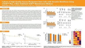 科学海报Culture of High-Quality Human Pluripotent Stem Cells with Versatile Workflows Using mTeSR™ Plus, a New Stabilized TeSR™ Maintenance Medium
 沪公网安备31010102008431号