A. M. Bujor et al. ( 2020)
Frontiers in immunology 11 800
Fli1 Downregulation in Scleroderma Myeloid Cells Has Profibrotic and Proinflammatory Effects.
Scleroderma (SSc) is an autoimmune connective tissue disease characterized by immune dysregulation,vasculopathy,and fibrosis. We have previously demonstrated that low Fli1 expression in SSc fibroblasts and endothelial cells plays an important role in SSc pathogenesis. Cells of myeloid and lymphoid origin also express Fli1 and are dysregulated in patients with SSc,playing key roles in disease pathogenesis. However,the role for immune Fli1 in SSc is not yet clear. Our aim was to elucidate whether Fli1 contributes to the immune dysregulation seen in SSc. Comparison of the expression of Fli1 in monocytes,B- and T-cell fractions of PBMCs isolated from SSc patients and healthy controls (HC),showed an increase in Fli1 levels in monocytes. We used siRNA transfected human myeloid cells and mouse peritoneal macrophages obtained from Fli1 flox/flox LysMCre+/+ mice,and found that markers of alternative macrophage activation were increased with Fli1 deletion. Coculture of Fli1-deficient myeloid cells and primary human or mouse fibroblasts resulted in a potent induction of collagen type I,independent of TGF$\beta$ upregulation. We next analyzed global gene expression profile in response to Fli1 downregulation,to gain further insight into the molecular mechanisms of this process and to identify differentially expressed genes in myeloid cells. Of relevance to SSc,the top most upregulated pathways were hallmark IFN-$\gamma$ and IFN-$\alpha$ response. Additionally,several genes previously linked to SSc pathogenesis and fibrosis in general were also induced,including CCL2,CCL7,MMP12,and CXCL10. ANKRD1,a profibrotic transcription co-regulator was the top upregulated gene in our array. Our results show that Fli1-deficient myeloid cells share key features with cells from SSc patients,with higher expression of profibrotic markers and activation of interferon responsive genes,thus suggesting that dysregulation of Fli1 in myeloid cells may contribute to SSc pathogenesis.
View Publication
T. N. Burn et al. (Dec 2025)
Nature Immunology 27 1
Antigen reactivity defines tissue-resident memory and exhausted T cells in tumors
CD8+ T cells are an important weapon in the therapeutic armamentarium against cancer. While CD8+CD103+ T cells with a tissue-resident memory T (TRM) cell phenotype are associated with favorable prognoses,the tumor microenvironment also contains dysfunctional exhausted T (TEX) cells that exhibit a variety of TRM-like features. Here we deconvolute TRM and TEX cells across human cancers,ascribing markers and gene signatures that distinguish these populations and enable their functional distinction. Although TRM cells have superior functionality and are associated with long-term survival post-tumor resection,they are not associated with responsiveness to immune checkpoint blockade. Tumor-associated TEX and TRM cells are clonally distinct,with the latter comprising tumor-independent bystanders and tumor-specific cells segregated from cognate antigen. Intratumoral TRM cells can be forced toward an exhausted fate when chronic antigen stimulation occurs,indicating that the presence or absence of continuous antigen exposure within the microenvironment is the key distinction between tumor-associated TEX and TRM populations. These results highlight unique functions for TRM and TEX cells in tumor control,underscoring the need for distinct strategies to harness these populations for cancer therapies. Here the authors show that tissue-resident memory and exhausted T cells in tumors are distinct populations that are shaped by relative presence or absence of TCR signals,suggesting that a tailored therapeutic strategy is needed to target each subset.
View Publication
Duan X et al. (JAN 2011)
Journal of cellular physiology 226 1 150--7
Application of induced pluripotent stem (iPS) cells in periodontal tissue regeneration
Tissue engineering provides a new paradigm for periodontal tissue regeneration in which proper stem cells and effective cellular factors are very important. The objective of this study was,for the first time,to investigate the capabilities and advantages of periodontal tissue regeneration using induced pluripotent stem (iPS) cells and enamel matrix derivatives (EMD). In this study the effect of EMD gel on iPS cells in vitro was first determined,and then tissue engineering technique was performed to repair periodontal defects in three groups: silk scaffold only; silk scaffold + EMD; and silk scaffold + EMD + iPS cells. EMD greatly enhanced the mRNA expression of Runx2 but inhibited the mRNA expression of OC and mineralization nodule formation in vitro. Transplantation of iPS cells showed higher expression levels of OC,Osx,and Runx2 genes,both 12 and 24 days postsurgery. At 24 days postsurgery in the iPS cell group,histological analysis showed much more new alveolar bone and cementum formation with regenerated periodontal ligament between them. The results showed the commitment role that EMD contributes in mesenchymal progenitors to early cells in the osteogenic lineage. iPS cells combined with EMD provide a valuable tool for periodontal tissue engineering,by promoting the formation of new cementum,alveolar bone,and normal periodontal ligament.
View Publication
产品类型:
产品号#:
85850
85857
产品名:
mTeSR™1
mTeSR™1
Downes A et al. (OCT 2011)
Journal of Raman Spectroscopy 42 10 1864--1870
Raman spectroscopy and CARS microscopy of stem cells and their derivatives
The characterisation of stem cells is of vital importance to regenerative medicine. Failure to separate out all stem cells from differentiated cells before therapies can result in teratomas - tumours of multiple cell types. Typically,characterisation is performed in a destructive manner with fluorescent assays. A truly non-invasive method of characterisation would be a major breakthrough in stem cell-based therapies. Raman spectroscopy has revealed that DNA and RNA levels drop when a stem cell differentiates into other cell types,which we link to a change in the relative sizes of the nucleus and cytoplasm. We also used Raman spectroscopy to investigate the biochemistry within an early embryo,or blastocyst,which differs greatly from colonies of embryonic stem cells. Certain cell types that differentiate from stem cells can be identified by directly imaging the biochemistry with CARS microscopy; examples presented are hydroxyapatite - a precursor to bone,and lipids in adipocytes.
View Publication
产品类型:
产品号#:
85850
85857
产品名:
mTeSR™1
mTeSR™1
N. Miura et al. (jun 2019)
BMC cancer 19 1 587
miR-520d-5p can reduce the mutations in hepatoma cancer cells and iPSCs-derivatives.
BACKGROUND Human microRNAs (miRNAs) have diverse functions in biology,and play a role in nearly every biological process. Here we report that miR-520d-5p (520d-5p) causes undifferentiated cancer cells to adopt benign or normal status in vivo in immunodeficient mice via demethylation and P53 upregulation. Further we found that 520-5p causes normal cells to elongate cellular lifetime and mesenchymal stem cell-like status with CD105 positivity. We hypothesized that ectopic 520d-5p expression reduced mutations in undifferentiated type of hepatoma (HLF) cells through synergistic modulation of methylation-related enzymatic expression. METHODS To examine whether there were any changes in mutation status in cells treated with 520d-5p,we performed next generation sequencing (NGS) in HLF cells and human iPSC-derivative cells in pre-mesenchymal stem cell status. We analyzed the data using both genome-wide and individual gene function approaches. RESULTS 520d-5p induced a shift towards a wild type or non-malignant phenotype,which was regulated by nucleotide mutations in both HLF cells and iPSCs. Further,520d-5p reduced mutation levels in both the whole genome and genomic fragment assemblies. CONCLUSIONS Cancer cell genomic mutations cannot be repaired in most contexts. However,these findings suggest that applied development of 520d-5p would allow new approaches to cancer research and improve the quality of iPSCs used in regenerative medicine.
View Publication
产品类型:
产品号#:
05240
产品名:
STEMdiff™ 间充质祖细胞试剂盒
Kim MY et al. (MAR 2017)
Oncology letters 13 3 1767--1774
Accumulation of low-dose BIX01294 promotes metastatic potential of U251 glioblastoma cells.
BIX01294 (Bix) is known to be a euchromatic histone-lysine N-methyltransferase 2 inhibitor and treatment with Bix suppresses cancer cell survival and proliferation. In the present study,it was observed that sequential treatment with low-dose Bix notably increases glioblastoma cell migration and metastasis. It was demonstrated that U251 cells sequentially treated with low-dose Bix exhibited induced characteristic changes in critical epithelial-mesenchymal transition (EMT) markers,including E-cadherin,N-cadherin,β-catenin and zinc finger protein SNAI2. Notably,sequential treatment with Bix also increased the expression of cancer stem cell-associated markers,including sex determining region Y-box 2,octamer-binding transcription factor 4 and cluster of differentiation 133. Neurosphere formation was significantly enhanced in cells sequentially treated with Bix,compared with control cells (control: P=0.011; single treatment of Bix,P=0.045). The results of the present study suggest that accumulation of low-dose Bix enhanced the migration and metastatic potential of glioblastoma cells by regulating EMT-associated gene expression,which may be the cause of the altered properties of glioblastoma stem cells.
View Publication
产品类型:
产品号#:
05750
产品名:
NeuroCult™ NS-A 基础培养基(人)
Stipcevic T et al. (DEC 2013)
Acta Neurologica Belgica 113 4 501--506
Stimulation of adult neural stem cells with a novel glycolipid biosurfactant
Glycolipids are amphipathic molecules which are highly expressed on cell membranes in skin and brain where they mediate several key cellular processes. Neural stem cells are defined as undifferentiated,proliferative,multipotential cells with extensive self-renewal and are responsive to brain injury. Di-rhamnolipid: α-L-rhamnopyranosyl-(1-2)α-L-rhamnopyranosyl-3-hydroxydecanoyl-3-hydroxydecanoic acid,also referred to as di-rhamnolipid BAC-3,is a glycolipid isolated from the bacteria Pseudomonas aeruginosa. In the previous studies,di-rhamnolipid enhanced dermal tissue healing and regeneration. The present study provides the first assessment of di-rhamnolipid,and glycolipid biosurfactants in general,on the nervous system. Treatment of neural stem cells isolated from the lateral ventricle of adult mice and cultured in defined media containing growth factors at 0.5 and 1 μg/ml of di-rhamnolipid increased the number of neurospheres (2.7- and 2.8-fold,respectively) compared to controls and this effect remained even after passaging in the absence of di-rhamnolipid. In addition,neural stem cells treated with di-rhamnolipid at 50 and 100 μg/ml in defined media supplemented with fetal calf serum and without growth factors exhibited increased cell viability,indicating an interaction between di-rhamnolipid and serum components in the regulation of neural stem cells and neuroprogenitors. Intracerebroventricular administration of di-rhamnolipid at 300 and 120 ng/day increased the number of neurospheres (1.3- and 1.63-fold,respectively) that could be derived from the anterior lateral ventricles of adult mice. These results indicate that di-rhamnolipid stimulates proliferation of neural stem cells and increases their endogenous pools which may have therapeutic potential in managing neurodegenerative or neuropsychiatric disorders and promoting nervous tissue regeneration following injury.
View Publication
产品类型:
产品号#:
05700
产品名:
NeuroCult™ 基础培养基(小鼠&大鼠)
Baud A et al. (FEB 2017)
Analytical chemistry 89 4 2440--2448
Induced pluripotent stem cells have great potential as a human model system in regenerative medicine,disease modeling,and drug screening. However,their use in medical research is hampered by laborious reprogramming procedures that yield low numbers of induced pluripotent stem cells. For further applications in research,only the best,competent clones should be used. The standard assays for pluripotency are based on genomic approaches,which take up to 1 week to perform and incur significant cost. Therefore,there is a need for a rapid and cost-effective assay able to distinguish between pluripotent and nonpluripotent cells. Here,we describe a novel multiplexed,high-throughput,and sensitive peptide-based multiple reaction monitoring mass spectrometry assay,allowing for the identification and absolute quantitation of multiple core transcription factors and pluripotency markers. This assay provides simpler and high-throughput classification into either pluripotent or nonpluripotent cells in 7 min analysis while being more cost-effective than conventional genomic tests.
View Publication
产品类型:
产品号#:
85850
85857
产品名:
mTeSR™1
mTeSR™1
Carrera Silva EA et al. ( 2017)
Blood 130 17 1898--1902
CD207+CD1a+ cells circulate in pediatric patients with active Langerhans cell histiocytosis.
Langerhans cell histiocytosis (LCH) is a rare disease with an unknown etiology characterized by heterogeneous lesions containing CD207+CD1a+ cells that can arise in almost any tissue and cause significant morbidity and mortality. Precursors of pathological Langerhans cells have yet to be defined. Our aim was to identify circulating CD207+CD1a+ cells and their inducers in LCH. Expression of CD207 and CD1a in the blood myeloid compartment as well as thymic stromal lymphopoietin (TSLP) and transforming growth factor β (TGF-β) plasma levels were measured in 22 pediatric patients with active disease (AD) or nonactive disease (NAD). In patients with AD vs those with NAD,the myeloid compartment showed an increased CD11b (CD11bhigh plus CD11b+) fraction (39.7 ± 3.6 vs 18.6 ± 1.9),a higher percentage of circulating CD11bhighCD11c+CD207+ cells (44.5 ± 11.3 vs 3.2 ± 0.5),and the presence of CD11chighCD207+CD1a+ cells (25.0 ± 9.1 vs 2.3 ± 0.5). Blood CD207+CD1a+ cells were not observed in adult controls or umbilical cord. Increased TSLP and TGF-β levels were detected in patients with AD. Interestingly,plasma from patients with AD induces CD207 expression on CD14+ monocytes. We conclude that CD207+CD1a+ cells are circulating in patients with active LCH,and TSLP and TGF-β are potential drivers of Langerhans-like cells in vivo.
View Publication

 EasySep™小鼠TIL(CD45)正选试剂盒


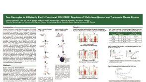 科学海报Cell Isolation of Functional CD4+CD25+ Regulatory T Cells from Mouse Strains


 沪公网安备31010102008431号