Mali P et al. (APR 2010)
Stem cells (Dayton,Ohio) 28 4 713--20
Butyrate greatly enhances derivation of human induced pluripotent stem cells by promoting epigenetic remodeling and the expression of pluripotency-associated genes.
We report here that butyrate,a naturally occurring fatty acid commonly used as a nutritional supplement and differentiation agent,greatly enhances the efficiency of induced pluripotent stem (iPS) cell derivation from human adult or fetal fibroblasts. After transient butyrate treatment,the iPS cell derivation efficiency is enhanced by 15- to 51-fold using either retroviral or piggyBac transposon vectors expressing 4 to 5 reprogramming genes. Butyrate stimulation is more remarkable (textgreater100- to 200-fold) on reprogramming in the absence of either KLF4 or MYC transgene. Butyrate treatment did not negatively affect properties of iPS cell lines established by either 3 or 4 retroviral vectors or a single piggyBac DNA transposon vector. These characterized iPS cell lines,including those derived from an adult patient with sickle cell disease by either the piggyBac or retroviral vectors,show normal karyotypes and pluripotency. To gain insights into the underlying mechanisms of butyrate stimulation,we conducted genome-wide gene expression and promoter DNA methylation microarrays and other epigenetic analyses on established iPS cells and cells from intermediate stages of the reprogramming process. By days 6 to 12 during reprogramming,butyrate treatment enhanced histone H3 acetylation,promoter DNA demethylation,and the expression of endogenous pluripotency-associated genes,including DPPA2,whose overexpression partially substitutes for butyrate stimulation. Thus,butyrate as a cell permeable small molecule provides a simple tool to further investigate molecular mechanisms of cellular reprogramming. Moreover,butyrate stimulation provides an efficient method for reprogramming various human adult somatic cells,including cells from patients that are more refractory to reprogramming.
View Publication
产品类型:
产品号#:
72212
产品名:
RG108
Emre N et al. (JAN 2010)
PLoS ONE 5 8 e12148
The ROCK inhibitor Y-27632 improves recovery of human embryonic stem cells after fluorescence-activated cell sorting with multiple cell surface markers
BACKGROUND: Due to the inherent sensitivity of human embryonic stem cells (hESCs) to manipulations,the recovery and survival of hESCs after fluorescence-activated cell sorting (FACS) can be low. Additionally,a well characterized and robust methodology for performing FACS on hESCs using multiple-cell surface markers has not been described. The p160-Rho-associated coiled kinase (ROCK) inhibitor,Y-27632,previously has been identified as enhancing survival of hESCs upon single-cell dissociation,as well as enhancing recovery from cryopreservation. Here we examined the application of Y-27632 to hESCs after FACS to improve survival in both feeder-dependent and feeder-independent growth conditions. METHODOLOGY/PRINCIPAL FINDINGS: HESCs were sorted using markers for SSEA-3,TRA-1-81,and SSEA-1. Cells were plated after sorting for 24 hours in either the presence or the absence of Y-27632. In both feeder-dependent and feeder-independent conditions,cell survival was greater when Y-27632 was applied to the hESCs after sort. Specifically,treatment of cells with Y-27632 improved post-sort recovery up to four fold. To determine the long-term effects of sorting with and without the application of Y-27632,hESCs were further analyzed. Specifically,hESCs sorted with and without the addition of Y-27632 retained normal morphology,expressed hESC-specific markers as measured by immunocytochemistry and flow cytometry,and maintained a stable karyotype. In addition,the hESCs could differentiate into three germ layers in vitro and in vivo in both feeder-dependent and feeder-independent growth conditions. CONCLUSIONS/SIGNIFICANCE: The application of Y-27632 to hESCs after cell sorting improves cell recovery with no observed effect on pluripotency,and enables the consistent recovery of hESCs by FACS using multiple surface markers. This improved methodology for cell sorting of hESCs will aid many applications such as removal of hESCs from secondary cell types,identification and isolation of stem cell subpopulations,and generation of single cell clones. Finally,these results demonstrate an additional application of ROCK inhibition to hESC research.
View Publication
Type I interferons increase expression of endogenous retrovirus K102 and envelope protein in myeloid cells from patients with autoimmune disease
Autoantibodies against envelope (Env) protein encoded by human endogenous retrovirus group K (HERV-K) are prevalent in rheumatoid arthritis (RA) and systemic lupus erythematosus (SLE),but it remains unclear which proviruses are responsible for this autoantigen. It also remains poorly understood how the transcription of HERV-K loci is regulated in cells that can produce Env.ResultsWe aligned our neutrophil RNA sequencing data to the new telomere-to-telomere reference genome and found uniquely mapping transcripts from HERV-K101,K102,K104,K108,K109,K117 and ERVK5,of which only K102,K108,and K109 encode an intact Env. Expression of K102 and K108 were higher in SLE than in healthy donors or RA (padj < 0.05). Transcripts from these proviruses increased in response to interferon-α in monocytes and neutrophils from RA patients and healthy donors,but not in SLE,presumably because they have chronically elevated type I interferons in vivo. Indeed,HERV-K expression was significantly higher in SLE patients with high type I interferon gene signature. Tumor necrosis factor-α and other cytokines and TLR ligands also induced HERV-K102 and K108 transcripts. Interferon-α also increased detectable Env protein in monocytes,macrophages,and neutrophils from RA patients. Among the genes for epigenetic silencers of HERV-K,only TRIM28 was significantly decreased in SLE patients with high interferons (padj = 0.00024).ConclusionsOur data establish a role for interferons in maintaining increased HERV-K expression in SLE and suggest that interferons or other cytokines can upregulate HERV-K to similar levels in RA. A transient increase may also accompany normal immune responses,suggesting that endogenous retroviruses may have been co-opted for efficient immune responses.Supplementary InformationThe online version contains supplementary material available at 10.1186/s13100-025-00371-y.
View Publication
产品类型:
产品号#:
100-1525
19054
19054RF
19058
19058RF
19059
19059RF
产品名:
EasySep™人单核细胞富集试剂盒(不去除CD16)
EasySep™人B细胞富集试剂盒
RoboSep™ 人B细胞富集试剂盒含滤芯吸头
EasySep™人单核细胞富集试剂盒(不去除CD16)
RoboSep™ 人单核细胞富集试剂盒(不去除CD16)含滤芯吸头
EasySep™人单核细胞富集试剂盒
RoboSep™ 人单核细胞富集试剂盒含滤芯吸头
A. Ferrelli et al. (Aug 2025)
HemaSphere 9 8
Mesenchymal stromal cells from JAK2 V617F myeloproliferative neoplasms support healthy and malignant hematopoiesis in a humanized scaffold model in vivo
Myeloproliferative Neoplasms (MPN) are malignancies of hematopoietic stem and progenitor cells (HSPCs) that lead to the overproduction of mature blood cells. These disorders include Essential Thrombocythemia (ET),Polycythemia Vera (PV),and Primary Myelofibrosis (PMF),primarily driven by somatic mutations such as JAK2 V617F . Research indicates that mesenchymal stromal cells (MSCs) support fibrosis in PMF,though their role in ET and PV remains less clear. Furthermore,in vivo studies of ET/PV HSPCs remain a challenge due to low engraftment levels in xenograft models. We employed a 3D scaffold model to create an MPN humanized xenograft mouse model,enabling in vivo functional studies of primary MPN progenitor cells and the supportive role of human MSCs. Using this model,we first demonstrated robust hematopoietic support of healthy (HD) HSPCs by PV and ET MSCs. We then investigated the role of MSCs in sustaining JAK2 V617F mutant cells by using a CRISPR‐Cas9 editing model,along with primary PV and ET HSPCs. Our results showed consistent engraftment of CRISPR‐edited JAK2 V617F mutant HSPCs and PV and ET patient‐derived HSPCs in scaffolds seeded with HD,PV,and ET stroma,providing the first in vivo evidence that PV and ET MSCs can sustain both healthy and MPN‐associated hematopoiesis. Furthermore,HD MSCs were also capable of sustaining PV and ET HSPCs in vivo. Overall,we present the first humanized MPN xenograft model that offers valuable insights into how human BM MSCs interact with JAK2 V617F mutant clones.
View Publication
Jing W et al. (OCT 2017)
Cancer research 77 20 5676--5686
T Cells Deficient in Diacylglycerol Kinase ζ Are Resistant to PD-1 Inhibition and Help Create Persistent Host Immunity to Leukemia.
Efforts to improve the efficacy of adoptive T-cell therapies and immune checkpoint therapies in myelogenous leukemia are desired. In this study,we evaluated the antileukemia activity of adoptively transferred polyclonal cancer antigen-reactive T cells deficient in the regulator diacylglycerol kinase zeta (DGKζ) with or without PD-1/PD-L1 blockade. In the C1498 mouse model of myeloid leukemia,we showed that leukemia was eradicated more effectively in DGKζ-deficient (DGKζ-/-) mice than wild-type mice. T cells transferred from DGKζ-deficient mice to wild-type tumor-bearing recipients conferred this benefit. Leukemia clearance was similar to mice treated with anti-PD-L1. Strikingly,we found that the activity of adoptively transferred DGKζ-/- T cells relied partly on induction of sustainable host T-cell immunity. Transferring DGKζ-deficient T cells increased the levels of IFNγ and other cytokines in recipient mice,especially with coadministration of anti-PD-L1. Overall,our results offered evidence that targeting DGKζ may leverage the efficacy of adoptive T-cell and immune checkpoint therapies in leukemia treatment. Furthermore,they suggest that DGKζ targeting might decrease risks of antigen escape or resistance to immune checkpoint blockade. Cancer Res; 77(20); 5676-86. textcopyright2017 AACR.
View Publication
产品类型:
产品号#:
19851
19851RF
产品名:
EasySep™小鼠T细胞分选试剂盒
RoboSep™ 小鼠T细胞分选试剂盒
Kharas MG et al. (SEP 2008)
The Journal of clinical investigation 118 9 3038--50
Ablation of PI3K blocks BCR-ABL leukemogenesis in mice, and a dual PI3K/mTOR inhibitor prevents expansion of human BCR-ABL+ leukemia cells.
Some cases of pre-B cell acute lymphoblastic leukemia (pre-B-ALL) are caused by the Philadelphia (Ph) chromosome-encoded BCR-ABL oncogene,and these tend to have a poor prognosis. Inhibitors of the PI3K/AKT pathway reduce BCR-ABL-mediated transformation in vitro; however,the specific PI3K isoforms involved are poorly defined. Using a murine model of Ph+ pre-B-ALL,we found that deletion of both Pik3r1 and Pik3r2,genes encoding class IA PI3K regulatory isoforms,severely impaired transformation. BCR-ABL-dependent pre/pro-B cell lines could be established at low frequency from progenitors that lacked these genes,but the cells were smaller,proliferated more slowly,and failed to cause leukemia in vivo. These cell lines displayed nearly undetectable PI3K signaling function and were resistant to the PI3K inhibitor wortmannin. However,they maintained activation of mammalian target of rapamycin (mTOR) and were more sensitive to rapamycin. Treatment with rapamycin caused feedback activation of AKT in WT cell lines but not PI3K-deficient lines. A dual inhibitor of PI3K and mTOR,PI-103,was more effective than rapamycin at suppressing proliferation of mouse pre-B-ALL and human CD19+CD34+)Ph+ ALL leukemia cells treated with the ABL kinase inhibitor imatinib. Our findings provide mechanistic insights into PI3K dependency in oncogenic networks and provide a rationale for targeting class IA PI3K,alone or together with mTOR,in the treatment of Ph+ ALL.
View Publication
产品类型:
产品号#:
03630
产品名:
MethoCult™M3630
Azevedo RI et al. (MAR 2009)
Blood 113 13 2999--3007
IL-7 sustains CD31 expression in human naive CD4+ T cells and preferentially expands the CD31+ subset in a PI3K-dependent manner.
The CD31(+) subset of human naive CD4(+) T cells is thought to contain the population of cells that have recently emigrated from the thymus,while their CD31(-) counterparts have been proposed to originate from CD31(+) cells after homeostatic cell division. Naive T-cell maintenance is known to involve homeostatic cytokines such as interleukin-7 (IL-7). It remains to be investigated what role this cytokine has in the homeostasis of naive CD4(+) T-cell subsets defined by CD31 expression. We provide evidence that IL-7 exerts a preferential proliferative effect on CD31(+) naive CD4(+) T cells from adult peripheral blood compared with the CD31(-) subset. IL-7-driven proliferation did not result in loss of CD31 expression,suggesting that CD31(+) naive CD4(+) T cells can undergo cytokine-driven homeostatic proliferation while preserving CD31. Furthermore,IL-7 sustained or increased CD31 expression even in nonproliferating cells. Both proliferation and CD31 maintenance were dependent on the activation of phosphoinositide 3-kinase (PI3K) signaling. Taken together,our data suggest that during adulthood CD31(+) naive CD4(+) T cells are maintained by IL-7 and that IL-7-based therapies may exert a preferential effect on this population.
View Publication
产品类型:
产品号#:
19052
19052RF
产品名:
EasySep™人CD4+ T细胞富集试剂盒
RoboSep™ 人CD4+ T细胞富集试剂盒含滤芯吸头
Zhang Y et al. (MAR 2015)
Molecular cancer 14 1 56
Sp1 and c-Myc modulate drug resistance of leukemia stem cells by regulating survivin expression through the ERK-MSK MAPK signaling pathway.
BACKGROUND Acute myeloid leukemia (AML) is initiated and maintained by a subset of self-renewing leukemia stem cells (LSCs),which contribute to the progression,recurrence and therapeutic resistance of leukemia. However,the mechanisms underlying the maintenance of LSCs drug resistance have not been fully defined. In this study,we attempted to elucidate the mechanisms of LSCs drug resistance. METHODS We performed reverse phase protein arrays to analyze the expression of anti-apoptotic proteins in the LSC-enriched leukemia cell line KG-1a. Immuno-blotting,cell viability and clinical AML samples were evaluated to verify the micro-assay results. The characteristics and transcriptional regulation of survivin were analyzed with the relative luciferase reporter assay,mutant constructs,chromatin immuno-precipitation (ChIP),quantitative real-time reverse transcription polymerase chain reaction (RT-qPCR),and western blotting. The levels of Sp1,c-Myc,phospho-extracellular signal-regulated kinase (p-ERK),phospho-mitogen and stress-activated protein kinase (p-MSK) were investigated in paired CD34+ and CD34- AML patient samples. RESULTS Survivin was highly over-expressed in CD34 + CD38- KG-1a cells and paired CD34+ AML patients compared with their differentiated counterparts. Functionally,survivin contributes to the drug resistance of LSCs,and Sp1 and c-Myc concurrently regulate levels of survivin transcription. Clinically,Sp1 and c-Myc were significantly up-regulated and positively correlated with survivin in CD34+ AML patients. Moreover,Sp1 and c-Myc were further activated by the ERK/MSK mitogen-activated protein kinase (MAPK) signaling pathway,modulating survivin levels. CONCLUSION Our findings demonstrated that ERK/MSK/Sp1/c-Myc axis functioned as a critical regulator of survivin expression in LSCs,offering a potential new therapeutic strategy for LSCs therapy.
View Publication
产品类型:
产品号#:
07930
07931
07940
07952
07955
07959
100-1061
产品名:
CryoStor® CS10
CryoStor® CS10
CryoStor® CS10
CryoStor® CS10
CryoStor® CS10
CryoStor® CS10
CryoStor® CS10
Cao Y et al. (MAR 2016)
Journal of Immunology 196 5 2075--84
Autoreactive T Cells from Patients with Myasthenia Gravis Are Characterized by Elevated IL-17, IFN-γ, and GM-CSF and Diminished IL-10 Production.
Myasthenia gravis (MG) is a prototypical autoimmune disease that is among the few for which the target Ag and the pathogenic autoantibodies are clearly defined. The pathology of the disease is affected by autoantibodies directed toward the acetylcholine receptor (AChR). Mature,Ag-experienced B cells rely on the action of Th cells to produce these pathogenic Abs. The phenotype of the MG Ag-reactive T cell compartment is not well defined; thus,we sought to determine whether such cells exhibit both a proinflammatory and a pathogenic phenotype. A novel T cell library assay that affords multiparameter interrogation of rare Ag-reactive CD4(+) T cells was applied. Proliferation and cytokine production in response to both AChR and control Ags were measured from 3120 T cell libraries derived from 11 MG patients and paired healthy control subjects. The frequency of CCR6(+) memory T cells from MG patients proliferating in response to AChR-derived peptides was significantly higher than that of healthy control subjects. Production of both IFN-γ and IL-17,in response to AChR,was also restricted to the CCR6(+) memory T cell compartment in the MG cohort,indicating a proinflammatory phenotype. These T cells also included an elevated expression of GM-CSF and absence of IL-10 expression,indicating a proinflammatory and pathogenic phenotype. This component of the autoimmune response in MG is of particular importance when considering the durability of MG treatment strategies that eliminate B cells,because the autoreactive T cells could renew autoimmunity in the reconstituted B cell compartment with ensuing clinical manifestations.
View Publication
产品类型:
产品号#:
17952
17952RF
100-0696
产品名:
EasySep™人CD4+ T细胞分选试剂盒
RoboSep™ 人CD4+ T细胞分选试剂盒
EasySep™人CD4+ T细胞分离试剂盒
Nakamura S et al. (NOV 2010)
Carcinogenesis 31 11 2012--21
The FOXM1 transcriptional factor promotes the proliferation of leukemia cells through modulation of cell cycle progression in acute myeloid leukemia.
FOXM1 is an important cell cycle regulator and regulates cell proliferation. In addition,FOXM1 has been reported to contribute to oncogenesis in various cancers. However,it is not clearly understood how FOXM1 contributes to acute myeloid leukemia (AML) cell proliferation. In this study,we investigated the cellular and molecular function of FOXM1 in AML cells. The FOXM1 messenger RNA (mRNA) expressed in AML cell lines was predominantly the FOXM1B isoform,and its levels were significantly higher than in normal high aldehyde dehydrogenase activity (ALDH(hi)) cells. Reduction of FOXM1 expression in AML cells inhibited cell proliferation compared with control cells,through induction of G(2)/M cell cycle arrest,a decrease in the protein expression of Aurora kinase B,Survivin,Cyclin B1,S-phase kinase-associated protein 2 and Cdc25B and an increase in the protein expression of p21(Cip1) and p27(Kip1). FOXM1 messenger RNA (mRNA) was overexpressed in all 127 AML clinical specimens tested (n = 21,56,32 and 18 for M1,M2,M4 and M5 subtypes,respectively). Compared with normal ALDH(hi) cells,FOXM1 gene expression was 1.65- to 2.26-fold higher in AML cells. Moreover,the FOXM1 protein was more strongly expressed in AML-derived ALDH(hi) cells compared with normal ALDH(hi) cells. In addition,depletion of FOXM1 reduced colony formation of AML-derived ALDH(hi) cells due to inhibition of Cdc25B and Cyclin B1 expression. In summary,we found that FOXM1B mRNA is predominantly expressed in AML cells and that aberrant expression of FOXM1 induces AML cell proliferation through modulation of cell cycle progression. Thus,inhibition of FOXM1 expression represents an attractive target for AML therapy.
View Publication

 EasySep™小鼠TIL(CD45)正选试剂盒


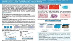 科学海报In Vitro and in Vivo Characterization of Human Bone Marrow-Derived Mesenchymal Stromal Cells in a Novel Animal Component-Free Culture Medium

 沪公网安备31010102008431号