X. Kang et al. ( 2022)
Journal of immunology research 2022 8118577
Deletion of Mettl3 at the Pro-B Stage Marginally Affects B Cell Development and Profibrogenic Activity of B Cells in Liver Fibrosis.
N6-methyladenosine (m6A) modification plays a pivotal role in cell fate determination. Previous studies show that eliminating m6A using Mb1-Cre dramatically impairs B cell development. However,whether disturbing m6A modification at later stages affects B cell development and function remains elusive. Here,we deleted m6A methyltransferase Mettl3 from the pro-B stage on using Cd19-Cre (Mettl3 cKO) and found that the frequency of total B cells in peripheral blood,peritoneal cavity,and liver is comparable between Mettl3 cKO mice and wild-type (WT) littermates,while the percentage of whole splenic B cells slightly increases in Mettl3 cKO individuals. The proportion of pre-pro-B,pro-B,pre-B,immature,and mature B cells in the bone marrow were minimally affected. Loss of Mettl3 resulted in increased apoptosis but barely affected B cells' proliferation and IgG production upon LPS,CD40L,anti-IgM,or TNF-$\alpha$ stimulation. Different stimuli had different effects on B cell activation. In addition,B cell-specific Mettl3 knockout had no influence on the pro-fibrogenic activity of B cells in liver fibrosis,evidenced by comparable fibrosis in carbon tetrachloride- (CCl4-) treated Mettl3 cKO mice and WT controls. In summary,our study demonstrated that deletion of Mettl3 from the pro-B stage on has minimal effects on B cell development and function,as well as profibrogenic activity of B cells in liver fibrosis,revealing a stage-specific dependence on Mettl3-mediated m6A of B cell development.
View Publication
产品类型:
产品号#:
18000
18954
18954RF
产品名:
EasySep™磁极
EasySep™ 小鼠CD19正选试剂盒 II
RoboSep™ 小鼠CD19正选试剂盒II
F. Ozmen et al. (Aug 2025)
NPJ Breast Cancer 11
Single-cell RNA sequencing reveals different cellular states in malignant cells and the tumor microenvironment in primary and metastatic ER-positive breast cancer
Metastatic breast cancer remains largely incurable,and the mechanisms driving the transition from primary to metastatic breast cancer remain elusive. We analyzed the complex landscape of estrogen receptor (ER)-positive breast cancer primary and metastatic tumors using scRNA-seq data from twenty-three female patients with either primary or metastatic disease. By employing single-cell transcriptional profiling of unpaired patient samples,we sought to elucidate the genetic and molecular mechanisms underlying changes in the metastatic tumor ecosystem. We identified specific subtypes of stromal and immune cells critical to forming a pro-tumor microenvironment in metastatic lesions,including CCL2+ macrophages,exhausted cytotoxic T cells,and FOXP3+ regulatory T cells. Analysis of cell-cell communication highlights a marked decrease in tumor-immune cell interactions in metastatic tissues,likely contributing to an immunosuppressive microenvironment. In contrast,primary breast cancer samples displayed increased activation of the TNF-α signaling pathway via NF-kB,indicating a potential therapeutic target. Our study comprehensively characterizes the transcriptional landscape encompassing primary and metastatic breast cancer.
View Publication
产品类型:
产品号#:
17899
产品名:
EasySep™ 死细胞去除 (Annexin V) 试剂盒
A. Wu et al. (Jul 2025)
International Journal of Molecular Sciences 26 13
Identification of a PAK6-Mediated MDM2/p21 Axis That Modulates Survival and Cell Cycle Control of Drug-Resistant Stem/Progenitor Cells in Chronic Myeloid Leukemia
Chronic myeloid leukemia (CML) is a leading example of a malignancy where a molecular targeted therapy revolutionized treatment but has rarely led to cures. Overcoming tyrosine kinase inhibitor (TKI) drug resistance remains a challenge in the treatment of CML. We have recently identified miR-185 as a predictive biomarker where reduced expression in CD34 + treatment-naïve CML cells was associated with TKI resistance. We have also identified PAK6 as a target gene of miR-185 that was upregulated in CD34 + TKI-nonresponder cells. However,its role in regulating TKI resistance remains largely unknown. In this study,we specifically targeted PAK6 in imatinib (IM)-resistant cells and CD34 + stem/progenitor cells from IM-nonresponders using a lentiviral-mediated PAK6 knockdown strategy. Interestingly,the genetic and pharmacological suppression of PAK6 significantly reduced proliferation and increased apoptosis in TKI-resistant cells. Cell survivability was further diminished when IM was combined with PAK6 knockdown. Importantly,PAK6 inhibition in TKI-resistant cells induced cell cycle arrest in the G2-M phase and cellular senescence,accompanied by increased levels of DNA damage-associated senescence markers. Mechanically,we identified a PAK6-mediated MDM2-p21 axis that regulates cell cycle arrest and senescence. Thus,PAK6 plays a critical role in determining alternative cell fates in leukemic cells,and targeting PAK6 may offer a therapeutic strategy to selectively eradicate TKI-resistant cells.
View Publication
产品类型:
产品号#:
04230
产品名:
MethoCult™H4230
Penicka M et al. (JUL 2007)
Heart (British Cardiac Society) 93 7 837--41
One-day kinetics of myocardial engraftment after intracoronary injection of bone marrow mononuclear cells in patients with acute and chronic myocardial infarction.
OBJECTIVE: To investigate the kinetics of myocardial engraftment of bone marrow-derived mononuclear cells (BMNCs) after intracoronary injection using 99mTc-d,l-hexamethylpropylene amine oxime (99mTc-HMPAO) nuclear imaging in patients with acute and chronic anterior myocardial infarction. DESIGN: Nuclear imaging-derived tracking of BMNCs at 2 and 20 h after injection in the left anterior descending (LAD) coronary artery. SETTING: Academical cardiocentre. PATIENTS: Five patients with acute (mean (SD) age 58 (11) years; ejection fraction range 33-45%) and five patients with chronic (mean (SD) age 50 (6) years; ejection fraction range 28-34%) anterior myocardial infarction. INTERVENTIONS: A total of 24.2 x 10(8)-57.0 x 10(8) BMNCs (20% labelled with 700-1000 MBq 99mTc-HMPAO) were injected in the LAD coronary artery. RESULTS: At 2 h after BMNC injection,myocardial activity was observed in all patients with acute (range 1.31-5.10%) and in all but one patient with chronic infarction (range 1.10-3.0%). At 20 h,myocardial engraftment was noted only in three patients with acute myocardial infarction,whereas no myocardial activity was noted in any patient with chronic infarction. CONCLUSIONS: Engraftment of BMNCs shows dynamic changes within the first 20 h after intracoronary injection. Persistent myocardial engraftment was noted only in a subset of patients with acute myocardial infarction.
View Publication
产品类型:
产品号#:
04434
04444
产品名:
MethoCult™H4434经典
MethoCult™H4434经典
R. A. Wilcox et al. (OCT 2009)
Blood 114 14 2936--44
Monocytes promote tumor cell survival in T-cell lymphoproliferative disorders and are impaired in their ability to differentiate into mature dendritic cells.
A variety of nonmalignant cells present in the tumor microenvironment promotes tumorigenesis by stimulating tumor cell growth and metastasis or suppressing host immunity. The role of such stromal cells in T-cell lymphoproliferative disorders is incompletely understood. Monocyte-derived cells (MDCs),including professional antigen-presenting cells such as dendritic cells (DCs),play a central role in T-cell biology. Here,we provide evidence that monocytes promote the survival of malignant T cells and demonstrate that MDCs are abundant within the tumor microenvironment of T cell-derived lymphomas. Malignant T cells were observed to remain viable during in vitro culture with autologous monocytes,but cell death was significantly increased after monocyte depletion. Furthermore,monocytes prevent the induction of cell death in T-cell lymphoma lines in response to either serum starvation or doxorubicin,and promote the engraftment of these cells in nonobese diabetic/severe combined immunodeficient mice. Monocytes are actively recruited to the tumor microenvironment by CCL5 (RANTES),where their differentiation into mature DCs is impaired by tumor-derived interleukin-10. Collectively,the data presented demonstrate a previously undescribed role for monocytes in T-cell lymphoproliferative disorders.
View Publication
产品类型:
产品号#:
19058
19058RF
100-1525
产品名:
EasySep™人单核细胞富集试剂盒(不去除CD16)
RoboSep™ 人单核细胞富集试剂盒(不去除CD16)含滤芯吸头
EasySep™人单核细胞富集试剂盒(不去除CD16)
Neumeister V et al. (MAY 2010)
The American journal of pathology 176 5 2131--8
In situ identification of putative cancer stem cells by multiplexing ALDH1, CD44, and cytokeratin identifies breast cancer patients with poor prognosis.
A subset of cells,tentatively called cancer stem cells (CSCs),in breast cancer have been associated with tumor initiation,drug resistance,and tumor persistence or aggressiveness. They are characterized by CD44 positivity,CD24 negativity,and/or ALDH1 positivity in flow cytometric studies. We hypothesized that the frequency or density of these cells may be associated with more aggressive tumor behavior. We borrowed these multiplexed,flow-based methods to develop an in situ method to define CSCs in formalin-fixed paraffin-embedded breast cancer tissue,with the goal of assessing the prognostic value of the presence of CSCs in breast cancer. Using a retrospective collection of 321 node-negative and 318 node-positive patients with a mean follow-up time of 12.6 years,we assessed TMAs using the AQUA method for quantitative immunofluorescence. Using a multiplexed assay for ALDH1,CD44,and cytokeratin to measure the coexpression of these proteins,putative CSCs appear in variable sized clusters and in 27 cases (of 490),which showed significantly worse outcome (log rank P = 0.0003). Multivariate analysis showed that this marker combination is independent of tumor size,histological grade,nodal status,ER-,PR,- and HER2-status. In this cohort,ALDH1 expression alone does not significantly predict outcome. We conclude that the multiplexed method of in situ identification of putative CSCs identifies high risk patients in breast cancer.
View Publication
产品类型:
产品号#:
01700
01705
01702
产品名:
ALDEFLUOR™ 试剂盒
ALDEFLUOR™ DEAB试剂
ALDEFLUOR™测定缓冲液
Kim S-J et al. (AUG 2010)
Neuroscience letters 479 3 292--6
Omega-3 and omega-6 fatty acids suppress ER- and oxidative stress in cultured neurons and neuronal progenitor cells from mice lacking PPT1.
Reactive oxygen species (ROS) damage brain lipids,carbohydrates,proteins,as well as DNA and may contribute to neurodegeneration. We previously reported that ER- and oxidative stress cause neuronal apoptosis in infantile neuronal ceroid lipofuscinosis (INCL),a lethal neurodegenerative storage disease,caused by palmitoyl-protein thioesterase-1 (PPT1) deficiency. Polyunsaturated fatty acids (PUFA) are essential components of cell membrane phospholipids in the brain and excessive ROS may cause oxidative damage of PUFA leading to neuronal death. Using cultured neurons and neuroprogenitor cells from mice lacking Ppt1,which mimic INCL,we demonstrate that Ppt1-deficient neurons and neuroprogenitor cells contain high levels of ROS,which may cause peroxidation of PUFA and render them incapable of providing protection against oxidative stress. We tested whether treatment of these cells with omega-3 or omega-6 PUFA protects the neurons and neuroprogenitor cells from oxidative stress and suppress apoptosis. We report here that both omega-3 and omega-6 fatty acids protect the Ppt1-deficient cells from ER- as well as oxidative stress and suppress apoptosis. Our results suggest that PUFA supplementation may have neuroprotective effects in INCL.
View Publication
产品类型:
产品号#:
05700
05701
05702
产品名:
NeuroCult™ 基础培养基(小鼠&大鼠)
NeuroCult™ 扩增添加物 (小鼠&大鼠)
NeuroCult™ 扩增试剂盒 (小鼠&大鼠)
Qiu W et al. (SEP 2011)
Biochemical and biophysical research communications 413 1 98--104
Activation of non-canonical Wnt/JNK pathway by Wnt3a is associated with differentiation fate determination of human bone marrow stromal (mesenchymal) stem cells.
The canonical Wnt signaling pathway can determine human bone marrow stromal (mesenchymal) stem cell (hMSC) differentiation fate into osteoblast or adipocyte lineages. However,its downstream targets in MSC are not well characterized. Thus,using DNA microarrays,we compared global gene expression patterns induced by Wnt3a treatment in two hMSC lines: hMSC-LRP5(T253) and hMSC-LRP5(T244) cells carrying known mutations of Wnt co-receptor LRP5 (T253I or T244M) that either enhances or represses canonical Wnt signaling,respectively. Wnt3a treatment of hMSC activated not only canonical Wnt signaling,but also the non-canonical Wnt/JNK pathway through upregulation of several non-canonical Wnt components e.g. naked cuticle 1 homolog (NKD1) and WNT11. Activation of the non-canonical Wnt/JNK pathway by anisomycin enhanced osteoblast differentiation whereas its inhibition by SP600125 enhanced adipocyte differentiation of hMSC. In conclusion,canonical and non-canonical Wnt signaling cooperate in determining MSC differentiation fate.
View Publication
Targeting integrated stress response with ISRIB combined with imatinib treatment attenuates RAS/RAF/MAPK and STAT5 signaling and eradicates chronic myeloid leukemia cells.
The integrated stress response (ISR) facilitates cellular adaptation to unfavorable conditions by reprogramming the cellular response. ISR activation was reported in neurological disorders and solid tumors; however,the function of ISR and its role as a possible therapeutic target in hematological malignancies still remain largely unexplored. Previously,we showed that the ISR is activated in chronic myeloid leukemia (CML) cells and correlates with blastic transformation and tyrosine kinase inhibitor (TKI) resistance. Moreover,the ISR was additionally activated in response to imatinib as a type of protective internal signaling. Here,we show that ISR inhibition combined with imatinib treatment sensitized and more effectively eradicated leukemic cells both in vitro and in vivo compared to treatment with single agents. The combined treatment specifically inhibited the STAT5 and RAS/RAF/MEK/ERK pathways,which are recognized as drivers of resistance. Mechanistically,this drug combination attenuated both interacting signaling networks,leading to BCR-ABL1- and ISR-dependent STAT5 activation. Consequently,leukemia engraftment in patient-derived xenograft mice bearing CD34+ TKI-resistant CML blasts carrying PTPN11 mutation responsible for hyperactivation of the RAS/RAF/MAPK and JAK/STAT5 pathways was decreased upon double treatment. This correlated with the downregulation of genes related to the RAS/RAF/MAPK,JAK/STAT5 and stress response pathways and was associated with lower expression of STAT5-target genes regulating proliferation,viability and the stress response. Collectively,these findings highlight the effect of imatinib plus ISRIB in the eradication of leukemic cells resistant to TKIs and suggest potential clinical benefits for leukemia patients with TKI resistance related to RAS/RAF/MAPK or STAT5 signaling. We propose that personalized treatment based on the genetic selection of patients carrying mutations that cause overactivation of the targeted pathways and therefore make their sensitivity to such treatment probable should be considered as a possible future direction in leukemia treatment.
View Publication
产品类型:
产品号#:
产品名:
Barbui AM et al. (APR 2006)
Experimental hematology 34 4 475--85
Clinical grade expansion of CD45RA, CD45RO, and CD62L-positive T-cell lines from HLA-compatible donors: high cytotoxic potential against AML and ALL cells.
OBJECTIVE: Identification of a clinical grade method for the ex vivo generation of donor-derived T cells cytotoxic against both myeloid and lymphoblastic cells still remains elusive. We investigated rapid generation and expansion of donor derived-allogeneic T-cell lines cytotoxic against patient leukemic cells. MATERIALS AND METHODS: Acute myelogenous leukemia (AML) and acute lymphoblastic leukemia (ALL) blasts were cultured 5 days in Stem Span,granulocyte macrophage colony-stimulating factor,interleukin-4,and calcium ionophore. All B-precursor ALL (N22) and AML (N13),but not T-cell ALL (N3),differentiated into mature leukemia-derived antigen-presenting cells (LD-APC). All but one LD-APC generated cytotoxic T lymphocyte (CTL) from adult human leukocyte antigen (HLA)-identical (N8) or unrelated donors (N2). RESULTS: Upon in vitro culture,donor-derived CTL acquired a memory T phenotype,showing concomitant high CD45RA,CD45RO,CD62L expression. CD8(+) cells,but not CD4(+) cells,were granzyme,perforine,and interferon-gamma-positive. Pooled CD4(+) and CD8(+) cells were cytotoxic against leukemic blasts (32%,30:1 E:T ratio),but not against autologous or patient-derived phytohemagglutinin blasts. LD-APC from five ALL patients were used to generate CTL from cord blood. A mixed population of CD4(+) and CD8(+) cells was documented in 54% of wells. T cells acquired classical effector memory phenotype and showed a higher cytotoxicity against leukemia blasts (47%,1:1 E:T ratio). Adult and cord blood CTL showed a skewing from a complete T-cell receptor repertoire to an oligo-clonal/clonal pattern. CONCLUSIONS: Availability of these cells should allow clinical trials for salvage treatment of leukemia patients relapsing after allogeneic stem cell transplantation.
View Publication
产品类型:
产品号#:
09600
09650
产品名:
StemSpan™ SFEM
StemSpan™ SFEM
Yoshimoto K et al. (JUL 2006)
International immunology 18 7 1189--96
Aberrant expression of BAFF in T cells of systemic lupus erythematosus, which is recapitulated by a human T cell line, Loucy.
B cell-activating factor of the tumor necrosis factor (TNF) family,or BAFF,is mainly produced in monocytes and dendritic cells,and indispensable for proliferation,differentiation and survival of B cells. BAFF is a type II membrane-bound protein and the extracellular C-terminal fragment is released from the cells as soluble BAFF (sBAFF),which binds to specific receptors on B cells. Accumulating evidence suggests that BAFF plays an important role in the pathogenesis of autoimmune diseases,such as systemic lupus erythematosus (SLE). In this study,we developed a sensitive sandwich ELISA system to quantify the amount of sBAFF using our own mAb. Treatment of peripheral T cells of SLE patients with an anti-CD3 antibody triggered robust expression of BAFF and subsequent release of sBAFF from the cells. On the other hand,the stimulus induced only marginal elevation of sBAFF from normal T cells. These data indicate that BAFF is expressed in T cells upon stimulation at least under pathological conditions. Expression of BAFF was also largely induced in a human T cell line,Loucy (American Type Tissue Collection CRL-2629),in response to several stimuli,while other T cell lines so far examined produced the cytokine almost constitutively. These data suggest that Loucy recapitulates some of the characteristics of SLE T cells. Investigation of molecular and cellular mechanisms of production of BAFF in Loucy demonstrated that expression of BAFF was regulated through a signal transduction pathway which involves c-jun NH2-terminal kinase and p38,and that shedding of BAFF was catalyzed by a membrane-bound protease,furin.
View Publication

 EasySep™小鼠TIL(CD45)正选试剂盒


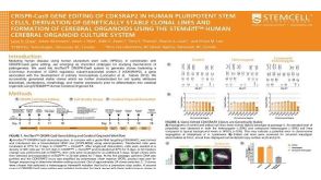 科学海报CRISPR-Cas9 Gene Editing Of CDK5RAP2 In Human Pluripotent Stem Cells, Derivation Of Genetically Stable Clonal Lines And Formation Of Cerebral Organoids
 沪公网安备31010102008431号