Yeo C et al. (SEP 2009)
Regenerative Medicine 4 5 689--696
Ficoll-Paque™ versus Lymphoprep™: a comparative study of two density gradient media for therapeutic bone marrow mononuclear cell preparations
AIMS Contradictory outcomes from recent clinical trials investigating the transplantation of autologous bone marrow mononuclear cell (BM-MNC) fraction containing stem/progenitor cells to damaged myocardium,following acute myocardial infarction,may be,in part,due to the different cell isolation protocols used. We compared total BM-MNC numbers and its cellular subsets obtained following isolation using Ficoll-Paque and Lymphoprep - two different density gradient media used in the clinical trials. MATERIALS & METHODS Bone marrow samples were taken from patients entered into the REGENERATE-IHD clinical trial after 5 days of subcutaneous granulocyte colony-stimulating factor injections. Each sample was divided equally for BM-MNC isolation using Ficoll-Paque and Lymphoprep,keeping all other procedural steps constant. Isolated fractions were characterized for hematopoietic stem cells,endothelial progenitor cells,T lymphocytes,B lymphocytes and NK cells using cell surface markers CD34(+),CD133(+)VEGFR2(+),CD45(+)CD3(+),CD45(+)CD19(+) and CD45(+)CD16(+)CD56(+),respectively. There were no significant differences in the absolute numbers and percentage cell recovery of various mononuclear cell types recovered following separation using either density gradient media. Cell viability and the proportion of various cell phenotypes investigated were similar between the two media. They were also equally efficient in excluding unwanted red blood cells,granulocytes and platelets from the final cell products. CONCLUSION We demonstrated that the composition and quantity of cell types found within therapeutic BM-MNC preparations for use in clinical trials of cardiac stem cell transplantation are not influenced by the type of density gradient media used when comparing Ficoll-Paque and Lymphoprep.
View Publication
产品类型:
产品号#:
07801
07811
07851
07861
18060
18061
产品名:
Lymphoprep™
Lymphoprep™
Lymphoprep™
Lymphoprep™
Qin J et al. (NOV 2016)
Scientific reports 6 37388
Connexin 32-mediated cell-cell communication is essential for hepatic differentiation from human embryonic stem cells.
Gap junction-mediated cell-cell interactions are highly conserved and play essential roles in cell survival,proliferation,differentiation and patterning. We report that Connexin 32 (Cx32)-mediated gap junctional intercellular communication (GJIC) is necessary for human embryonic stem cell-derived hepatocytes (hESC-Heps) during step-wise hepatic lineage restriction and maturation. Vitamin K2,previously shown to promote Cx32 expression in mature hepatocytes,up-regulated Cx32 expression and GJIC activation during hepatic differentiation and maturation,resulting in significant increases of hepatic markers expression and hepatocyte functions. In contrast,negative Cx32 regulator 2-aminoethoxydiphenyl borate blocked hESC-to-hepatocyte maturation and muted hepatocyte functions through disruption of GJIC activities. Dynamic gap junction organization and internalization are phosphorylation-dependent and the p38 mitogen-activated protein kinases pathway (MAPK) can negatively regulate Cxs through phosphorylation-dependent degradation of Cxs. We found that p38 MAPK inhibitor SB203580 improved maturation of hESC-Heps correlating with up-regulation of Cx32; by contrast,the p38 MAPK activator,anisomycin,blocked hESC-Heps maturation correlating with down-regulation of Cx32. These results suggested that Cx32 is essential for cell-cell interactions that facilitate driving hESCs through hepatic-lineage maturation. Regulators of both Cx32 and other members of its pathways maybe used as a promising approach on regulating hepatic lineage restriction of pluripotent stem cells and optimizing their functional maturation.
View Publication
D. R. McHugh et al. ( 2018)
PloS one 13 6 e0199573
A G542X cystic fibrosis mouse model for examining nonsense mutation directed therapies.
Nonsense mutations are present in 10{\%} of patients with CF,produce a premature termination codon in CFTR mRNA causing early termination of translation,and lead to lack of CFTR function. There are no currently available animal models which contain a nonsense mutation in the endogenous Cftr locus that can be utilized to test nonsense mutation therapies. In this study,we create a CF mouse model carrying the G542X nonsense mutation in Cftr using CRISPR/Cas9 gene editing. The G542X mouse model has reduced Cftr mRNA levels,demonstrates absence of CFTR function,and displays characteristic manifestations of CF mice such as reduced growth and intestinal obstruction. Importantly,CFTR restoration is observed in G542X intestinal organoids treated with G418,an aminoglycoside with translational readthrough capabilities. The G542X mouse model provides an invaluable resource for the identification of potential therapies of CF nonsense mutations as well as the assessment of in vivo effectiveness of these potential therapies targeting nonsense mutations.
View Publication
产品类型:
产品号#:
06005
产品名:
IntestiCult™ 类器官生长培养基 (小鼠)
Zhang J et al. (FEB 2007)
The Journal of clinical investigation 117 2 473--81
Primitive hematopoietic cells resist HIV-1 infection via p21.
Hematopoietic stem cells are resistant to HIV-1 infection. Here,we report a novel mechanism by which the cyclin-dependent kinase inhibitor (CKI) p21(Waf1/Cip1/Sdi1) (p21),a known regulator of stem cell pool size,restricts HIV-1 infection of primitive hematopoietic cells. Modifying p21 expression altered HIV-1 infection prior to changes in cell cycling and was selective for p21 since silencing the related CKIs,p27(Kip1) and p18(INK4C),had no effect on HIV-1. We show that p21 blocked viral infection by complexing with HIV-1 integrase and aborting chromosomal integration. A closely related lentivirus with a distinct integrase,SIVmac-251,and the other cell-intrinsic inhibitors of HIV-1,Trim5alpha,PML,Murr1,and IFN-alpha,were unaffected by p21. Therefore,p21 is an endogenous cellular component in stem cells that provides a unique molecular barrier to HIV-1 infection and may explain how these cells remain an uninfected sanctuary" in HIV disease."
View Publication
产品类型:
产品号#:
09850
产品名:
R. O. Bak et al. (FEB 2018)
Nature protocols 13 2 358--376
CRISPR/Cas9 genome editing in human hematopoietic stem cells.
Genome editing via homologous recombination (HR) (gene targeting) in human hematopoietic stem cells (HSCs) has the power to reveal gene-function relationships and potentially transform curative hematological gene and cell therapies. However,there are no comprehensive and reproducible protocols for targeting HSCs for HR. Herein,we provide a detailed protocol for the production,enrichment,and in vitro and in vivo analyses of HR-targeted HSCs by combining CRISPR/Cas9 technology with the use of rAAV6 and flow cytometry. Using this protocol,researchers can introduce single-nucleotide changes into the genome or longer gene cassettes with the precision of genome editing. Along with our troubleshooting and optimization guidelines,researchers can use this protocol to streamline HSC genome editing at any locus of interest. The in vitro HSC-targeting protocol and analyses can be completed in 3 weeks,and the long-term in vivo HSC engraftment analyses in immunodeficient mice can be achieved in 16 weeks. This protocol enables manipulation of genes for investigation of gene functions during hematopoiesis,as well as for the correction of genetic mutations in HSC transplantation-based therapies for diseases such as sickle cell disease,$\beta$-thalassemia,and primary immunodeficiencies.
View Publication
Rebel VI et al. (NOV 2002)
Proceedings of the National Academy of Sciences of the United States of America 99 23 14789--94
Distinct roles for CREB-binding protein and p300 in hematopoietic stem cell self-renewal.
Hematopoietic stem cells (HSC) are tightly regulated through,as yet,undefined mechanisms that balance self-renewal and differentiation. We have identified a role for the transcriptional coactivators CREB-binding protein (CBP) and p300 in such HSC fate decisions. A full dose of CBP,but not p300,is crucial for HSC self-renewal. Conversely,p300,but not CBP,is essential for proper hematopoietic differentiation. Furthermore,in chimeric mice,hematologic malignancies emerged from both CBP(-/-) and p300(-/-) cell populations. Thus,CBP and p300 play essential but distinct roles in maintaining normal hematopoiesis,and,in mice,both are required for preventing hematologic tumorigenesis.
View Publication
产品类型:
产品号#:
06902
06952
00321
00322
00323
00324
00325
产品名:
Lidonnici MR et al. (MAY 2008)
Blood 111 9 4771--9
Requirement of c-Myb for p210(BCR/ABL)-dependent transformation of hematopoietic progenitors and leukemogenesis.
The c-Myb gene encodes a transcription factor required for proliferation and survival of normal myeloid progenitors and leukemic blast cells. Targeting of c-Myb by antisense oligodeoxynucleotides has suggested that myeloid leukemia blasts (including chronic myelogenous leukemia [CML]-blast crisis cells) rely on c-Myb expression more than normal progenitors,but a genetic approach to assess the requirement of c-Myb by p210(BCR/ABL)-transformed hematopoietic progenitors has not been taken. We show here that loss of a c-Myb allele had modest effects (20%-28% decrease) on colony formation of nontransduced progenitors,while the effect on p210(BCR/ABL)-expressing Lin(-) Sca-1(+) and Lin(-) Sca-1(+)Kit(+) cells was more pronounced (50%-80% decrease). Using a model of CML-blast crisis,mice (n = 14) injected with p210(BCR/ABL)-transduced p53(-/-)c-Myb(w/w) marrow cells developed leukemia rapidly and had a median survival of 26 days,while only 67% of mice (n = 12) injected with p210(BCR/ABL)-transduced p53(-/-)c-Myb(w/d) marrow cells died of leukemia with a median survival of 96 days. p210(BCR/ABL)-transduced c-Myb(w/w) and c-Myb(w/d) marrow progenitors expressed similar levels of the c-Myb-regulated genes c-Myc and cyclin B1,while those of Bcl-2 were reduced. However,ectopic Bcl-2 expression did not enhance colony formation of p210(BCR/ABL)-transduced c-Myb(w/d) Lin(-)Sca-1(+)Kit(+) cells. Together,these studies support the requirement of c-Myb for p210(BCR/ABL)-dependent leukemogenesis.
View Publication
产品类型:
产品号#:
04230
产品名:
MethoCult™ H4230
Sugii S et al. (FEB 2010)
Proceedings of the National Academy of Sciences of the United States of America 107 8 3558--63
Human and mouse adipose-derived cells support feeder-independent induction of pluripotent stem cells.
Although adipose tissue is an expandable and readily attainable source of proliferating,multipotent stem cells,its potential for use in regenerative medicine has not been extensively explored. Here we report that adult human and mouse adipose-derived stem cells can be reprogrammed to induced pluripotent stem (iPS) cells with substantially higher efficiencies than those reported for human and mouse fibroblasts. Unexpectedly,both human and mouse iPS cells can be obtained in feeder-free conditions. We discovered that adipose-derived stem cells intrinsically express high levels of pluripotency factors such as basic FGF,TGFbeta,fibronectin,and vitronectin and can serve as feeders for both autologous and heterologous pluripotent cells. These results demonstrate a great potential for adipose-derived cells in regenerative therapeutics and as a model for studying the molecular mechanisms of feeder-free iPS generation and maintenance.
View Publication

 EasySep™小鼠TIL(CD45)正选试剂盒


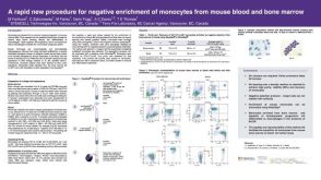 科学海报Procedure for Negative Enrichment of Monocytes from Mouse Blood and Bone Marrow
 产品手册SmartDish™ and STEMgrid™-6 Meniscus-Free Cultureware for More Accurate Counting of Hematopoietic Colonies
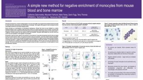 科学海报Method for Negative Enrichment of Monocytes from Mouse Blood and Bone Marrow

 沪公网安备31010102008431号