Daynac M et al. (DEC 2014)
STEM CELLS 32 12 3257--3265
TGFβ Lengthens the G1 Phase of Stem Cells in Aged Mouse Brain
Neurogenesis decreases during aging causing a progressive cognitive decline but it is still controversial whether proliferation defects in neurogenic niches result from a loss of neural stem cells or from an impairment of their progression through the cell cycle. Using an accurate fluorescence-activated cell sorting technique,we show that the pool of neural stem cells is maintained in the subventricular zone of middle-aged mice while they have a reduced proliferative potential eventually leading to the subsequent decrease of their progeny. In addition,we demonstrate that the G1 phase is lengthened during aging specifically in activated stem cells,but not in transit-amplifying cells,and directly impacts on neurogenesis. Finally,we report that inhibition of TGFβ signaling restores cell cycle progression defects in stem cells. Our data highlight the significance of cell cycle dysregulation in stem cells in the aged brain and provide an attractive foundation for the development of anti-TGFβ regenerative therapies based on stimulating endogenous neural stem cells.
View Publication
产品类型:
产品号#:
05700
05701
05702
产品名:
NeuroCult™ 基础培养基(小鼠和大鼠)
NeuroCult™ 扩增添加物(小鼠和大鼠)
NeuroCult™扩增试剂盒(小鼠和大鼠)
W. Yang et al. (Aug 2025)
Cancers 17 17
A Polyomavirus-Positive Merkel Cell Carcinoma Mouse Model Supports a Unified Origin for Somatic and Germ Cell Cancers
Cancer research has long focused on mutations in normal body cells,but this approach has not produced major breakthroughs for most cancers. Our study explores a different concept that some aggressive cancers may actually arise from early reproductive cells called primordial germ cells,which normally develop into eggs and sperm. We created a new experimental model showing how a virus can transform human primordial germ cell-like cells into virus-positive Merkel cell carcinoma,a rare but deadly skin cancer. This model shows that cancers can emerge through changes in developmental states rather than relying solely on genetic mutations. By linking cancer development to early germ cells,our findings suggest a unifying explanation for both germ cell cancers and body cancers. This new perspective may guide more effective approaches to study,diagnose,and treat cancer by focusing on early human development rather than only DNA mutations and later developmental stages.
View Publication
产品类型:
产品号#:
100-0483
100-0484
产品名:
Hausser Scientificᵀᴹ 明线血球计数板
ReLeSR™
Khalfallah O et al. (JUL 2009)
Stem cells (Dayton,Ohio) 27 7 1529--37
Dax-1 knockdown in mouse embryonic stem cells induces loss of pluripotency and multilineage differentiation.
Dax-1 (Nr0b1) is an orphan member of the nuclear hormone receptor superfamily that has a key role in adrenogonadal development and function. Recent studies have also implicated Dax-1 in the transcriptional network controlling embryonic stem (ES) cell pluripotency. Here,we show that Dax-1 expression is affected by differentiating treatments and pharmacological activation of beta-catenin-dependent transcription in mouse ES cells. Furthermore,Dax-1 knockdown induced upregulation of multilineage differentiation markers,and produced enhanced differentiation and defects in ES viability and proliferation. Through RNA interference and transcriptome analysis,we have identified genes regulated by Dax-1 in mouse ES cells at 24 and 48 hours after knockdown. Strikingly,the great majority of these genes are upregulated,showing that the prevalent function of Dax-1 is to act as a transcriptional repressor in mouse ES cells,as confirmed by experiments using the Gal4 system. Genes involved in tissue differentiation and control of proliferation are significantly enriched among Dax-1-regulated transcripts. These data show that Dax-1 is an essential element in the molecular circuit involved in the maintenance of ES cell pluripotency and have implications for the understanding of stem cell function in both physiological (adrenal gland) and clinical (Ewing tumors) settings where Dax-1 plays a pivotal role in development and pathogenesis,respectively.
View Publication
产品类型:
产品号#:
06902
06952
00321
00322
00323
00324
00325
产品名:
J.-H. Kim et al. (mar 2020)
International journal of molecular sciences 21 6
Thymosin $\beta$4-Enhancing Therapeutic Efficacy of Human Adipose-Derived Stem Cells in Mouse Ischemic Hindlimb Model.
Thymosin $\beta$4 (T$\beta$4) is a G-actin sequestering protein that contributes to diverse cellular activities,such as migration and angiogenesis. In this study,the beneficial effects of combined cell therapy with T$\beta$4 and human adipose-derived stem cells (hASCs) in a mouse ischemic hindlimb model were investigated. We observed that exogenous treatment with T$\beta$4 enhanced endogenous TMSB4X mRNA expression and promoted morphological changes (increased cell length) in hASCs. Interestingly,T$\beta$4 induced the active state of hASCs by up-regulating intracellular signaling pathways including the PI3K/AKT/mTOR and MAPK/ERK pathways. Treatment with T$\beta$4 significantly increased cell migration and sprouting from microbeads. Moreover,additional treatment with T$\beta$4 promoted the endothelial differentiation potential of hASCs by up-regulating various angiogenic genes. To evaluate the in vivo effects of the T$\beta$4-hASCs combination on vessel recruitment,dorsal window chambers were transplanted,and the co-treated mice were found to have a significantly increased number of microvessel branches. Transplantation of hASCs in combination with T$\beta$4 was found to improve blood flow and attenuate limb or foot loss post-ischemia compared to transplantation with hASCs alone. Taken together,the therapeutic application of hASCs combined with T$\beta$4 could be effective in enhancing endothelial differentiation and vascularization for treating hindlimb ischemia.
View Publication
Heterotopically transplanted CVO neural stem cells generate neurons and migrate with SVZ cells in the adult mouse brain.
Production of new neurons throughout adulthood has been well characterized in two brain regions,the subventricular zone (SVZ) of the anterolateral ventricle and the subgranular zone (SGZ) of the hippocampus. The neurons produced from these regions arise from neural stem cells (NSCs) found in highly regulated stem cell niches. We recently showed that midline structures called circumventricular organs (CVOs) also contain NSCs capable of neurogenesis and/or astrogliogenesis in vitro and in situ (Bennett et al.). The present study demonstrates that NSCs derived from two astrogliogenic CVOs,the median eminence and organum vasculosum of the lamina terminalis of the nestin-GFP mouse,possess the potential to integrate into the SVZ and differentiate into cells with a neuronal phenotype. These NSCs,following expansion and BrdU-labeling in culture and heterotopic transplantation into a region proximal to the SVZ in adult mice,migrate caudally to the SVZ and express early neuronal markers (TUC-4,PSA-NCAM) as they migrate along the rostral migratory stream. CVO-derived BrdU(+) cells ultimately reach the olfactory bulb where they express early (PSA-NCAM) and mature (NeuN) neuronal markers. Collectively,these data suggest that although NSCs derived from the ME and OVLT CVOs are astrogliogenic in situ,they produce cells phenotypic of neurons in vivo when placed in a neurogenic environment. These findings may have implications for neural repair in the adult brain.
View Publication
产品类型:
产品号#:
05700
05701
05702
产品名:
NeuroCult™ 基础培养基(小鼠和大鼠)
NeuroCult™ 扩增添加物(小鼠和大鼠)
NeuroCult™扩增试剂盒(小鼠和大鼠)
Giebel B et al. (MAR 2006)
Blood 107 5 2146--52
Primitive human hematopoietic cells give rise to differentially specified daughter cells upon their initial cell division.
It is often predicted that stem cells divide asymmetrically,creating a daughter cell that maintains the stem-cell capacity,and 1 daughter cell committed to differentiation. While asymmetric stem-cell divisions have been proven to occur in model organisms (eg,in Drosophila),it remains illusive whether primitive hematopoietic cells in mammals actually can divide asymmetrically. In our experiments we have challenged this question and analyzed the developmental capacity of separated offspring of primitive human hematopoietic cells at a single-cell level. We show for the first time that the vast majority of the most primitive,in vitro-detectable human hematopoietic cells give rise to daughter cells adopting different cell fates; 1 inheriting the developmental capacity of the mother cell,and 1 becoming more specified. In contrast,approximately half of the committed progenitor cells studied gave rise to daughter cells,both of which adopted the cell fate of their mother. Although our data are compatible with the model of asymmetric cell division,other mechanisms of cell fate specification are discussed. In addition,we describe a novel human hematopoietic progenitor cell that has the capacity to form natural killer (NK) cells as well as macrophages,but not cells of other myeloid lineages.
View Publication
产品类型:
产品号#:
05150
产品名:
MyeloCult™ H5100
Gori JL et al. (SEP 2012)
Blood 120 13 e35--44
Efficient generation, purification, and expansion of CD34(+) hematopoietic progenitor cells from nonhuman primate-induced pluripotent stem cells.
Induced pluripotent stem cell (iPSC) therapeutics are a promising treatment for genetic and infectious diseases. To assess engraftment,risk of neoplastic formation,and therapeutic benefit in an autologous setting,testing iPSC therapeutics in an appropriate model,such as the pigtail macaque (Macaca nemestrina; Mn),is crucial. Here,we developed a chemically defined,scalable,and reproducible specification protocol with bone morphogenetic protein 4,prostaglandin-E2 (PGE2),and StemRegenin 1 (SR1) for hematopoietic differentiation of Mn iPSCs. Sequential coculture with bone morphogenetic protein 4,PGE2,and SR1 led to robust Mn iPSC hematopoietic progenitor cell formation. The combination of PGE2 and SR1 increased CD34(+)CD38(-)Thy1(+)CD45RA(-)CD49f(+) cell yield by 6-fold. CD34(+)CD38(-)Thy1(+)CD45RA(-)CD49f(+) cells isolated on the basis of CD34 expression and cultured in SR1 expanded 3-fold and maintained this long-term repopulating HSC phenotype. Purified CD34(high) cells exhibited 4-fold greater hematopoietic colony-forming potential compared with unsorted hematopoietic progenitors and had bilineage differentiation potential. On the basis of these studies,we calculated the cell yields that must be achieved at each stage to meet a threshold CD34(+) cell dose that is required for engraftment in the pigtail macaque. Our protocol will support scale-up and testing of iPSC-derived CD34(high) cell therapies in a clinically relevant nonhuman primate model.
View Publication
Miyake N et al. (MAR 2006)
Stem cells (Dayton,Ohio) 24 3 653--61
HOXB4-induced self-renewal of hematopoietic stem cells is significantly enhanced by p21 deficiency.
Enforced expression of the HOXB4 transcription factor and downregulation of p21(Cip1/Waf) (p21) can each independently increase proliferation of murine hematopoietic stem cells (HSCs). We asked whether the increase in HSC self-renewal generated by overexpression of HOXB4 is enhanced in p21-deficient HSCs. HOXB4 was overexpressed in hematopoietic cells from wild-type (wt) and p21-/- mice. Bone marrow (BM) cells were transduced with a retroviral vector expressing HOXB4 together with GFP (MIGB4),or a control vector containing GFP alone (MIG) and maintained in liquid culture for up to 11 days. At day 11 of the expansion culture,the number of primary CFU-GM (colony-forming unit granulocyte-macrophage) colonies and the repopulating ability were significantly increased in MIGB4 p21-/- BM (p21B4) cells compared with MIGB4-transduced wt BM (wtB4) cells. To test proliferation of HSCs in vivo,we performed competitive repopulation experiments and obtained significantly higher long-term engraftment of expanded p21B4 cells compared with wtB4 cells. The 5-day expansion of p21B4 HSCs generated 100-fold higher numbers of competitive repopulating units compared with wtMIG and threefold higher numbers compared with wtB4. The findings demonstrate that increased expression of HOXB4,in combination with suppression of p21 expression,could be a useful strategy for effective and robust expansion of HSCs.
View Publication
产品类型:
产品号#:
03534
产品名:
MethoCult™ GF M3534
Tchernychev B et al. (DEC 2010)
Proceedings of the National Academy of Sciences of the United States of America 107 51 22255--9
Discovery of a CXCR4 agonist pepducin that mobilizes bone marrow hematopoietic cells.
The G protein-coupled receptor (GPCR),chemokine CXC-type receptor 4 (CXCR4),and its ligand,CXCL12,mediate the retention of polymorphonuclear neutrophils (PMNs) and hematopoietic stem and progenitor cells (HSPCs) in the bone marrow. Agents that disrupt CXCL12-mediated chemoattraction of CXCR4-expressing cells mobilize PMNs and HSPCs into the peripheral circulation and are therapeutically useful for HSPC collection before autologous bone marrow transplantation (ABMT). Our aim was to develop unique CXCR4-targeted therapeutics using lipopeptide GPCR modulators called pepducins. A pepducin is a synthetic molecule composed of a peptide derived from the amino acid sequence of one of the intracellular (IC) loops of a target GPCR coupled to a lipid tether. We prepared and screened a small CXCR4-targeted pepducin library and identified several pepducins with in vitro agonist activity,including ATI-2341,whose peptide sequence derives from the first IC loop. ATI-2341 induced CXCR4- and G protein-dependent signaling,receptor internalization,and chemotaxis in CXCR4-expressing cells. It also induced dose-dependent peritoneal recruitment of PMNs when administered i.p. to mice. However,when administered systemically by i.v. bolus,ATI-2341 acted as a functional antagonist and dose-dependently mediated release of PMNs from the bone marrow of both mice and cynomolgus monkeys. ATI-2341-mediated release of granulocyte/macrophage progenitor cells from the bone marrow was confirmed by colony-forming assays. We conclude that ATI-2341 is a potent and efficacious mobilizer of bone marrow PMNs and HSPCs and could represent a previously undescribed therapeutic approach for the recruitment of HSPCs before ABMT.
View Publication
产品类型:
产品号#:
03534
产品名:
MethoCult™ GF M3534
Trowbridge JJ et al. (SEP 2006)
Proceedings of the National Academy of Sciences of the United States of America 103 38 14134--9
Hedgehog modulates cell cycle regulators in stem cells to control hematopoietic regeneration.
The signals that control the regenerative ability of hematopoietic stem cells (HSCs) in response to damage are unknown. Here,we demonstrate that downstream activation of the Hedgehog (Hh) signaling pathway induces cycling and expansion of primitive bone marrow hematopoietic cells under homeostatic conditions and during acute regeneration. However,this effect is at the expense of HSC function,because continued Hh activation during regeneration represses expression of specific cell cycle regulators,leading to HSC exhaustion. In vivo treatment with an inhibitor of the Hh pathway rescues these transcriptional and functional defects in HSCs. Our study establishes Hh signaling as a regulator of the HSC cell cycle machinery that balances hematopoietic homeostasis and regeneration in vivo.
View Publication
产品类型:
产品号#:
03434
03444
产品名:
MethoCult™ GF M3434
MethoCult™ GF M3434
Van Meter MEM et al. (MAY 2007)
Blood 109 9 3945--52
K-RasG12D expression induces hyperproliferation and aberrant signaling in primary hematopoietic stem/progenitor cells.
Defining how cancer-associated mutations perturb signaling networks in stem/progenitor populations that are integral to tumor formation and maintenance is a fundamental problem with biologic and clinical implications. Point mutations in RAS genes contribute to many cancers,including myeloid malignancies. We investigated the effects of an oncogenic Kras(G12D) allele on phosphorylated signaling molecules in primary c-kit(+) lin(-/low) hematopoietic stem/progenitor cells. Comparison of wild-type and Kras(G12D) c-kit(+) lin(-/low) cells shows that K-Ras(G12D) expression causes hyperproliferation in vivo and results in abnormal levels of phosphorylated STAT5,ERK,and S6 under basal and stimulated conditions. Whereas Kras(G12D) cells demonstrate hyperactive signaling after exposure to granulocyte-macrophage colony-stimulating factor,we unexpectedly observe a paradoxical attenuation of ERK and S6 phosphorylation in response to stem cell factor. These studies provide direct biochemical evidence that cancer stem/progenitor cells remodel signaling networks in response to oncogenic stress and demonstrate that multi-parameter flow cytometry can be used to monitor the effects of targeted therapeutics in vivo. This strategy has broad implications for defining the architecture of signaling networks in primary cancer cells and for implementing stem cell-targeted interventions.
View Publication

 EasySep™小鼠TIL(CD45)正选试剂盒


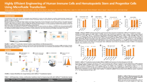 科学海报Highly Efficient Engineering of Human Immune Cells and Hematopoietic Stem and Progenitor Cells Using Microfluidic Transfection
 沪公网安备31010102008431号