Lin S and Talbot P (JAN 2011)
Methods in molecular biology (Clifton,N.J.) 690 31--56
Methods for culturing mouse and human embryonic stem cells
Mouse embryonic stem cells (mESCs) were first derived and cultured almost 30 years ago and ever since have been valuable tools for creating knockout mice and for studying early mammalian development. More recently (1998),human embryonic stem cells (hESCs) have been derived from blastocysts,and numerous methods have evolved to culture hESCs in vitro in both complex and defined media. hESCs are especially important at this time as they could potentially be used to treat degenerative diseases and to access the toxicity of new drugs and environmental chemicals. For both human and mouse ESCs,fibroblast feeder layers are often used at some phase in the culturing protocol. The feeders - often mouse embryonic fibroblasts (mEFs) - provide a substrate that increases plating efficiency,helps maintain pluripotency,and facilitates survival and growth of the stem cells. Various protocols for culturing embryonic stem cells from both species are available with newer trends moving toward feeder-free and serum-free culture. The purpose of this chapter is to provide basic protocol information on the isolation of mouse embryonic fibroblasts and establishment of feeder layers,the culture of mESCs on both mEFs and on gelatin in serum-containing medium,and the culture of hESCs in defined media on both mEFs (hESC culture medium) and Matrigel (mTeSR). These basic protocols are intended for researchers wanting to develop stem cell research in their labs. These protocols have been tested in our laboratory and work well. They can be modified and adapted for any relevant user's particular purpose.
View Publication
产品类型:
产品号#:
05850
05857
05870
05875
85850
85857
85870
85875
产品名:
mTeSR™1
mTeSR™1
Stier S et al. (AUG 2003)
Blood 102 4 1260--6
Ex vivo targeting of p21Cip1/Waf1 permits relative expansion of human hematopoietic stem cells.
Relative quiescence is a defining characteristic of hematopoietic stem cells. Reasoning that inhibitory tone dominates control of stem cell cycling,we previously showed that mice engineered to be deficient in the cyclin-dependent kinase inhibitor,p21Cip1/Waf1 (p21),have an increased stem cell pool under homeostatic conditions. Since p21 was necessary to maintain stem cell quiescence and its absence sufficient to permit increased murine stem cell cycling,we tested whether reduction of p21 alone in human adult-derived stem cells could affect stem cell proliferation. We demonstrate here that interrupting p21 expression ex vivo resulted in expanded stem cell number and in vivo stem cell function compared with control,manipulated cells. Further,we demonstrate full multilineage reconstitution capability in cells where p21 expression was knocked down. Therefore,lifting the brake on cell proliferation by altering cell cycle checkpoints provides an alternative paradigm for increasing hematopoietic stem cell numbers. This approach may be useful for relative ex vivo human stem cell expansion.
View Publication
产品类型:
产品号#:
05150
04435
04445
产品名:
MyeloCult™ H5100
MethoCult™ H4435 Enriched
MethoCult™ H4435 Enriched
Boussaad I et al. (AUG 2011)
Journal of virology 85 15 7710--8
Wild-type measles virus interferes with short-term engraftment of human CD34+ hematopoietic progenitor cells.
Transient lymphopenia is a hallmark of measles virus (MV)-induced immunosuppression. To address to what extent replenishment of the peripheral lymphocyte compartment from bone marrow (BM) progenitor/stem cells might be affected,we analyzed the interaction of wild-type MV with hematopoietic stem and progenitor cells (HS/PCs) and stroma cells in vitro. Infection of human CD34(+) HS/PCs or stroma cells with wild-type MV is highly inefficient yet noncytolytic. It occurs independently of CD150 in stroma cells but also in HS/PCs,where infection is established in CD34(+) CD150(-) and CD34(+) CD150(+) (in humans representing HS/PC oligopotent precursors) subsets. Stroma cells and HS/PCs can mutually transmit MV and may thereby create a possible niche for continuous viral exchange in the BM. Infected lymphocytes homing to this compartment may serve as sources for HS/PC or stroma cell infection,as reflected by highly efficient transmission of MV from both populations in cocultures with MV-infected B or T cells. Though MV exposure does not detectably affect the viability,expansion,and colony-forming activity of either CD150(+) or CD150(-) HS/PCs in vitro,it efficiently interferes with short- but not long-term hematopoietic reconstitution in NOD/SCID mice. Altogether,these findings support the hypothesis that MV accession of the BM compartment by infected lymphocytes may contribute to peripheral blood mononuclear cell lymphopenia at the level of BM suppression.
View Publication
产品类型:
产品号#:
04434
04444
产品名:
MethoCult™ H4434 Classic
MethoCult™ H4434 Classic
Gottschling S et al. (MAR 2007)
Stem cells (Dayton,Ohio) 25 3 798--806
Human mesenchymal stromal cells regulate initial self-renewing divisions of hematopoietic progenitor cells by a beta1-integrin-dependent mechanism.
In previous reports,we have demonstrated that only direct cell-cell contact with stromal cells,such as the murine stromal cell line AFT024,was able to alter the cell division kinetics and self-renewing capacity of hematopoietic progenitor cells (HPC). Because beta(1)-integrins were shown to be crucial for the interaction of HPC with the bone marrow microenvironment,we have studied the role of beta(1)-integrins in the regulation of self-renewing cell divisions. For this purpose,we used primary human mesenchymal stromal (MS) cells as in vitro surrogate niche and monitored the division history and subsequent functional fate of individually plated CD34(+)133(+) cells in the absence or presence of an anti-beta(1)-integrin blocking antibody by time-lapse microscopy and subsequent long-term culture-initiating cell (LTC-IC) assays. beta(1)-Integrin-mediated contact with MS cells significantly increased the proportion of asymmetrically dividing cells and led to a substantial increase of LTC-IC. Provided that beta(1)-integrin-mediated contact was available within the first 72 hours,human MS cells were able to recruit HPC into cell cycle and accelerate their division kinetics without loss of stem cell function. Activation of beta(1)-integrins by ligands alone (e.g.,fibronectin and vascular cell adhesion molecule-1) was not sufficient to alter the cell division symmetry and promote self-renewal of HPC,thus indicating an indirect effect. These results have provided evidence that primary human MS cells are able to induce self-renewing divisions of HPC by a beta(1)-integrin-dependent mechanism.
View Publication
产品类型:
产品号#:
04434
04444
产品名:
MethoCult™ H4434 Classic
MethoCult™ H4434 Classic
Jensen H et al. ( 2017)
Journal of immunology (Baltimore,Md. : 1950) 199 6 1967--1972
Cutting Edge: IL-2-Induced Expression of the Amino Acid Transporters SLC1A5 and CD98 Is a Prerequisite for NKG2D-Mediated Activation of Human NK Cells.
Priming of human NK cells with IL-2 is necessary to render them functionally competent upon NKG2D engagement. We examined the underlying mechanisms that control NKG2D responsiveness in NK cells and found that IL-2 upregulates expression of the amino acid transporters SLC1A5 and CD98. Using specific inhibitors to block SLC1A5 and CD98 function,we found that production of IFN-γ and degranulation by CD56bright and CD56dim NK cells following NKG2D stimulation were dependent on both transporters. IL-2 priming increased the activity of mTORC1,and inhibition of mTORC1 abrogated the ability of the IL-2-primed NK cells to produce IFN-γ in response to NKG2D-mediated stimulation. This study identifies a series of IL-2-induced cellular changes that regulates the NKG2D responsiveness in human NK cells.
View Publication
产品类型:
产品号#:
19055
19055RF
产品名:
EasySep™人NK细胞富集试剂盒
RoboSep™ 人NK细胞富集试剂盒含滤芯吸头
M. A. DeWitt et al. (OCT 2016)
Science translational medicine 8 360 360ra134
Selection-free genome editing of the sickle mutation in human adult hematopoietic stem/progenitor cells.
Genetic diseases of blood cells are prime candidates for treatment through ex vivo gene editing of CD34+ hematopoietic stem/progenitor cells (HSPCs),and a variety of technologies have been proposed to treat these disorders. Sickle cell disease (SCD) is a recessive genetic disorder caused by a single-nucleotide polymorphism in the $\beta$-globin gene (HBB). Sickle hemoglobin damages erythrocytes,causing vasoocclusion,severe pain,progressive organ damage,and premature death. We optimize design and delivery parameters of a ribonucleoprotein (RNP) complex comprising Cas9 protein and unmodified single guide RNA,together with a single-stranded DNA oligonucleotide donor (ssODN),to enable efficient replacement of the SCD mutation in human HSPCs. Corrected HSPCs from SCD patients produced less sickle hemoglobin RNA and protein and correspondingly increased wild-type hemoglobin when differentiated into erythroblasts. When engrafted into immunocompromised mice,ex vivo treated human HSPCs maintain SCD gene edits throughout 16 weeks at a level likely to have clinical benefit. These results demonstrate that an accessible approach combining Cas9 RNP with an ssODN can mediate efficient HSPC genome editing,enables investigator-led exploration of gene editing reagents in primary hematopoietic stem cells,and suggests a path toward the development of new gene editing treatments for SCD and other hematopoietic diseases.
View Publication
产品类型:
产品号#:
02697
02692
09600
09650
09605
09655
产品名:
StemSpan™ CC110
StemSpan™红系扩增添加物 (100X)
StemSpan™ SFEM
StemSpan™ SFEM
StemSpan™ SFEM II
StemSpan™ SFEM II
Bagó et al. (FEB 2017)
Science Translational Medicine 9 375 eaah6510
Tumor-homing cytotoxic human induced neural stem cells for cancer therapy
Engineered neural stem cells (NSCs) are a promising approach to treating glioblastoma (GBM). The ideal NSC drug carrier for clinical use should be easily isolated and autologous to avoid immune rejection. We transdifferentiated (TD) human fibroblasts into tumor-homing early-stage induced NSCs (h-iNSC(TE)),engineered them to express optical reporters and different therapeutic gene products,and assessed the tumor-homing migration and therapeutic efficacy of cytotoxic h-iNSC(TE) in patient-derived GBM models of surgical and nonsurgical disease. Molecular and functional analysis revealed that our single-factor SOX2 TD strategy converted human skin fibroblasts into h-iNSC(TE) that were nestin(+) and expressed pathways associated with tumor-homing migration in 4 days. Time-lapse motion analysis showed that h-iNSC(TE) rapidly migrated to human GBM cells and penetrated human GBM spheroids,a process inhibited by blockade of CXCR4. Serial imaging showed that h-iNSC(TE) delivery of the proapoptotic agent tumor necrosis factor-α-related apoptosis-inducing ligand (TRAIL) reduced the size of solid human GBM xenografts 250-fold in 3 weeks and prolonged median survival from 22 to 49 days. Additionally,h-iNSC(TE) thymidine kinase/ganciclovir enzyme/prodrug therapy (h-iNSC(TE)-TK) reduced the size of patient-derived GBM xenografts 20-fold and extended survival from 32 to 62 days. Mimicking clinical NSC therapy,h-iNSC(TE)-TK therapy delivered into the postoperative surgical resection cavity delayed the regrowth of residual GBMs threefold and prolonged survival from 46 to 60 days. These results suggest that TD of human skin into h-iNSC(TE) is a platform for creating tumor-homing cytotoxic cell therapies for cancer,where the potential to avoid carrier rejection could maximize treatment durability in human trials.
View Publication
Csaszar E et al. (FEB 2012)
Cell stem cell 10 2 218--29
Rapid expansion of human hematopoietic stem cells by automated control of inhibitory feedback signaling.
Clinical hematopoietic transplantation outcomes are strongly correlated with the numbers of cells infused. Anticipated novel therapeutic implementations of hematopoietic stem cells (HSCs) and their derivatives further increase interest in strategies to expand HSCs ex vivo. A fundamental limitation in all HSC-driven culture systems is the rapid generation of differentiating cells and their secreted inhibitory feedback signals. Herein we describe an integrated computational and experimental strategy that enables a tunable reduction in the global levels and impact of paracrine signaling factors in an automated closed-system process by employing a controlled fed-batch media dilution approach. Application of this system to human cord blood cells yielded a rapid (12-day) 11-fold increase of HSCs with self-renewing,multilineage repopulating ability. These results highlight the marked improvements that control of feedback signaling can offer primary stem cell culture and demonstrate a clinically relevant rapid and relatively low culture volume strategy for ex vivo HSC expansion.
View Publication
产品类型:
产品号#:
72342
72344
72352
72354
产品名:
StemRegenin 1
StemRegenin 1
StemRegenin 1(盐酸盐)
StemRegenin 1(盐酸盐)
Bruserud O et al. (JUN 2005)
Journal of cancer research and clinical oncology 131 6 377--84
In vitro culture of human osteosarcoma cell lines: a comparison of functional characteristics for cell lines cultured in medium without and with fetal calf serum.
PURPOSE: Experimental in vitro models including well-characterised cell lines can be used to identify possible new therapeutic targets for the treatment of osteosarcoma. Culture media including inactivated serum is often recommended for in vitro culture of osteosarcoma cells,but the serum component then represents a nonstandardised parameter including a wide range of unidentified mediators. To improve the standardisation we have investigated whether serum-free culture media can be used in experimental in vitro studies of osteosarcoma cell lines. METHODS: The seven osteosarcoma cell lines Cal72,SJSA-1,Saos-2,SK-ES-1,U2OS,143.98.2,and KHOS-32IH were cultured in vitro in various serum-free media and media supplemented with 10% heat-inactivated fetal calf serum (FCS). RESULTS: Although proliferation often was relatively low in serum-free media (X-vivo 10,X-vivo 15,X-vivo 20,Stem Span SFEM),some cell lines (Cal72,KHOS-32IH,Saos-2) showed proliferation comparable with the recommended FCS-containing media even when using serum-free conditions. The optimal serum-free medium then varied between cell lines. We also compared 6 different FCS-containing media (including Stem Span with 10% FCS) and the optimal FCS-containing medium varied between cell lines. However,all cell lines proliferated well in Stem Span with FCS,and this medium was regarded as optimal for four of the lines. FCS could not be replaced by fatty acids or low density lipoprotein when testing the Stem Span medium. The release of a wide range of soluble mediators showed only minor differences when using serum-free and FCS-containing media (including Stem Span with and without FCS),and serum-free Stem Span could also be used for in vitro studies of mitogen-stimulated T cell activation in the presence of accessory osteosarcoma cells. The use of Stem Span with 10% FCS allowed the release of a wide range of chemokines by osteosarcoma cell lines (Cal72,SJSA-1),and the chemokine release profile was very similar to the fibroblast lines Hs27 and HFL1. CONCLUSIONS: Serum-free culture media can be used for in vitro studies of several osteosarcoma cell lines,but the optimal medium varies between cell lines and thus depends on: (i) the cell lines to be investigated/compared; (ii) the functional characteristic that is evaluated (proliferation,cytokine release); and (iii) whether coculture experiments are included.
View Publication

 EasySep™小鼠TIL(CD45)正选试剂盒


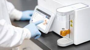 实验方案Gene Editing Human CD34+ Hematopoietic Stem and Progenitor Cells with the CellPore™ Transfection System


 沪公网安备31010102008431号